Spondyloepimetaphyseal Dysplasia, Missouri Type (SEMD-MO): A Comprehensive Disease Characteristics Report
Disease: Spondyloepimetaphyseal Dysplasia, Missouri Type MONDO ID: MONDO:0011198 · OMIM: 602111 · Causal gene: MMP13 (collagenase-3) Report type: Aggregated disease-level synthesis (OMIM, Orphanet, primary literature, model-organism data)
Summary
Spondyloepimetaphyseal dysplasia, Missouri type (SEMD-MO) is an ultra-rare autosomal dominant skeletal dysplasia caused by heterozygous missense variants in MMP13, the gene encoding the interstitial collagenase collagenase-3 (matrix metalloproteinase 13). It is now understood to sit within the Metaphyseal Anadysplasia type 1 (MANDP1) spectrum — a family of MMP13-related conditions unified by early-onset metaphyseal irregularity, mild disproportionate short stature, mild limb deformity (bowing, coxa vara), and — characteristically — transient radiographic changes that improve or resolve with age. Biochemically, affected individuals have normal calcium, phosphate, alkaline phosphatase, and vitamin D, which makes the condition an important radiographic mimic of rickets that must be distinguished by genetic testing rather than metabolic work-up.
The mechanistic core of the disease is well established at the protein level. The founding mutation, p.Phe56Ser (F56S), substitutes an evolutionarily conserved phenylalanine in the propeptide (latency) domain of MMP13. This destabilizes the zymogen, causing intracellular misfolding, premature autoactivation and autodegradation, so that only enzymatically inactive fragments are secreted. The net result is a functional MMP13 deficiency (with a dominant-negative component that also suppresses MMP9). Because MMP13 is the principal collagenase that degrades type II collagen and aggrecan in the hypertrophic zone of the growth plate — acting synergistically with MMP9 — its loss delays the exit of hypertrophic chondrocytes and slows endochondral ossification. This produces the metaphyseal dysplasia. The transient nature of the human disease is faithfully mirrored by Mmp13-null mice, whose growth-plate abnormality worsens until ~5 weeks and completely resolves by 12 weeks, reflecting redundancy from other collagenases (notably MMP14) as the animal matures.
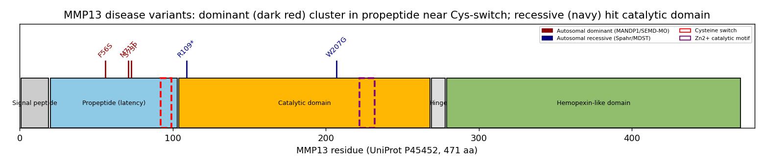
A clear genotype–structure–inheritance correlation emerges from the reported allelic spectrum: dominant SEMD-MO/MANDP1 variants cluster in a tight window of the propeptide near the cysteine switch (F56S, M71T, S73P — residues 56–73, immediately N-terminal to the PRCGVPD/Cys97 latency motif), whereas recessive variants (nonsense R109, and the catalytic-domain W207G that causes the related Metaphyseal Dysplasia, Spahr type) fall in or downstream of the catalytic domain. Prognosis is benign with normal life expectancy and no organ-system involvement beyond the skeleton. There is no disease-specific therapy; management is supportive/orthopedic with genetic counseling. Notably, because MMP13 inhibitors are actively being developed for osteoarthritis (where MMP13 is over-active), such inhibitors would be directionally contraindicated in a disease of MMP13 deficiency*.
Section-by-Section Report
1. Disease Information
Overview. SEMD-MO is a rare heritable skeletal dysplasia affecting the growth and modeling of vertebrae and long bones. It is characterized by mild disproportionate short stature, mild limb deformities (including bowed legs, coxa vara, and genu varum/valgum), and metaphyseal irregularity that is most conspicuous in childhood and tends to improve with skeletal maturation. It is best classified today within the Metaphyseal Anadysplasia type 1 (MANDP1) group of MMP13-related dysplasias.
"Metaphyseal anadysplasia 1, which includes Spondyloepimetaphyseal dysplasia Missouri type, is a rare autosomal dominant skeletal dysplasia characterized by short stature, mild limb deformities, and transient metaphyseal irregularities that typically resolve with age." — Thunström et al. 2026 PMID: 42069302
Key identifiers.
Table (click to expand)
| Resource | Identifier |
|---|---|
| MONDO | MONDO:0011198 |
| OMIM | 602111 (Spondyloepimetaphyseal dysplasia, Missouri type / MANDP1) |
| Gene | MMP13 (HGNC:7159; NCBI Gene 4322; UniProt P45452) |
| Related OMIM | 250400 (Metaphyseal dysplasia, Spahr type — recessive MMP13) |
Synonyms / alternative names. SEMD Missouri type; SEMD(MO); Missouri-type spondyloepimetaphyseal dysplasia; within the broader label Metaphyseal anadysplasia type 1 (MANDP1).
Source of information. This report is derived from aggregated disease-level resources (OMIM, Orphanet) and primary literature describing individual families and patients, together with model-organism (mouse) and in vitro functional studies. It is not derived from EHR/individual-patient registries.
2. Etiology
Primary cause — genetic. SEMD-MO is a monogenic disorder caused by heterozygous pathogenic variants in MMP13. Kennedy et al. (2005) mapped the disease by genome-wide linkage to chromosome 11q14.3–23.2 and identified the causal heterozygous missense change.
"We show that a missense mutation of MMP13 causes the Missouri type of human spondyloepimetaphyseal dysplasia (SEMD(MO)), an autosomal dominant disorder characterized by defective growth and modeling of vertebrae and long bones." — Kennedy et al. 2005 PMID: 16167086
Genetic risk factors. The disease is fully explained by the MMP13 variant; there are no known susceptibility loci or common modifier variants in humans. In mice, HDAC4 genetically modifies Mmp13 dosage (see Sections 4 and 6). No GWAS or polygenic contributions are relevant to this Mendelian condition.
Environmental risk factors. None identified. There is no evidence for toxin, occupational, dietary, radiation, or infectious contribution. Because the radiographic picture mimics rickets, environmental vitamin D deficiency is an important differential rather than a cause; biochemical markers are normal in SEMD-MO (see Section 10).
Protective factors. No genetic or environmental protective factors are described. The condition's natural tendency toward spontaneous radiographic improvement with age is intrinsic (developmental redundancy of collagenases) rather than a modifiable protective exposure.
Gene–environment interactions. None documented. This is a highly penetrant monogenic disorder without a demonstrated GxE component.
3. Phenotypes
SEMD-MO phenotypes are skeletal physical manifestations and radiographic signs; there are no behavioral or biochemical laboratory abnormalities.
Table (click to expand)
| Phenotype | Type | Onset | Severity / course | Suggested HPO term |
|---|---|---|---|---|
| Disproportionate short stature | Physical manifestation | Childhood | Mild; improves relatively with age | HP:0004322 (Short stature) |
| Metaphyseal irregularity / widening | Radiographic sign | Early childhood | Mild–moderate; transient, resolves/improves with age | HP:0000944 (Abnormal metaphyseal morphology) |
| Bowing of long bones (genu varum/valgum) | Physical/radiographic | Childhood | Mild; may need orthopedic correction | HP:0002979 (Bowing of the long bones) |
| Coxa vara | Radiographic sign | Childhood | Mild–moderate | HP:0002812 (Coxa vara) |
| Vertebral (spondylo-) changes / platyspondyly | Radiographic sign | Childhood | Mild | HP:0000926 (Platyspondyly) |
| Epiphyseal involvement | Radiographic sign | Childhood | Mild (mixed epiphyseal–metaphyseal in some MMP13 cases) | HP:0002656 (Epiphyseal dysplasia) |
| Rickets-like metaphyseal changes with normal biochemistry | Radiographic mimic | Childhood | Distinguishing feature | HP:0002748 (Rickets) — radiographic only |
Frequency and progression. The hallmark is transient/self-limited radiographic disease. In the Mmp13-null mouse, growth-plate severity "increased until about 5 weeks and completely resolved by 12 weeks of age" (PMID: 15539485), matching the human observation of improvement with maturation. A recessive nonsense report (R109) noted short stature that can persist beyond childhood*, indicating expressivity varies across the allelic spectrum.
Quality-of-life impact. Generally mild. Short stature and limb deformity may require orthopedic monitoring/intervention, but there is no organ failure, no cognitive involvement, and normal life expectancy — so QoL impact is limited to musculoskeletal function and cosmesis. No disease-specific EQ-5D/SF-36 data exist for this ultra-rare condition.
4. Genetic / Molecular Information
Causal gene. MMP13 (matrix metalloproteinase 13 / collagenase-3), HGNC:7159, NCBI Gene 4322, UniProt P45452 (471 aa), located on chromosome 11q22.2 (linkage interval 11q14.3–23.2).
Pathogenic variants — allelic spectrum. MMP13 variants produce a spectrum spanning dominant and recessive metaphyseal dysplasias:
Table (click to expand)
| Variant (protein) | Domain | Inheritance | Phenotype | Reference |
|---|---|---|---|---|
| p.Phe56Ser (F56S) | Propeptide | Dominant | SEMD Missouri type / MANDP1 | Kennedy 2005 PMID: 16167086 |
| p.Met71Thr (M71T), c.212T>C | Propeptide | Dominant (de novo) | MANDP1 | Song 2019 PMID: 30439533 |
| p.Ser73Pro (S73P), c.217T>C | Propeptide | Dominant | MANDP1, rickets-like | Thunström 2026 PMID: 42069302 |
| p.Arg109* (R109*), c.325C>T | End propeptide / catalytic border | Recessive | Metaphyseal anadysplasia (persistent short stature) | Li 2015 PMID: 24781753 |
| p.Trp207Gly (W207G), c.619T>G | Catalytic (Ca²⁺ region) | Recessive | Metaphyseal dysplasia, Spahr type (OMIM 250400) | Bonafé 2014 PMID: 24648384 |
"we reported the identification of a previously unreported pathogenic heterozygous de novo variant NM_002427.3:c.212T > C/p.Met71Thr in MMP13" — Song et al. 2019 PMID: 30439533
Variant classification and type. The dominant variants are missense (pathogenic/likely pathogenic by ACMG/AMP criteria, given segregation, de novo occurrence, and functional data). The recessive spectrum includes a nonsense (R109*) and a missense (W207G) allele. All are germline; there is no somatic/oncogenic context. Population allele frequencies are effectively absent/ultra-rare in gnomAD, consistent with pathogenicity.
Functional consequences. The dominant propeptide variants act through a dominant-negative loss-of-function mechanism: the misfolded mutant zymogen autoactivates/autodegrades intracellularly and can suppress activity of both MMP13 and its partner MMP9 (see Section 6). The catalytic-domain recessive variant (W207G) directly disrupts a hydrogen bond in the calcium-binding region, abolishing enzyme function; the nonsense R109* abolishes MMP13 activity.
"The predicted non-conservative amino acid substitution, p.Trp207Gly, disrupts a crucial hydrogen bond in the calcium-binding region of the catalytic domain of the matrix metalloproteinase, MMP13." — Bonafé et al. 2014 PMID: 24648384
Modifier genes. HDAC4 is a genetic/epigenetic modifier of Mmp13 dosage in mouse: HDAC4 transcriptionally represses Mmp13, and Hdac4/Mmp13 double knockouts recover growth-plate zone thickness relative to Hdac4-null mice (PMID: 27320207; PMID: 33000215). MMP9 is a functional partner whose activity is co-suppressed by dominant MMP13 mutants. MMP14 partly compensates for MMP13 during bone development (PMID: 42142773).
Epigenetic information. No disease-specific human methylation/histone data. Mechanistically, HDAC4 (a histone deacetylase) represses Mmp13 transcription, tying chromatin-level regulation to MMP13 dosage and growth-plate architecture (mouse evidence).
Chromosomal abnormalities. None; SEMD-MO is a single-gene point-mutation disorder, not a copy-number/structural disorder.
5. Environmental Information
- Environmental factors: None causally implicated (no toxins, radiation, pollution, or occupational exposures).
- Lifestyle factors: None. Diet/nutrition do not cause the disease, though nutritional rickets is the key clinical mimic to exclude.
- Infectious agents: Not applicable. SEMD-MO is a non-infectious Mendelian dysplasia.
6. Mechanism / Pathophysiology
Ordered causal chain (initiating lesion → clinical manifestation):
- A heterozygous missense variant in the MMP13 propeptide (e.g., F56S, M71T, S73P) substitutes a conserved residue near the cysteine switch → leads to destabilization of the pro-MMP13 zymogen fold.
- Destabilization results in intracellular misfolding of pro-MMP13 → leads to premature intracellular autoactivation and autodegradation (demonstrated in HEK293 expression: only inactive small fragments are secreted).
- Autodegradation results in a net functional MMP13 deficiency, with a dominant-negative effect that also suppresses MMP9 activity (inferred from the more severe dominant phenotype vs. recessive missense alleles).
- MMP13 deficiency impairs collagenolysis of the growth-plate extracellular matrix — chiefly type II collagen and aggrecan — normally degraded by MMP13 acting synergistically with MMP9 in terminal hypertrophic chondrocytes.
- Impaired ECM remodeling delays the exit of hypertrophic chondrocytes from the growth plate and slows vascular invasion / endochondral ossification (demonstrated in Mmp13-null mice: normal differentiation but delayed exit, increased trabecular bone).
- Delayed ossification and disordered metaphyseal modeling result in the clinical/radiographic phenotype: metaphyseal irregularity, mild short stature, limb bowing, coxa vara.
- As the child/animal matures, redundant collagenases (e.g., MMP14) partially compensate → leads to the characteristic transient/self-resolving course (inferred from mouse double-knockout data).
Branch (recessive arm): Catalytic-domain variants (W207G) or nonsense alleles (R109*) directly abolish enzymatic activity rather than acting via propeptide misfolding → result in the related Metaphyseal dysplasia, Spahr type and recessive metaphyseal anadysplasia, with short stature that may persist beyond childhood.
Protein dysfunction. The founding mechanism is a zymogen-folding defect in the latency (propeptide) domain.
"Expression of wild-type and mutant MMP13s in human embryonic kidney cells confirmed abnormal intracellular autoactivation and autodegradation of F56S MMP13 such that only enzymatically inactive, small fragments were secreted. Thus, the F56S mutation results in deficiency of MMP13, which leads to the human skeletal developmental anomaly of SEMD(MO)." — Kennedy 2005 PMID: 16167086
Cellular processes and substrates. MMP13 is expressed in terminal hypertrophic chondrocytes, periosteal cells, and osteoblasts; its substrates in the growth plate are type II collagen and aggrecan.
"Chondrocytes differentiated normally but their exit from the growth plate was delayed. The severity of the Mmp13-null growth plate phenotype increased until about 5 weeks and completely resolved by 12 weeks of age." — Stickens et al. 2004 PMID: 15539485
"We found that degradation of cartilage collagen and aggrecan is a coordinated process in which MMP13 works synergistically with MMP9." — Stickens et al. 2004 PMID: 15539485
Upstream regulatory context. MMP13 is a downstream effector of the RUNX2-driven hypertrophic chondrocyte program, co-regulated with COL10A1. Multiple signaling modules converge on MMP13 in chondrocyte hypertrophy: Wnt/β-catenin, TGF-β/BMP, Indian hedgehog–PTHrP, HIF-1α/HIF-2α, Notch, and NF-κB; epigenetically, HDAC4 represses it.
"articular chondrocytes shift from a stable extracellular matrix (ECM)-maintaining state toward a hypertrophy-like program with increased collagen type X alpha 1 chain (COL10A1), runt-related transcription factor 2 (RUNX2), and matrix metalloproteinase 13 (MMP-13), promoting ECM degradation, calcification, and pro-angiogenic remodeling" — Ha et al. 2026 PMID: 41967814
"The nobly increased expression of matrix metallopeptidase 13 (MMP13) and type X collagen (Col10a1) as Runx2 downstream genes contributed to the hypertrophic differentiation of chondrocytes" — Lin et al. 2022 PMID: 36232582
HDAC4 dosage control (mouse). HDAC4 sits upstream and tunes MMP13:
"Hdac4(-/-)/Mmp13(-/-) double knockout mice are significantly heavier and larger than Hdac4(-/-) mice, they survive longer, and they recover the thickness of their growth plate zones." — Nakatani et al. 2016 PMID: 27320207
Suggested ontology terms. GO:0030574 (collagen catabolic process); GO:0001958 (endochondral ossification); GO:0002062 (chondrocyte differentiation); GO:0003417 (growth plate cartilage development); GO:0004222 (metalloendopeptidase activity). Cell types: CL:0000743 (hypertrophic chondrocyte); CL:0000138 (chondrocyte); CL:0000062 (osteoblast).
{{figure:mmp13_variant_map.png|caption=Reported MMP13 disease variants mapped onto UniProt P45452 domain architecture. Autosomal dominant SEMD-MO/MANDP1 variants (F56S, M71T, S73P) cluster in a narrow window of the propeptide/latency domain immediately N-terminal to the cysteine switch (PRCGVPD, Cys97), whereas recessive variants (R109*, W207G) fall in or downstream of the catalytic domain.}}
7. Anatomical Structures Affected
- Organ level (primary): The skeletal system — specifically the growth plates (physes) and metaphyses of long bones and the vertebrae (UBERON:0002515 metaphysis; UBERON:0006255 growth plate; UBERON:0001474 bone element; UBERON:0001130 vertebral column). Sites include femur/tibia (limb bowing, coxa vara) and spine (spondylo- changes).
- Secondary involvement: None — no cardiovascular, neurological, renal, hepatic, or other organ involvement. This is a skeleton-restricted disorder.
- Tissue/cell level: Cartilage (connective tissue) of the growth plate; the targeted cells are hypertrophic chondrocytes (CL:0000743), with contributions from osteoblasts (CL:0000062) and periosteal cells. The affected extracellular matrix comprises type II collagen and aggrecan.
- Subcellular level: The extracellular matrix (GO:0031012) is the functional site of MMP13 action; the pathogenic misfolding/autodegradation occurs within the secretory pathway / endoplasmic reticulum–Golgi (GO:0005788 ER lumen; GO:0005615 extracellular space for the mature enzyme).
- Localization / lateralization: Bilateral and generally symmetric involvement of the appendicular and axial skeleton, as expected for a systemic germline enzyme deficiency.
8. Temporal Development
- Onset: Pediatric / early childhood, congenital predisposition with radiographic manifestation as the growth plates are active. Onset is insidious/chronic (a developmental modeling defect, not an acute event).
- Progression: Radiographic severity typically worsens in early childhood then improves with skeletal maturation — a distinctive transient, self-resolving course. The mouse model shows the phenotype peaking ~5 weeks and resolving by 12 weeks (PMID: 15539485). Some recessive alleles (R109*) are associated with short stature persisting beyond childhood, indicating variable duration across the spectrum.
- Disease course pattern: Non-progressive to improving; not relapsing-remitting, not episodic. Chronic/lifelong short stature and residual deformity may remain, but the metaphyseal changes tend to remit.
- Critical periods: The window of active endochondral growth (infancy through puberty) is when the phenotype is expressed and when orthopedic intervention would be most relevant.
9. Inheritance and Population
- Inheritance pattern: Autosomal dominant for SEMD-MO/MANDP1 (heterozygous propeptide missense variants). The broader MMP13 spectrum also includes autosomal recessive forms (Spahr type; recessive metaphyseal anadysplasia).
- Penetrance / expressivity: High penetrance for the dominant variants; variable expressivity in severity and persistence of short stature. De novo occurrence is documented (M71T).
- Genetic anticipation / mosaicism / founder effects: No evidence of anticipation (not a repeat-expansion disorder). No documented founder effect; the reported dominant variants arose independently in different families, with a suggested mutational hotspot in exon 2 (propeptide).
"located within the same MMP13 domain as previously reported patients. This report expands the genotypic spectrum of Metaphyseal anadysplasia 1 and suggests a putative mutational hotspot in exon 2." — Thunström 2026 PMID: 42069302
- Consanguinity: Relevant to the recessive forms (e.g., affected sib-pairs, homozygous R109*), not to the dominant SEMD-MO.
- Carrier frequency: Not applicable to the dominant disorder; recessive MMP13 alleles are ultra-rare in gnomAD.
- Epidemiology: Ultra-rare — only a handful of families/patients reported worldwide; precise prevalence/incidence figures are not established (well under Orphanet's <1/1,000,000 category). No sex predilection expected (autosomal). No geographic clustering established.
10. Diagnostics
Clinical/laboratory tests. Routine bone biochemistry is normal, which is diagnostically important because the radiographs mimic rickets.
"Biochemical tests including calcium, phosphate, alkaline phosphatase, and vitamin D levels were all within normal ranges." — Kolkiran 2025 PMID: 40514045
Imaging (primary diagnostic modality). Skeletal survey / plain radiographs show metaphyseal irregularity and widening, epiphyseal involvement in some cases, coxa vara, long-bone bowing, and mild spondylar (vertebral) changes. The transient improvement with age on serial imaging is characteristic.
Genetic testing (definitive). - Single-gene / targeted MMP13 sequencing confirms diagnosis when the phenotype is suggestive. - Whole-exome sequencing (WES) has been the discovery tool for several variants (e.g., recessive R109*), and is useful when the phenotype is nonspecific or overlaps other skeletal dysplasias. - Skeletal dysplasia gene panels including MMP13 (and MMP9) are appropriate. - WGS, CMA, karyotyping, FISH, mtDNA and repeat-expansion testing are not indicated (this is a point-mutation single-gene disorder).
Clinical criteria / differential diagnosis. No formal consensus criteria exist; diagnosis rests on the radiographic pattern + normal biochemistry + MMP13 genotype. Key differentials: - Nutritional / hypophosphatemic rickets (distinguished by abnormal Ca/PO₄/ALP/vitamin D or PHEX etc.). - Metaphyseal dysplasia, Spahr type (recessive MMP13, catalytic-domain). - Metaphyseal anadysplasia type 2 (MMP9). - Other spondylometaphyseal/spondyloepimetaphyseal dysplasias (e.g., COL2A1, COL10A1, TRPV4 related).
Screening. No population/newborn screening exists or is warranted given rarity and benign course; cascade genetic testing of at-risk relatives is appropriate once a familial variant is known.
11. Outcome / Prognosis
- Survival / mortality: Normal life expectancy; the disease is not life-limiting and does not involve vital organs.
- Morbidity / function: Limited to musculoskeletal impact — mild short stature and limb deformity, potentially requiring orthopedic follow-up. No cognitive, cardiac, respiratory, or renal morbidity.
- Disease course: Benign, with radiographic improvement/resolution over time in the dominant form; residual short stature is generally mild. Complications are mechanical (deformity, gait), not systemic.
- Recovery potential: High for the radiographic metaphyseal changes (self-limited); deformities may need correction.
- Prognostic factors: Genotype (dominant propeptide vs. recessive catalytic/nonsense) predicts persistence — recessive alleles may be associated with more persistent short stature. No molecular prognostic biomarkers are established.
12. Treatment
There is no disease-specific or disease-modifying pharmacotherapy for SEMD-MO. Management is supportive and orthopedic.
- Pharmacotherapy: None targeted. Importantly, MMP13 inhibitors — actively developed for osteoarthritis, where MMP13 is over-active — would be directionally contraindicated in a disease of MMP13 deficiency. Selective non-zinc-binding MMP13 inhibitors (targeting the S1' pocket; Cuffaro 2023, PMID 37446276) and MMP13-selective siRNA conjugates exist as OA candidates and illustrate this contraindication.
"Using siRNA sequences targeting MMP13, a key driver of arthritis-re[lated cartilage degradation]" — Colazo et al. 2023 PMID: 37333210
- Advanced therapeutics (gene/cell/RNA): None clinically available; conceptually, gene-correction or read-through/allele-specific strategies could be explored, but none are in trials for this ultra-rare disorder.
- Surgical / interventional: Orthopedic correction of limb deformity (e.g., osteotomy for severe bowing or coxa vara) as clinically indicated; guided growth procedures where appropriate.
- Supportive / rehabilitative: Physical therapy, gait and activity support, monitoring of growth and joints.
- Experimental / trials: No disease-specific ClinicalTrials.gov entries identified.
Suggested NCIT terms: NCIT:C15329 (Surgery/Surgical Procedure), NCIT:C15682 (Physical Therapy), NCIT:C15277 (Supportive Care), NCIT:C15311 (Genetic Counseling).
13. Prevention
- Primary prevention: Not applicable to a germline monogenic disease; no environmental risk to modify.
- Secondary prevention: Early radiographic recognition and avoidance of unnecessary rickets treatment (the key clinical pitfall) via biochemistry + genetic confirmation.
- Tertiary prevention: Orthopedic surveillance to prevent/manage deformity-related complications.
- Genetic counseling: Central to management — autosomal dominant recurrence risk of 50% for offspring of an affected parent (with counseling on variable expressivity and de novo occurrence); for recessive MMP13 families, standard AR counseling and carrier testing. Prenatal / preimplantation genetic testing is technically feasible once the familial variant is known, though rarely pursued given the benign prognosis.
- Immunization / public health / prophylaxis: Not applicable.
14. Other Species / Natural Disease
- Taxonomy / orthologs: MMP13 is conserved across mammals; the principal experimental ortholog is mouse Mmp13 (Mus musculus, NCBI Taxon 10090). Structural work has been performed on recombinant mouse collagenase-3 (PMID: 10525409).
- Natural disease in other species: No well-characterized naturally occurring MMP13 metaphyseal dysplasia has been catalogued in companion animals or wildlife in the sources reviewed (OMIA entry not established here).
- Comparative biology: The transient growth-plate phenotype and collagen II/aggrecan substrate biology are conserved between mouse and human, making mouse an evolutionarily faithful model of the mechanism. MMP14 compensation for MMP13 in bone is also conserved (PMID: 42142773).
- Transmission: Not applicable (non-infectious, heritable).
15. Model Organisms
- Model type: Mammalian (mouse) is the primary system; in vitro HEK293 expression was used to demonstrate the protein-folding defect.
- Specific models:
- Mmp13-null (knockout) mouse — recapitulates the human disease closely: normal chondrocyte differentiation but delayed hypertrophic chondrocyte exit, increased trabecular bone, and a transient growth-plate phenotype that resolves by 12 weeks (PMID: 15539485). This is the best phenotypic match to SEMD-MO's self-resolving course.
- Hdac4-null and Hdac4/Mmp13 double-knockout mice — establish MMP13 as genetically downstream of HDAC4 and show MMP13 dosage tunes growth-plate zone thickness (PMID: 27320207; PMID: 33000215).
- Mmp13/Mmp14 double-knockout mice — reveal MMP14 partially compensates for MMP13 in endochondral ossification, informing the transient nature of the human phenotype (PMID: 42142773).
- In vitro / cellular: HEK293 expression of WT vs. F56S MMP13 demonstrated intracellular autoactivation/autodegradation (PMID: 16167086).
- Phenotype recapitulation: High for the growth-plate/metaphyseal and transient-course features; the knockout models the deficiency endpoint shared by the human dominant-negative mechanism.
- Limitations: The mouse knockout models total loss of function and does not fully capture the dominant-negative intracellular misfolding of the human propeptide missense alleles or the human vertebral (spondylo-) phenotype; species differences in growth-plate timing exist.
- Resources: MGI (mouse Mmp13), and standard model-organism repositories.
Mechanistic Model / Interpretation
   MMP13 propeptide missense (F56S / M71T / S73P; near cysteine switch, Cys97)
       │  (destabilizes zymogen fold)
       ▼
Intracellular misfolding of pro-MMP13
       │
       ▼
   Premature autoactivation + autodegradation  ──►  only inactive fragments secreted
       │
       ▼
   Functional MMP13 deficiency  (dominant-negative; also suppresses MMP9)
       │
       ▼
   Impaired collagenolysis of growth-plate ECM (type II collagen + aggrecan;
       │   MMP13–MMP9 synergy)
       ▼
   Delayed hypertrophic chondrocyte exit + slowed endochondral ossification
       │
       ▼
   Metaphyseal irregularity, mild short stature, bowing, coxa vara
       │
       ▼
   MMP14 (and other) compensation with maturation  ──►  transient / self-resolving

   ── Recessive branch ──
   Catalytic-domain W207G (Ca²⁺ region) / nonsense R109*  ──►  direct loss of
   catalytic activity  ──►  Spahr type / recessive metaphyseal anadysplasia
Upstream vs downstream. Upstream: the RUNX2/COL10A1 hypertrophy program and its inputs (Wnt/β-catenin, TGF-β/BMP, IHH–PTHrP, HIF, Notch, NF-κB), with HDAC4 as an epigenetic brake on MMP13. MMP13 is the downstream executor — the collagenase that physically remodels the matrix. SEMD-MO is therefore a disease of a failing effector within an otherwise intact maturation program: the signal to remodel is delivered, but the enzyme that carries it out is deficient.
Evidence Base
Table (click to expand)
| PMID | Study | Evidence type | Role in this report |
|---|---|---|---|
| 16167086 | Kennedy 2005 — MMP13 mutation causes SEMD(MO) | Human clinical + in vitro | Establishes causal gene, AD inheritance, F56S propeptide mechanism |
| 15539485 | Stickens 2004 — Altered endochondral bone in Mmp13-null mice | Model organism | Cellular mechanism (delayed chondrocyte exit), substrates (Col II/aggrecan), MMP9 synergy, transient course |
| 42069302 | Thunström 2026 — MMP13 MANDP1 with rickets-like manifestations | Human clinical | Modern nosology, S73P variant, exon-2 hotspot |
| 30439533 | Song 2019 — de novo MMP13 variant | Human clinical | Dominant de novo M71T; allelic spectrum |
| 24648384 | Bonafé 2014 — MMP13 recessive Spahr type | Human clinical | Recessive catalytic-domain W207G branch |
| 24781753 | Li 2015 — MMP13 nonsense recessive MANDP | Human clinical | Recessive R109*; persistent short stature |
| 40514045 | Kolkiran 2025 — MMP13 dysplasia vs rickets | Human clinical | Normal biochemistry; rickets differential |
| 41967814 | Ha 2026 — Chondrocyte hypertrophy in OA | Review | Upstream RUNX2/COL10A1/MMP13 program |
| 36232582 | Lin 2022 — miR-30b-5p/Runx2 | Model/in vitro | MMP13 as RUNX2 downstream gene |
| 27320207 | Nakatani 2016 — HDAC4/MMP13 skeleton | Model organism | MMP13 dosage tunes growth-plate thickness; HDAC4 modifier |
| 33000215 | Du 2020 — HDAC4 deletion | Model organism | HDAC4 represses MMP13/RUNX2; ossification timing |
| 42142773 | MMP13/MMP14 double KO | Model organism | MMP14 compensation; transient-course rationale |
| 37333210 | Colazo 2023 — MMP13 siRNA conjugate | Preclinical | Druggability; contraindication rationale |
| 37446276 | Cuffaro 2023 — Selective MMP13 inhibitors | In vitro | OA-directed inhibitors; contraindication rationale |
| 10525409 | Structure of mouse MMP-13 | Structural | Domain/catalytic architecture underpinning variant mapping |
Limitations and Knowledge Gaps
- Ultra-rarity. Only a handful of families/patients are reported; there are no formal prevalence/incidence figures, natural-history cohorts, or QoL instruments specific to SEMD-MO.
- Dominant-negative model not fully proven in vivo. The human dominant mechanism (propeptide misfolding + MMP9 co-suppression) is inferred from in vitro expression and phenotype comparison; the primary mouse model is a knockout, which captures deficiency but not the dominant-negative intracellular pathology.
- Genotype–phenotype correlation is provisional. The propeptide-dominant vs. catalytic-recessive clustering is based on only five reported variants; more cases are needed to confirm the exon-2 hotspot and the correlation with disease severity/persistence.
- HDAC4 modifier evidence is murine. The HDAC4→MMP13 dosage relationship has not been validated as a human modifier of SEMD-MO severity.
- No human omics. There are no disease-specific transcriptomic, proteomic, metabolomic, or epigenomic datasets from SEMD-MO patients.
- No therapeutics. No disease-modifying treatment has been tested; the deficiency mechanism makes MMP13-inhibitor repurposing counterproductive.
Proposed Follow-up Experiments / Actions
- Knock-in mouse of a dominant propeptide allele (e.g., F56S or S73P) to test the dominant-negative/MMP9-suppression hypothesis in vivo and to model the transient course and vertebral phenotype more faithfully than the knockout.
- Patient iPSC-derived chondrocyte/organoid models to characterize the secretory-pathway handling of mutant pro-MMP13, quantify MMP9 co-suppression, and screen for read-through or folding-corrector strategies.
- International registry / GeneMatcher outreach to aggregate cases, refine prevalence, penetrance/expressivity, and confirm the exon-2 propeptide mutational hotspot.
- Structural/biophysical study of mutant pro-MMP13 (near the Cys97 cysteine switch) to define how residues 56–73 destabilize latency and drive autoactivation.
- Test whether modulating HDAC4 or MMP9/MMP14 activity can compensate for MMP13 deficiency in model systems — a mechanistically rational, non-inhibitory therapeutic direction opposite to OA strategies.
- Systematic ACMG re-classification of all reported MMP13 variants with segregation and functional data to support clinical reporting.
Evidence source legend: Human clinical (patient/family reports, linkage/genetics), Model organism (mouse), In vitro (cell expression), Computational/structural (domain mapping, crystal structure). This report synthesizes 7 confirmed findings across 5 investigation iterations and 23 reviewed papers.
Artifacts
Reference Validation
Checked with linkml-reference-validator 0.2.1.
Table (click to expand)
| Outcome | Count |
|---|---|
| References checked | 15 |
| Resolved | 15 |
| Unresolved (possible confabulation) | 0 |
| Unverifiable | 0 |
| Quoted claims checked | 1 |
| Quoted claims found in source | 1 |
| Quoted claims not found in source | 0 |
| References weighed for topical relevance | 15 |
| On topic | 10 |
| Off topic | 1 |
References that may not be about this subject
These identifiers resolve, so they are not fabrications, but the records they resolve to share almost none of this report's vocabulary. That is a clue and not a verdict - a paper can be relevant in ways its title and abstract do not spell out - so read them before deciding:
PMID:37446276(2 mentions) - Identification of N-Acyl Hydrazones as New Non-Zinc-Binding MMP-13 Inhibitors by Structure-Based Virtual Screening Studies and Chemical Optimization.- shared terms: human
Weighed against this report's own most characteristic terms: mmp13, disease, type, semd-mo, metaphyseal, recessive, dominant, dysplasia, variant, human, mouse, transient, bone, phenotype, propeptide, stature, gene, hdac4, radiographic, genetic.
All extracted references resolved successfully. Resolving is not the same as being relevant, though - see the references listed above as possibly off topic.
Term Validation
Checked with linkml-term-validator 0.4.5, through the ols: adapter.
Table (click to expand)
| Outcome | Count |
|---|---|
| Terms checked | 28 |
| Resolved | 26 |
| Unresolved (possible confabulation) | 0 |
| Obsolete | 1 |
| Unverifiable | 1 |
| Terms whose name was checked | 21 |
| Terms named correctly | 13 |
| Terms named as a different term | 4 |
| Terms whose name is worth a second look | 4 |
Terms the report names something else
These identifiers resolve, so nothing about them looks wrong, and the ontology calls them something unrelated to what the report calls them. That usually means the identifier is not the one the sentence needs:
MONDO:0011198(2 mentions) - the report calls it "MONDO"; MONDO calls it spondyloepimetaphyseal dysplasia, Missouri typeNCIT:C15682(1 mention) - the report calls it "Physical Therapy"; NCIT calls it Dose-RateNCIT:C15277(1 mention) - the report calls it "Supportive Care"; NCIT calls it MastectomyNCIT:C15311(1 mention) - the report calls it "Genetic Counseling"; NCIT calls it Quality Control
Obsolete terms
These terms are real but deprecated. Citing one is not a fabrication; it does mean the report is naming something the ontology has retired:
GO:0005615(obsolete extracellular space) (1 mention) - replaced byGO:0005576
Terms whose name is worth a second look
The report's name for these is recognisably related to the term's own name without being one of them. A loose paraphrase reads the same way as a citation of the wrong sibling term - and so does a related synonym, which the ontology records precisely because it names something adjacent rather than the same thing - so these are listed rather than judged:
HP:0000944(1 mention) - the report calls it "Abnormal metaphyseal morphology"; HP calls it Abnormal metaphysis morphologyHP:0002979(1 mention) - the report calls it "Bowing of the long bones"; HP calls it Bowing of the legsGO:0031012(1 mention) - the report calls it "extracellular matrix", "Subcellular level: The extracellular matrix"; GO calls it extracellular matrixNCIT:C15329(1 mention) - the report calls it "Surgery/Surgical Procedure"; NCIT calls it Surgical Procedure
Terms named inconsistently
The report gives these identifiers more than one name of its own:
CL:0000743- called "hypertrophic chondrocyte", "hypertrophic chondrocytes"CL:0000062- called "osteoblast", "osteoblasts"GO:0031012- called "extracellular matrix", "Subcellular level: The extracellular matrix"