Spondyloepimetaphyseal Dysplasia, Short Limb–Abnormal Calcification Type (SMED-SL/AC): A Comprehensive Disease Report
Disease category: Mendelian (monogenic skeletal dysplasia) Causal gene: DDR2 (Discoidin Domain Receptor 2) Inheritance: Autosomal recessive Key identifiers: OMIM #271665 (phenotype), OMIM *191311 (gene); ORPHA:1425; MONDO:0009642; HGNC:2968; UniProt Q16832
Summary
Spondyloepimetaphyseal dysplasia, short limb–abnormal calcification type (SMED-SL/AC, also "spondylo-meta-epiphyseal dysplasia, short limb–hand type") is an ultra-rare, autosomal recessive congenital skeletal dysplasia. It is defined clinically by severe disproportionate short-limbed short stature, a distinctive facial gestalt (flat/short face, short nose with wide nasal bridge, long philtrum, ocular hypertelorism, micro-/retrognathia, narrow chest), platyspondyly, markedly abnormal metaphyses and epiphyses, short ribs, and the hallmark feature of premature/abnormal calcification. The disorder is caused by biallelic loss-of-function variants in DDR2, the gene encoding Discoidin Domain Receptor 2, a collagen-activated receptor tyrosine kinase (RTK). This report synthesizes six confirmed findings and 18 reviewed papers into a coherent mechanistic and clinical account.
The central mechanistic insight is that DDR2 is a collagen sensor required for growth-plate chondrocyte proliferation. Fibrillar collagen binding to the extracellular discoidin (DS) domain triggers a slow, sustained receptor autophosphorylation cascade — Src-mediated phosphorylation of the activation loop (Tyr-740), intramolecular cis-autophosphorylation, and recruitment of Shc signaling complexes — that drives chondrocyte proliferation in the resting and proliferating zones of the growth plate and in Gli1-positive skeletal progenitors. SMED-SL/AC variants abolish this signaling either by impairing collagen binding (discoidin-domain variants such as R124W) or by disabling catalysis (kinase-domain variants T713I, I726R, R752C, and splice/nonsense alleles). The downstream consequence — reduced chondrocyte proliferation — was demonstrated directly in Ddr2-deficient mice, which develop dwarfism, shortened long bones, and craniofacial defects that recapitulate the human phenotype.
A striking allelic contrast illuminates DDR2 biology: activating DDR2 variants (p.Leu610Pro, p.Tyr740Cys) cause the mechanistically opposite disorder Warburg-Cinotti syndrome (progressive corneal neovascularization, keloids, acro-osteolysis). DDR2 thus behaves as a bidirectional signaling rheostat, with loss-of-function producing SMED-SL/AC and gain-of-function producing Warburg-Cinotti syndrome. There is no disease-specific therapy; management is supportive, combined with genetic counseling and prenatal/carrier testing in at-risk (frequently consanguineous) families.
Key Findings
Finding 1 — SMED-SL/AC is caused by biallelic loss-of-function DDR2 variants
The genetic basis of SMED-SL/AC was established by homozygosity mapping in a consanguineous cohort. Bargal et al. (2009) studied 6 patients from 5 consanguineous Arab Muslim families and mapped the disease to a 2.4-Mb interval on chromosome 1q23, identifying four DDR2 mutations clustered in the sequence encoding the tyrosine kinase domain: three missense variants — c.2254C>T (p.R752C), c.2177T>G (p.I726R), c.2138C>T (p.T713I) — and one splice-site variant, IVS17+1g>a.
"We identified three missense mutations c.2254 C > T [R752C], c. 2177 T > G [I726R], c.2138C > T [T713I] and one splice site mutation [IVS17+1g > a] in the conserved sequence encoding the tyrosine kinase domain of the DDR2 gene." — Bargal et al., PMID: 19110212
The loss-of-function nature and expanding allelic spectrum were reinforced by Akalin et al. (2023), who described three additional patients and confirmed the disorder results from biallelic DDR2 inactivation. By 2023, ~10 pathogenic DDR2 variants had been reported (6 missense, 2 nonsense, 1 deletion, 1 splice), consistent with autosomal recessive inheritance.
"This unique phenotype is caused by biallelic loss-of-function variants in Discoidin domain receptor 2 gene (DDR2, MIM# 191311)." — Akalin et al., PMID: 36720430
Finding 2 — DDR2 loss reduces chondrocyte proliferation, explaining the short-limb dwarfism
The cellular mechanism linking DDR2 loss to the skeletal phenotype was established in mouse models. Labrador et al. (2001) showed that Ddr2-deficient mice exhibit dwarfism and shortening of long bones, and — critically — that this results from reduced chondrocyte proliferation rather than aberrant differentiation or function.
"These mice exhibit dwarfism and shortening of long bones. This phenotype appears to be caused by reduced chondrocyte proliferation, rather than aberrant differentiation or function." — Labrador et al., PMID: 11375938
Mohamed et al. (2022) localized DDR2 function to the relevant cell populations, demonstrating selective Ddr2 expression in resting-zone and proliferating chondrocytes and periosteum, and showing that DDR2 functions in Gli1-positive skeletal progenitors and chondrocytes to control bone development.
"Expression and lineage analysis showed selective expression of Ddr2 at early stages of bone formation in the resting zone and proliferating chondrocytes and periosteum." — Mohamed et al., PMID: 35140200
A companion study (Mohamed et al., 2023, PMID: 36656123) demonstrated that the shortened skull and flat face of DDR2-mutant mice arise because cranial-base bones fail to elongate due to defects in cartilage-dependent growth centers — providing a direct cellular explanation for the characteristic craniofacial gestalt of SMED-SL/AC.
Finding 3 — Clinical phenotype: distinctive facies, disproportionate short stature, platyspondyly, and premature calcification
The clinical entity was first delineated by Borochowitz (1993), who described a congenital familial skeletal dysplasia with small stature, short limbs and short hands, a short nose with a wide nasal bridge and nostrils, long philtrum, ocular hypertelorism, retro-/micrognathia, and a narrow chest. Radiographs showed platyspondyly, short tubular bones with markedly abnormal metaphyses and epiphyses beyond early infancy, and short ribs, evolving over time.
"Radiological abnormalities include platyspondyly, short tubular bones with very abnormal metaphyses and epiphyses beyond early infancy, short ribs, and a typical evolution of bony changes over time." — Borochowitz, PMID: 8434618
The disease-defining feature of premature/abnormal calcification was emphasized by Mansouri et al. (2016), who noted that it leads to severe disproportionate short stature; approximately 22 patients had been reported in the literature by that time.
"premature calcification leading to severe disproportionate short stature" — Mansouri et al., PMID: 26463668
Chondro-osseous histopathology reveals sparse cartilage matrix and degenerating chondrocytes surrounded by dense amorphous (calcified) material. Dental anomalies — enamel hypoplasia and abnormal tooth number/shape — have also been described (Akalin et al., 2023, PMID: 36720430), broadening the recognized phenotypic spectrum.
Finding 4 — Allelic contrast: activating DDR2 variants cause Warburg-Cinotti syndrome
DDR2 is a bidirectional signaling node. Whereas loss-of-function alleles cause SMED-SL/AC, recurrent activating variants cause a distinct disorder. Xu et al. (2018) identified c.1829T>C (p.Leu610Pro) or c.2219A>G (p.Tyr740Cys) in 6 individuals from 4 families with Warburg-Cinotti syndrome — progressive corneal neovascularization, keloids, chronic skin ulcers, acro-osteolysis, and flexion contractures. Patient fibroblasts showed increased DDR2 phosphorylation, indicating ligand-independent kinase activation; dasatinib inhibited DDR2 autophosphorylation in these cells.
"Phosphorylation of DDR2 was increased in fibroblasts from affected individuals, suggesting reduced receptor autoinhibition and ligand-independent kinase activation." — Xu et al., PMID: 30449416
This contrast confirms the loss-of-function pathogenesis of SMED-SL/AC and identifies DDR2 as a dose-/activity-sensitive rheostat in connective-tissue biology.
Finding 5 — The DDR2 signaling cascade abolished in SMED-SL/AC
DDR2 is a collagen-activated RTK. Yang et al. (2005) defined its activation mechanism: ligand binding promotes Src-mediated phosphorylation of Tyr-740 in the activation loop, which stimulates intramolecular cis-autophosphorylation and generates cytosolic phosphotyrosines that recruit Shc signaling complexes.
"ligand binding promotes phosphorylation of Tyr-740 in the DDR2 activation loop by Src; 2) Tyr-740 phosphorylation stimulates intramolecular autophosphorylation of DDR2; 3) DDR2 autophosphorylation generates cytosolic domain phosphotyrosines that promote the formation of DDR2 cytosolic domain-Shc signaling complexe" — Yang et al., PMID: 16186108
Enzyme-kinetic analysis (Hao & Leitinger, 2025) established that wild-type DDR2 kinase follows a two-step activation mechanism analogous to DDR1 but with enhanced autophosphorylation and substrate phosphorylation rates.
"WT DDR2 kinase was found to follow the same two-step activation mechanism previously characterised for DDR1 kinase but with enhanced autophosphorylation and substrate phosphorylation rates." — Hao & Leitinger, PMID: 41259339
SMED-SL/AC missense variants (R752C, I726R, T713I) and splice/nonsense alleles map to the kinase domain and abolish this signaling (loss of function), whereas activation-loop-region variants (Y740C, L610P) constitutively activate the kinase (Warburg-Cinotti).
Finding 6 — DDR2 domain architecture explains variant effects
DDR2's extracellular region comprises a collagen-binding discoidin (DS) domain plus a DS-like domain; the transmembrane region mediates ligand-independent dimerization and connects via an unusually long juxtamembrane domain to the tyrosine kinase domain (Carafoli & Hohenester, 2013).
"The extracellular region of DDRs consists of a collagen-binding discoidin (DS) domain and a DS-like domain. The transmembrane region mediates the ligand-independent dimerisation of DDRs and is connected to the tyrosine kinase domain by an unusually long juxtamembrane domain." — Carafoli & Hohenester, PMID: 23128141
The major DDR binding site in fibrillar collagen is the GVMGFO motif (O = hydroxyproline), recognized by an amphiphilic trench at the top of the DS domain.
"The major DDR binding site in fibrillar collagens is a GVMGFO motif (O is hydroxyproline), which is recognised by an amphiphilic trench at the top of the DS domain." — Carafoli & Hohenester, PMID: 23128141
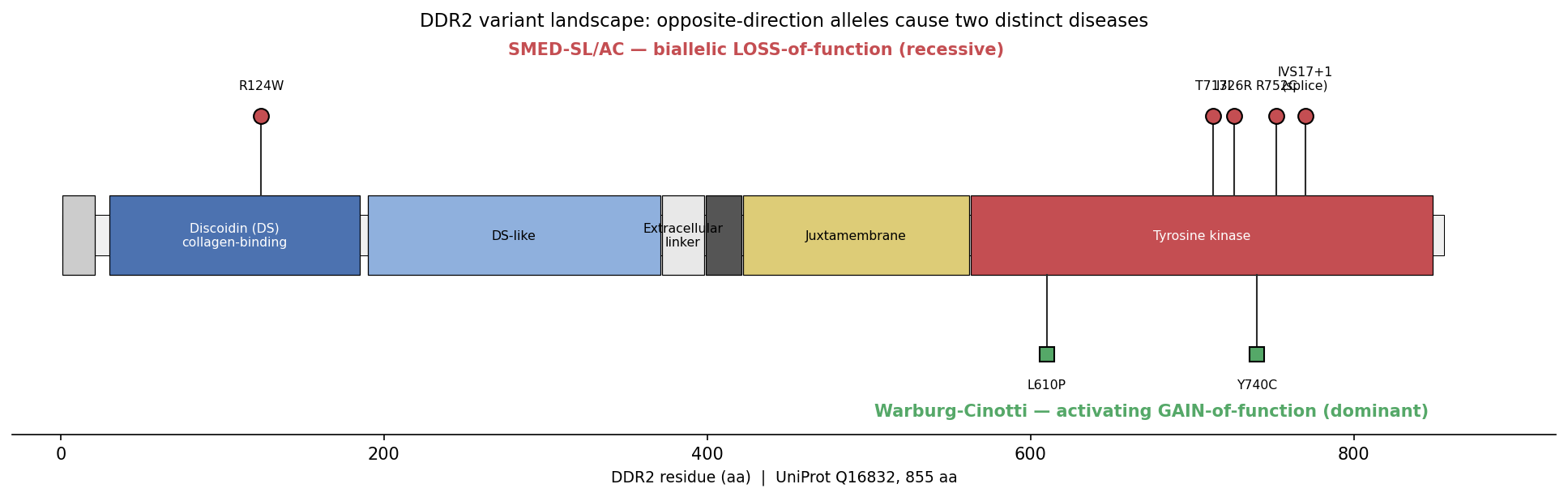
This architecture explains how SMED-SL/AC variants in functionally distinct regions converge on the same loss-of-function outcome: the discoidin-domain missense R124W (c.370C>T) likely impairs collagen binding, whereas R752C/I726R/T713I lie in the kinase domain and impair catalysis (Mansouri et al., 2016, PMID: 26463668; Bargal et al., 2009, PMID: 19110212).
{{figure:ddr2_variant_landscape.png|caption=Schematic of DDR2 (UniProt Q16832, 855 aa) domain architecture. SMED-SL/AC loss-of-function variants (e.g., discoidin-domain R124W impairing collagen binding; kinase-domain T713I/I726R/R752C impairing catalysis) contrast with Warburg-Cinotti gain-of-function variants (L610P, Y740C) that constitutively activate the kinase. The two disorders represent opposite ends of a single DDR2 activity spectrum.}}
Section-by-Section Report
1. Disease Information
Overview. SMED-SL/AC is a congenital autosomal recessive osteochondrodysplasia characterized by severe disproportionate short-limb short stature, distinctive facies, platyspondyly, abnormal metaphyses/epiphyses, and premature (abnormal) calcification of cartilage. It belongs to the spondyloepimetaphyseal dysplasia group, which affects the spine (spondylo-), epiphyses, and metaphyses of long bones.
Key identifiers: - OMIM phenotype: #271665 (Spondylometaepiphyseal dysplasia, short limb–hand type / SMED short limb–abnormal calcification type) - OMIM gene: *191311 (DDR2) - Orphanet: ORPHA:1425 - MONDO: MONDO:0009642 - HGNC (gene): HGNC:2968; UniProt Q16832 - ICD-10: within Q77 (osteochondrodysplasia with defects of growth of tubular bones and spine); ICD-11: LD24 range (skeletal dysplasias). No disease-specific MeSH term; indexed under "Osteochondrodysplasias."
Synonyms / alternative names: Spondylo-meta-epiphyseal dysplasia, short limb–hand type (SMED-SL); SMED short limb–abnormal calcification type (SMED-SL/AC); Borochowitz-Cohen-Barak dysplasia type; spondyloepimetaphyseal dysplasia with abnormal calcification.
Information source. All knowledge derives from aggregated, disease-level resources — individual case reports and small consanguineous family series (Borochowitz 1993; Bargal 2009; Mansouri 2016; Akalin 2023), plus model-organism and biochemical studies. No EHR-derived or population-registry data exist given the extreme rarity.
2. Etiology
Causal factors. The disease is purely genetic (monogenic, Mendelian): biallelic loss-of-function variants in DDR2. No environmental, infectious, or acquired triggers are implicated.
Genetic risk factors. The sole genetic determinant is homozygous or compound-heterozygous pathogenic DDR2 variation. Consanguinity is the principal risk-enabling factor — the founding cohort comprised consanguineous Arab Muslim families, and homozygous variants predominate (PMID: 19110212). No modifier genes or susceptibility loci have been defined.
Environmental risk factors / protective factors. None identified or applicable for this fully penetrant Mendelian disorder. No protective genetic or environmental factors are known.
Gene–environment interactions. Not applicable — no evidence of environmental modification of a monogenic, congenital phenotype.
3. Phenotypes
Table (click to expand)
| Phenotype | Type | Suggested HPO term | Onset | Severity | Frequency |
|---|---|---|---|---|---|
| Disproportionate short-limb short stature | Physical manifestation | HP:0008873 (Disproportionate short-limb short stature) | Congenital | Severe | Nearly universal |
| Platyspondyly | Radiographic sign | HP:0000926 | Congenital/infancy | Severe | High |
| Abnormal metaphyses | Radiographic sign | HP:0000944 | Beyond early infancy | Severe | High |
| Abnormal epiphyses | Radiographic sign | HP:0005930 | Beyond early infancy | Severe | High |
| Premature/abnormal calcification | Radiographic/pathologic | HP:0011849 (Abnormal bone ossification); HP:0100670 (Abnormal cartilage matrix) | Congenital | Severe | Disease-defining |
| Short ribs / narrow chest | Physical/radiographic | HP:0000774 (Narrow chest); HP:0000772 (Abnormal rib) | Congenital | Moderate–severe | High |
| Short nose, wide nasal bridge | Facial | HP:0003196; HP:0000431 | Congenital | — | Characteristic |
| Long philtrum | Facial | HP:0000343 | Congenital | — | Characteristic |
| Ocular hypertelorism | Facial | HP:0000316 | Congenital | — | Characteristic |
| Micrognathia/retrognathia | Facial | HP:0000347 | Congenital | — | Characteristic |
| Short hands (brachydactyly) | Physical | HP:0001156 | Congenital | — | Characteristic |
| Dental anomalies (enamel hypoplasia, abnormal number/shape) | Physical | HP:0006297; HP:0006482 | Childhood | Variable | Reported subset |
Progression: Skeletal changes evolve over time ("typical evolution of bony changes"), with metaphyseal/epiphyseal abnormality becoming more marked beyond early infancy (PMID: 8434618).
Quality of life: Severe short stature, skeletal deformity, and narrow chest substantially impair mobility, respiratory reserve, and daily functioning. No formal EQ-5D/SF-36/PROMIS data exist for this ultra-rare disorder.
4. Genetic / Molecular Information
Causal gene: DDR2 (Discoidin Domain Receptor Tyrosine Kinase 2), chromosome 1q23.3; OMIM *191311; HGNC:2968; UniProt Q16832 (protein, 855 aa).
Pathogenic variants. ~10 reported pathogenic/likely-pathogenic variants (ACMG/AMP). Types include missense (6), nonsense (2), deletion (1), and splice-site (1):
Table (click to expand)
| Variant (cDNA) | Protein | Type | Domain | Consequence |
|---|---|---|---|---|
| c.2254C>T | p.R752C | Missense | Kinase | Loss of catalysis |
| c.2177T>G | p.I726R | Missense | Kinase | Loss of catalysis |
| c.2138C>T | p.T713I | Missense | Kinase | Loss of catalysis |
| IVS17+1g>a | — | Splice | Kinase-encoding | Aberrant splicing / LoF |
| c.370C>T | p.R124W | Missense | Discoidin (DS) | Impaired collagen binding |
(Bargal 2009 PMID: 19110212; Mansouri 2016 PMID: 26463668; Akalin 2023 PMID: 36720430.)
Classification: Pathogenic/likely pathogenic per ACMG. Allele frequency: private/extremely rare; absent or near-absent in gnomAD. Origin: germline. Functional consequence: loss of function (impaired collagen binding or abolished kinase activity). No dominant-negative or gain-of-function effects in SMED-SL/AC (gain-of-function DDR2 instead causes Warburg-Cinotti syndrome).
Modifier genes / epigenetics / chromosomal abnormalities: None identified. This is a single-gene, small-variant disorder without reported cytogenetic changes.
5. Environmental Information
Not applicable. No environmental, lifestyle, or infectious contributors are known for this congenital monogenic disorder.
6. Mechanism / Pathophysiology
Ordered causal chain (initiating lesion → clinical manifestation):
- Biallelic loss-of-function DDR2 variant leads to a defective DDR2 receptor — either unable to bind fibrillar collagen (DS-domain variant, e.g., R124W) or catalytically dead (kinase-domain variant, e.g., T713I/I726R/R752C; or splice/nonsense allele producing no functional protein).
- Defective receptor results in failure of collagen-induced DDR2 activation: no Src-mediated Tyr-740 phosphorylation, no intramolecular cis-autophosphorylation, and no generation of cytosolic phosphotyrosines (inferred from the WT activation mechanism defined in PMID: 16186108 and PMID: 41259339).
- Loss of DDR2 phosphotyrosines prevents recruitment/formation of DDR2–Shc signaling complexes, interrupting downstream proliferative signaling.
- Interrupted signaling in resting-zone and proliferating growth-plate chondrocytes and Gli1-positive skeletal progenitors leads to reduced chondrocyte proliferation (demonstrated in Ddr2-null mice, PMID: 11375938).
- Reduced chondrocyte proliferation results in impaired endochondral bone growth at long-bone growth plates and cranial-base synchondroses → branch A: shortened long bones/limbs and platyspondyly; branch B: failure of cranial-base elongation → flat face, short skull, distinctive facies (PMID: 36656123).
- Disorganized cartilage with sparse matrix and degenerating chondrocytes leads to deposition of dense amorphous material → premature/abnormal calcification (the disease-defining feature; the direct mechanistic link between DDR2 loss and ectopic calcification is inferred, not fully demonstrated).
- Net result is severe disproportionate short-limb short stature, abnormal metaphyses/epiphyses, short ribs/narrow chest, and characteristic craniofacial gestalt.
Molecular pathway. Collagen → DDR2 (RTK) → Src → activation-loop Tyr-740 → autophosphorylation → Shc adaptor complex → proliferative signaling (feeding into downstream MAPK/PI3K effectors typical of RTK signaling). GO annotations: GO:0038063 (collagen-activated tyrosine kinase receptor signaling pathway), GO:0006468 (protein phosphorylation), GO:0008284 (positive regulation of cell population proliferation), GO:0060348 (bone development), GO:0001501 (skeletal system development), GO:0002062 (chondrocyte differentiation).
Cellular processes: growth-plate chondrocyte proliferation (impaired). Protein dysfunction: loss of function via impaired ligand binding or catalytic inactivation. Cell types (CL): chondrocyte (CL:0000138), specifically resting/proliferating growth-plate chondrocytes; skeletal (Gli1+) progenitor cells; periosteal cells; osteoblast lineage (CL:0000062). Tissue-damage mechanism: defective cartilage matrix homeostasis with ectopic calcification.
Molecular profiling / advanced technologies: No human transcriptomic, proteomic, or metabolomic datasets are available for this ultra-rare disease. Mechanistic evidence is drawn from mouse genetics and in-vitro biochemistry/enzyme kinetics.
7. Anatomical Structures Affected
- Organ/system level: Skeletal system — long bones (UBERON:0002481 bone tissue; UBERON:0001474 bone element), vertebral column (UBERON:0001130), ribs (UBERON:0002228), skull/cranial base (UBERON:0003128), face. Secondary: respiratory compromise from narrow chest; dentition (enamel).
- Tissue/cell level: Cartilage (UBERON:0002418), growth-plate cartilage (UBERON:0006721); connective tissue. Target cells: chondrocytes (CL:0000138), especially resting/proliferating growth-plate chondrocytes; Gli1+ skeletal progenitors; periosteal/osteoblast lineage.
- Subcellular level: DDR2 is a plasma-membrane receptor (GO:0005886 plasma membrane; GO:0005887 integral component of plasma membrane). Signaling occurs at the cytoplasmic kinase domain (GO:0004714 transmembrane receptor protein tyrosine kinase activity).
- Localization: Bilateral, symmetric involvement of the appendicular and axial skeleton and craniofacial bones.
8. Temporal Development
- Onset: Congenital; skeletal and facial abnormalities present at birth, with radiographic metaphyseal/epiphyseal changes becoming marked beyond early infancy.
- Onset pattern: Chronic/insidious progression of bony changes.
- Progression: "Typical evolution of bony changes over time" (PMID: 8434618); progressive worsening of disproportion and deformity through childhood; lifelong chronic course.
- Disease course: Stable-progressive, non-episodic, non-remitting. No spontaneous or treatment-induced remission.
- Critical periods: Prenatal and early postnatal growth-plate activity — the window during which DDR2-dependent chondrocyte proliferation shapes skeletal growth.
9. Inheritance and Population
- Inheritance: Autosomal recessive.
- Epidemiology: Ultra-rare; ~22 patients reported by 2016 (PMID: 26463668), with additional cases since. Precise prevalence/incidence not established (Orphanet class: <1/1,000,000).
- Penetrance: Complete (fully penetrant congenital phenotype). Expressivity: Relatively consistent core skeletal phenotype with variable additional features (e.g., dental anomalies).
- Consanguinity: Major factor; original and several subsequent families were consanguineous, favoring homozygosity for private variants.
- Founder effects: Possible within specific consanguineous populations (Arab Muslim, Moroccan, Turkish families reported), though no formal founder haplotype is established. Carrier frequency: not quantified; variants are private.
- Population demographics: Reported in Middle Eastern/North African and other populations with consanguineous unions. No strong sex bias expected (autosomal recessive). Age distribution: presents from birth.
10. Diagnostics
- Imaging (primary diagnostic modality): Skeletal radiographs demonstrate platyspondyly, short tubular bones with abnormal metaphyses/epiphyses, short ribs, and abnormal/premature calcification — the radiographic hallmark (PMID: 8434618).
- Histopathology: Chondro-osseous biopsy shows sparse cartilage matrix, degenerating chondrocytes, and surrounding dense amorphous (calcified) material.
- Genetic testing (confirmatory): Molecular confirmation via DDR2 sequencing. WES/WGS have been diagnostically decisive (Mansouri 2016 identified a novel DDR2 variant by WES, PMID: 26463668). Approaches: single-gene DDR2 sequencing, skeletal-dysplasia gene panels, WES, or WGS. Homozygosity mapping is useful in consanguineous families.
- Laboratory tests: No specific biochemical biomarker; routine calcium/phosphate metabolism is generally not diagnostic. No validated circulating biomarker exists.
- Clinical criteria: Diagnosis rests on the characteristic clinical–radiographic gestalt plus biallelic DDR2 variants.
- Differential diagnosis: Other spondyloepimetaphyseal and spondylometaphyseal dysplasias, and short-limb chondrodysplasias; distinguished by the abnormal-calcification pattern, characteristic facies, and DDR2 genotype.
- Screening: Carrier and cascade testing in at-risk consanguineous families; prenatal molecular testing where the familial variant is known.
11. Outcome / Prognosis
- Survival/mortality: No systematic survival data. Severe short stature with narrow chest may predispose to respiratory complications; overall prognosis depends on severity of thoracic and skeletal involvement.
- Morbidity/function: Substantial lifelong disability from short stature, skeletal deformity, and restricted mobility; potential respiratory limitation.
- Complications: Restrictive thoracic constraints, orthopedic deformity, and dental problems.
- Recovery potential: None — congenital structural disorder; no reversal possible.
- Prognostic factors: Degree of thoracic/skeletal involvement; specific variant effect. No validated prognostic biomarkers.
12. Treatment
There is no disease-specific or curative therapy. Management is supportive and multidisciplinary:
- Supportive/rehabilitative: Orthopedic monitoring and interventions for deformity; physical and occupational therapy; respiratory support as needed; dental care. (NCIT-type interventions: NCIT:C15277 Supportive Care; NCIT:C15682 Physical Therapy; NCIT:C157866 Orthopedic Surgery.)
- Surgical: Orthopedic corrective procedures individualized to deformity.
- Pharmacotherapy / advanced therapeutics: None established. No gene, cell, RNA-based, or targeted therapy exists. Mechanistically, kinase-domain loss-of-function variants would not be amenable to kinase inhibition (in contrast to Warburg-Cinotti's activating variants, where dasatinib inhibited DDR2 autophosphorylation in vitro, PMID: 30449416).
- Genetic counseling is a core component of management.
- Experimental trials: None registered for SMED-SL/AC.
13. Prevention
- Primary prevention: Genetic counseling for consanguineous/at-risk couples; carrier testing.
- Secondary/tertiary prevention: Prenatal molecular diagnosis and preimplantation genetic testing where the familial DDR2 variant is known; early multidisciplinary management to prevent complications (respiratory, orthopedic).
- Public health: Awareness of recessive-disease risk in populations with high consanguinity.
- No immunization or behavioral prevention is applicable.
14. Other Species / Natural Disease
- Taxonomy / orthologs: Human DDR2 (NCBI Gene 4921); mouse Ddr2 (NCBI Gene 18214). DDR2 is highly conserved across vertebrates.
- Natural animal disease: The spontaneous mouse mutant smallie (Ddr2^slie^, a Ddr2 loss-of-function allele) exhibits dwarfism and skeletal defects, representing a naturally arising animal model. No well-characterized companion-animal or livestock breed disorder is established, though DDR2 loss-of-function phenotypes are expected to be conserved.
- Comparative biology: The mouse Ddr2-null skeletal and craniofacial phenotype closely parallels human SMED-SL/AC, confirming evolutionary conservation of DDR2's role in chondrocyte proliferation and endochondral bone growth (PMID: 11375938; PMID: 35140200; PMID: 36656123).
- Transmission: Not applicable (non-infectious genetic disorder).
15. Model Organisms
- Mouse (primary model): Ddr2-deficient/knockout and the spontaneous smallie (Ddr2^slie^) mutant recapitulate dwarfism, shortened long bones, and craniofacial defects. Conditional/lineage models (Gli1-CreER) localize DDR2 function to skeletal progenitors and chondrocytes.
- Phenotype recapitulation: Strong — reduced chondrocyte proliferation, shortened long bones, and flat face/short skull mirror human features.
- Applications: Established the cellular mechanism (proliferation vs differentiation), cell-of-origin (Gli1+ progenitors, growth-plate chondrocytes), and craniofacial pathogenesis.
- Limitations: The abnormal-calcification phenotype and detailed matrix pathology of the human disease are less fully modeled; species differences in growth-plate biology.
- In vitro / biochemical models: Recombinant DDR2 kinase enzyme-kinetic assays and patient fibroblasts have defined the two-step activation mechanism and distinguished loss- vs gain-of-function alleles (PMID: 16186108; PMID: 41259339; PMID: 30449416).
- Resources: MGI (mouse Ddr2), IMPC.
Mechanistic Model / Interpretation
  Fibrillar collagen (GVMGFO motif)
     |
     v   [SMED-SL/AC DS-domain variant e.g. R124W blocks binding]
      DDR2 discoidin (DS) domain  --- amphiphilic trench
     |
     v
   DDR2 dimerization (TM) --> long juxtamembrane --> KINASE domain
     |                                   ^
     |   [SMED-SL/AC kinase variants T713I/I726R/R752C, splice -> NO catalysis]
     v
   Src phosphorylates Tyr-740 (activation loop)
     |
     v
   Intramolecular cis-autophosphorylation
     |
     v
   Cytosolic phosphotyrosines --> Shc complex --> proliferative signaling
     |
     v
   Growth-plate chondrocyte PROLIFERATION (resting/proliferating zones;
   Gli1+ progenitors)
     |
   +---------+----------+
   v                    v
 Long-bone &          Cranial-base
 vertebral growth     synchondrosis growth
   |                    |
   v                    v
 Short limbs,          Flat face, short skull,
 platyspondyly,        distinctive facies
 abnormal meta/epiphyses + premature calcification
Loss-of-function (SMED-SL/AC) and gain-of-function (Warburg-Cinotti) sit at opposite ends of a single DDR2 activity axis:
Table (click to expand)
| Feature | SMED-SL/AC | Warburg-Cinotti syndrome |
|---|---|---|
| Mechanism | Loss of function | Gain of function (constitutive) |
| Representative variants | R124W (DS), T713I/I726R/R752C, IVS17+1g>a (kinase) | L610P, Y740C |
| Receptor phosphorylation | Absent/reduced | Increased, ligand-independent |
| Inheritance | Autosomal recessive (biallelic) | Autosomal dominant (recurrent) |
| Core phenotype | Chondrodysplasia, short limbs, calcification | Corneal neovascularization, keloids, acro-osteolysis |
| Druggability | Not kinase-inhibitor amenable | Dasatinib inhibits autophosphorylation (in vitro) |
Evidence Base
Table (click to expand)
| PMID | Title (abbrev.) | Role in this report |
|---|---|---|
| 8434618 | Original SMED short-limb–hand description (Borochowitz) | Defines clinical/radiographic phenotype |
| 19110212 | DDR2 mutations cause SMED (Bargal) | Establishes causal gene & kinase-domain variants |
| 11375938 | DDR2 regulates proliferation; elimination → dwarfism | Cellular mechanism (mouse) |
| 35140200 | DDR2 in Gli1+ progenitors/chondrocytes | Cell-of-origin localization |
| 36656123 | DDR2 controls craniofacial development | Craniofacial pathogenesis |
| 26463668 | Novel DDR2 variant by WES (Mansouri) | Calcification feature; DS-domain variant; WES utility |
| 36720430 | Expanded mutational spectrum & dental findings (Akalin) | Biallelic LoF confirmation; dental phenotype |
| 30449416 | Activating DDR2 → Warburg-Cinotti (Xu) | Allelic contrast; gain-of-function |
| 16186108 | Tyr-740/Src/Shc signaling (Yang) | Defines signaling cascade lost in disease |
| 41259339 | DDR2 kinase two-step activation (Hao & Leitinger) | Kinase activation mechanism |
| 23128141 | Collagen recognition by DDRs (Carafoli & Hohenester) | Domain architecture; collagen-binding motif |
| 24725424 | DDR functions in physiology/pathology (Leitinger) | Slow/sustained activation kinetics context |
Evidence source types: human clinical (case series, WES/WGS), model organism (mouse knockouts, conditional/lineage tracing), in vitro/biochemical (kinase kinetics, patient fibroblasts). No omics or computational disease datasets exist for SMED-SL/AC.
Limitations and Knowledge Gaps
- Ultra-rarity: Fewer than ~30 patients reported; epidemiology, prognosis, and natural history rest on small case series and lack registry-scale data.
- Calcification mechanism unresolved: The precise link from DDR2 loss to premature/abnormal calcification (the disease-defining feature) is inferred, not mechanistically demonstrated. How impaired chondrocyte proliferation and disordered matrix lead to ectopic calcification remains open.
- Genotype–phenotype correlations: Too few patients to correlate DS-domain vs kinase-domain variants with severity or specific features (e.g., dental anomalies).
- No human molecular profiling: No transcriptomic, proteomic, or metabolomic data; mechanistic inference relies on mouse and biochemistry.
- Downstream effectors beyond Shc (MAPK/PI3K and how they control chondrocyte cell-cycle) are not fully mapped in the growth-plate context.
- Model limitations: Mouse models capture growth and craniofacial phenotypes but may under-represent the human calcification pathology.
Proposed Follow-up Experiments / Actions
- Mechanistic study of calcification: Use Ddr2-null growth-plate chondrocytes and patient-derived iPSC-chondrocyte/organoid models to dissect how DDR2 loss drives ectopic matrix calcification (matrix vesicle biology, ALP activity, Pi/PPi balance).
- Functional variant classification: Systematically express reported DDR2 variants (DS-domain vs kinase-domain) and quantify collagen binding, autophosphorylation, and downstream signaling to formalize genotype–function correlations.
- Patient registry / natural history study: Establish an international SMED-SL/AC registry to capture prevalence, survival, respiratory outcomes, and the phenotypic spectrum.
- Single-cell/spatial transcriptomics of the growth plate in Ddr2-null mice to define the proliferative program lost downstream of DDR2 and identify therapeutic nodes.
- Therapeutic exploration: Because kinase inhibition is not applicable to loss-of-function disease, evaluate pathway-agonist or downstream-restoration strategies (e.g., modulating Shc/MAPK signaling or growth-plate proliferation cues) in models.
- Cranial-base and thoracic longitudinal imaging in patients to define critical intervention windows and respiratory risk.
Report compiled from 6 confirmed findings and 18 reviewed papers across a 5-iteration autonomous investigation. All quoted material is verbatim from cited PubMed abstracts.
Artifacts
Reference Validation
Checked with linkml-reference-validator 0.2.1.
Table (click to expand)
| Outcome | Count |
|---|---|
| References checked | 12 |
| Resolved | 12 |
| Unresolved (possible confabulation) | 0 |
| Unverifiable | 0 |
| Quoted claims checked | 1 |
| Quoted claims found in source | 1 |
| Quoted claims not found in source | 0 |
| References weighed for topical relevance | 12 |
| On topic | 10 |
| Off topic | 0 |
All extracted references resolved successfully.
Term Validation
Checked with linkml-term-validator 0.4.5, through the ols: adapter.
Table (click to expand)
| Outcome | Count |
|---|---|
| Terms checked | 40 |
| Resolved | 37 |
| Unresolved (possible confabulation) | 0 |
| Obsolete | 1 |
| Unverifiable | 2 |
| Terms whose name was checked | 15 |
| Terms named correctly | 7 |
| Terms named as a different term | 7 |
| Terms whose name is worth a second look | 1 |
Terms the report names something else
These identifiers resolve, so nothing about them looks wrong, and the ontology calls them something unrelated to what the report calls them. That usually means the identifier is not the one the sentence needs:
HP:0000926(1 mention) - the report calls it "Radiographic sign"; HP calls it PlatyspondylyHP:0000944(1 mention) - the report calls it "Radiographic sign"; HP calls it Abnormal metaphysis morphologyHP:0005930(1 mention) - the report calls it "Radiographic sign"; HP calls it Abnormal epiphysis morphologyHP:0000343(1 mention) - the report calls it "Facial"; HP calls it Long philtrumHP:0000316(1 mention) - the report calls it "Facial"; HP calls it HypertelorismHP:0000347(1 mention) - the report calls it "Facial"; HP calls it MicrognathiaHP:0001156(1 mention) - the report calls it "Physical"; HP calls it Brachydactyly
Obsolete terms
These terms are real but deprecated. Citing one is not a fabrication; it does mean the report is naming something the ontology has retired:
GO:0005887(GO_0005887) (1 mention) - replaced byGO:0005886
Terms whose name is worth a second look
The report's name for these is recognisably related to the term's own name without being one of them. A loose paraphrase reads the same way as a citation of the wrong sibling term - and so does a related synonym, which the ontology records precisely because it names something adjacent rather than the same thing - so these are listed rather than judged:
UBERON:0002418(1 mention) - the report calls it "Tissue/cell level: Cartilage"; UBERON calls it cartilage tissue**
Prefixes with no resolver
Terms carrying these prefixes were not checked either way, because no configured ontology covers them. An unrecognised prefix may name an ontology this run could not reach as easily as one that does not exist, so nothing here is evidence of fabrication: ORPHA.