GM1 Gangliosidosis Type 1
GM1 gangliosidosis type 1 (infantile GM1 gangliosidosis) is an ultra-rare, rapidly progressive neuronopathic lysosomal storage disorder caused by biallelic loss-of-function variants in GLB1 encoding lysosomal beta-galactosidase. Near-absent enzyme activity prevents cleavage of the terminal galactose from GM1 ganglioside, so GM1 ganglioside and other beta-galactose-terminal glycoconjugates (GA1, oligosaccharides, keratan sulfate) accumulate in neurons and somatic tissues. The infantile form presents in early infancy (mean first-symptom onset ~2.8 months) with rapidly progressive neurodegeneration, hypotonia, developmental regression, hepatosplenomegaly, dysostosis multiplex, coarse facial features, and a macular cherry-red spot, with a life expectancy of two to three years (mean age at death ~18.9 months). No FDA-approved disease-modifying therapy exists; AAV-mediated GLB1 gene therapy is in clinical trials.
Ask OpenScientist
Ask a research question about GM1 Gangliosidosis Type 1. OpenScientist will conduct autonomous deep research using the Disorder Mechanisms Knowledge Base and PubMed literature (typically 10-30 minutes).
Do not include personal health information in your question. Questions and results are cached in your browser's local storage.
Classifications
Mappings
Definitions
1Show evidence (1 reference)
Inheritance
1Show evidence (2 references)
Pathophysiology
5Show evidence (3 references)
Show evidence (2 references)
Show evidence (1 reference)
Show evidence (1 reference)
Show evidence (1 reference)
Histopathology
1Show evidence (1 reference)
Pathograph
Phenotypes
19Cardiovascular 3
Show evidence (1 reference)
Show evidence (1 reference)
Show evidence (1 reference)
Digestive 1
Show evidence (1 reference)
Ear 1
Show evidence (1 reference)
Eye 1
Show evidence (1 reference)
Head and Neck 2
Show evidence (1 reference)
Show evidence (2 references)
Integument 1
Show evidence (1 reference)
Metabolism 1
Show evidence (1 reference)
Musculoskeletal 4
Show evidence (2 references)
Show evidence (1 reference)
Show evidence (1 reference)
Show evidence (1 reference)
Nervous System 4
Show evidence (1 reference)
Show evidence (1 reference)
Show evidence (1 reference)
Show evidence (2 references)
Growth 1
Show evidence (1 reference)
Genetic Associations
1Show evidence (3 references)
External Assertions
1Medical Actions
4Show evidence (1 reference)
Show evidence (2 references)
Show evidence (1 reference)
Show evidence (2 references)
Biochemical Markers
3Show evidence (1 reference)
Show evidence (1 reference)
Show evidence (1 reference)
Diagnosis
3Show evidence (1 reference)
Show evidence (1 reference)
Show evidence (1 reference)
Progression
2Show evidence (1 reference)
Show evidence (4 references)
Prevalence
1Show evidence (2 references)
Differential Diagnoses
1Conditions with similar clinical presentations that must be differentiated from GM1 Gangliosidosis Type 1:
- Later symptom onset and a more protracted course with higher residual beta-galactosidase activity.
- Coarse facies, cherry-red spots, and visceromegaly occur only in the infantile (type 1) form.
Show evidence (2 references)
Clinical Trials
4Show evidence (1 reference)
Show evidence (1 reference)
Show evidence (1 reference)
Show evidence (1 reference)
Source YAML
click to showname: GM1 Gangliosidosis Type 1
creation_date: "2026-06-13T00:00:00Z"
description: >-
GM1 gangliosidosis type 1 (infantile GM1 gangliosidosis) is an ultra-rare, rapidly
progressive neuronopathic lysosomal storage disorder caused by biallelic
loss-of-function variants in GLB1 encoding lysosomal beta-galactosidase. Near-absent
enzyme activity prevents cleavage of the terminal galactose from GM1 ganglioside, so
GM1 ganglioside and other beta-galactose-terminal glycoconjugates (GA1, oligosaccharides,
keratan sulfate) accumulate in neurons and somatic tissues. The infantile form presents
in early infancy (mean first-symptom onset ~2.8 months) with rapidly progressive
neurodegeneration, hypotonia, developmental regression, hepatosplenomegaly, dysostosis
multiplex, coarse facial features, and a macular cherry-red spot, with a life expectancy
of two to three years (mean age at death ~18.9 months). No FDA-approved disease-modifying
therapy exists; AAV-mediated GLB1 gene therapy is in clinical trials.
category: Mendelian
disease_term:
preferred_term: GM1 gangliosidosis type 1
term:
id: MONDO:0009260
label: GM1 gangliosidosis type 1
mappings:
mondo_mappings:
- term:
id: MONDO:0009260
label: GM1 gangliosidosis type 1
mapping_predicate: skos:exactMatch
mapping_source: MONDO
mapping_justification: Primary MONDO disease identifier for this infantile GM1 gangliosidosis entry.
external_assertions:
- name: OMIM GM1-gangliosidosis, type I record
source: OMIM
assertion_type: disease_record
external_id: OMIM:230500
description: >-
OMIM phenotype entry (MIM #230500) for GM1-gangliosidosis, type I, the infantile form
of GLB1-related beta-galactosidase deficiency.
synonyms:
- Infantile GM1 gangliosidosis
- GM1 gangliosidosis, type I
- Beta-galactosidase deficiency, infantile
- Landing disease
- Norman-Landing disease
references:
- reference: PMID:24156116
title: "GLB1-Related Disorders."
tags:
- GeneReviews
parents:
- Sphingolipidosis
- Lysosomal Storage Disorder
- Neurodegenerative Disease
classifications:
harrisons_chapter:
- classification_value: ENDOCRINOLOGY_METABOLISM
notes: >-
An inborn error of metabolism (lysosomal storage disorder) with a dominant
neurodegenerative phenotype.
lysosomal_storage_category:
classification_value: sphingolipidosis
notes: >-
GM1 gangliosidosis is a sphingolipidosis: beta-galactosidase deficiency blocks
catabolism of the GM1 ganglioside glycosphingolipid, which accumulates in neurons.
evidence:
- reference: PMID:34539759
reference_title: "GM1 Gangliosidosis-A Mini-Review."
supports: SUPPORT
evidence_source: HUMAN_CLINICAL
snippet: "GM1 gangliosidosis is a progressive, neurosomatic, lysosomal storage disorder caused by mutations in the GLB1 gene encoding the enzyme β-galactosidase."
explanation: "Supports classification as a lysosomal storage disorder (sphingolipidosis) of GM1 ganglioside catabolism."
isds_skeletal_category:
- classification_value: lysosomal_storage_with_skeletal_involvement
notes: >-
ISDS Nosology and Classification of Genetic Skeletal Disorders, 2019
revision (Mortier et al., PMID:31633310), Table 1 group 27 "Lysosomal
storage diseases with skeletal involvement (dysostosis multiplex group)";
listed as "GM1 Gangliosidosis, several forms".
pathophysiology:
- name: GLB1 Loss of Function and Beta-Galactosidase Deficiency
conforms_to: "lysosomal_substrate_accumulation#Lysosomal Hydrolase or Cofactor Deficiency"
biological_scale: MOLECULAR
description: >-
Biallelic loss-of-function variants in GLB1 abolish lysosomal beta-galactosidase
activity. The enzyme normally cleaves the terminal beta-1,4-linked galactose from GM1
ganglioside and also degrades oligosaccharides and keratan sulfate; its loss is the
primary biochemical lesion. Disease severity tracks residual enzyme activity, which is
nearly absent in type 1-associated mutations.
gene:
preferred_term: GLB1
term:
id: hgnc:4298
label: GLB1
molecular_functions:
- preferred_term: beta-galactosidase activity
term:
id: GO:0004565
label: beta-galactosidase activity
modifier: DECREASED
biological_processes:
- preferred_term: ganglioside catabolic process
modifier: DECREASED
term:
id: GO:0006689
label: ganglioside catabolic process
evidence:
- reference: PMID:33859490
reference_title: "GM1 Gangliosidosis: Mechanisms and Management."
supports: SUPPORT
evidence_source: HUMAN_CLINICAL
snippet: "Mutation of the GLB1 gene, which codes for β-gal, prevents cleavage of the terminal β-1,4-linked galactose residue from GM1 ganglioside."
explanation: "GLB1 mutation causes beta-galactosidase deficiency that blocks GM1 ganglioside degradation."
- reference: PMID:31937438
reference_title: "The natural history of Type 1 infantile GM1 gangliosidosis: A literature-based meta-analysis."
supports: SUPPORT
evidence_source: HUMAN_CLINICAL
snippet: "The ubiquitous lysosomal enzyme βgal is responsible for the degradation of GM1 ganglioside, oligosaccharides, and keratan sulfate"
explanation: "Beta-galactosidase degrades GM1 ganglioside plus oligosaccharides and keratan sulfate, explaining the multisystem substrate burden."
- reference: PMID:31937438
reference_title: "The natural history of Type 1 infantile GM1 gangliosidosis: A literature-based meta-analysis."
supports: SUPPORT
evidence_source: HUMAN_CLINICAL
snippet: "Disease severity is associated with the residual activity of the mutant βgal enzyme, with activity nearly absent in Type 1-associated mutations"
explanation: "Near-absent residual beta-galactosidase activity defines the severe infantile (type 1) end of the spectrum."
downstream:
- target: Lysosomal GM1 Ganglioside Accumulation in Neurons
description: Loss of enzyme activity allows GM1 ganglioside to accumulate in lysosomes.
- name: Lysosomal GM1 Ganglioside Accumulation in Neurons
conforms_to: "lysosomal_substrate_accumulation#Lysosomal Substrate Accumulation"
biological_scale: CELLULAR
description: >-
GM1 ganglioside and other beta-galactose-terminal glycoconjugates accumulate in
neuronal lysosomes (and, via keratan sulfate/oligosaccharide storage, in somatic
tissues such as heart, bone, liver, and spleen), impairing cell physiology.
cell_types:
- preferred_term: neuron
term:
id: CL:0000540
label: neuron
cellular_components:
- preferred_term: lysosome
term:
id: GO:0005764
label: lysosome
biological_processes:
- preferred_term: glycosphingolipid metabolic process
modifier: ABNORMAL
term:
id: GO:0006687
label: glycosphingolipid metabolic process
evidence:
- reference: PMID:34539759
reference_title: "GM1 Gangliosidosis-A Mini-Review."
supports: SUPPORT
evidence_source: HUMAN_CLINICAL
snippet: "Absent or reduced β-galactosidase activity leads to the accumulation of β-linked galactose-containing glycoconjugates including the glycosphingolipid (GSL) GM1-ganglioside in neuronal tissue."
explanation: "Beta-galactosidase deficiency causes neuronal GM1 ganglioside accumulation."
- reference: PMID:31937438
reference_title: "The natural history of Type 1 infantile GM1 gangliosidosis: A literature-based meta-analysis."
supports: SUPPORT
evidence_source: HUMAN_CLINICAL
snippet: "Systemic disease manifestations are also common due to keratan sulfate and oligosaccharide buildup in peripheral organs, such as the heart, bone, liver, and spleen"
explanation: "Somatic storage of keratan sulfate/oligosaccharides underlies the visceral, skeletal, and cardiac involvement."
downstream:
- target: Neuronal Apoptosis
description: Toxic lysosomal storage of undegraded substrates precipitates neuronal cell death.
- target: Multisystem Somatic Storage
description: >-
Keratan sulfate and oligosaccharide storage in peripheral organs produces the
visceral, skeletal, and cardiac disease, in parallel with the CNS arm.
- name: Neuronal Apoptosis
conforms_to: "lysosomal_substrate_accumulation#Storage-Cell Cytotoxicity and Neuroinflammation"
biological_scale: CELLULAR
description: >-
Toxic lysosomal storage of GM1 ganglioside and related substrates precipitates
neuronal apoptotic cell death, a key route to the progressive neuron loss of the
disease. (Model-system work in beta-gal-deficient mice further implicates GM1-driven
ER-stress/unfolded-protein-response and mitochondrial calcium dysregulation upstream
of this apoptosis, but that specific cascade is model-derived and not asserted here
as a human-established mechanism.)
cell_types:
- preferred_term: neuron
term:
id: CL:0000540
label: neuron
biological_processes:
- preferred_term: apoptotic process
modifier: INCREASED
term:
id: GO:0006915
label: apoptotic process
evidence:
- reference: PMID:31937438
reference_title: "The natural history of Type 1 infantile GM1 gangliosidosis: A literature-based meta-analysis."
supports: SUPPORT
evidence_source: HUMAN_CLINICAL
snippet: "Deficiency of βgal results in toxic lysosomal storage of these substrates, leading to cellular apoptosis."
explanation: "Toxic lysosomal substrate storage precipitates cellular apoptosis."
downstream:
- target: Progressive Neurodegeneration
description: Progressive neuronal apoptotic loss produces the neurodegenerative disease.
- name: Progressive Neurodegeneration
conforms_to: "lysosomal_substrate_accumulation#Progressive Multisystem and Neurodegenerative Disease"
biological_scale: ORGANISM
description: >-
Progressive neuronal loss produces the central-nervous-system arm of the disease:
rapidly progressive neurodegeneration with developmental regression, hypotonia,
spasticity, seizures, visual and hearing loss, bulbar dysfunction, and, in the retina,
the macular cherry-red spot.
cell_types:
- preferred_term: neuron
term:
id: CL:0000540
label: neuron
- preferred_term: retinal ganglion cell
term:
id: CL:0000740
label: retinal ganglion cell
evidence:
- reference: PMID:33859490
reference_title: "GM1 Gangliosidosis: Mechanisms and Management."
supports: SUPPORT
evidence_source: HUMAN_CLINICAL
snippet: "Subsequent accumulation of GM1 ganglioside and other substrates in the lysosome impairs cell physiology and precipitates dysfunction of the nervous system."
explanation: "Lysosomal GM1 storage drives nervous-system dysfunction and neurodegeneration."
downstream:
- target: Neurodegeneration
description: Neuronal storage drives progressive neurodegeneration.
- target: Developmental regression
description: Progressive neuronal storage and dysfunction cause loss of acquired developmental milestones.
- target: Global developmental delay
description: Progressive neuronal dysfunction causes failure to attain developmental milestones.
- target: Hypotonia
description: Central nervous system storage produces early, near-universal hypotonia.
- target: Seizures
description: Progressive cortical neuronal storage produces later-onset seizures.
- target: Spasticity
description: Progressive central nervous system storage and dysfunction produce spasticity.
- target: Decerebrate rigidity
description: End-stage central nervous system dysfunction produces decerebrate rigidity.
- target: Blindness
description: Cortical and retinal storage/dysfunction leads to visual loss.
- target: Hearing impairment
description: Progressive central nervous system involvement produces deafness.
- target: Feeding difficulties
description: Progressive bulbar dysfunction produces feeding difficulties and aspiration risk.
- target: Drooling
description: Bulbar dysfunction produces increased oral secretions/drooling.
- target: Cherry red spot of the macula
description: Retinal ganglion-cell storage produces the macular cherry-red spot.
- name: Multisystem Somatic Storage
conforms_to: "lysosomal_substrate_accumulation#Progressive Multisystem and Neurodegenerative Disease"
biological_scale: ORGANISM
description: >-
Keratan sulfate and oligosaccharide storage in peripheral organs produces the somatic
arm of the disease: visceral organomegaly, skeletal dysostosis, coarse facies, cardiac
involvement, and prenatal-onset manifestations.
evidence:
- reference: PMID:31937438
reference_title: "The natural history of Type 1 infantile GM1 gangliosidosis: A literature-based meta-analysis."
supports: SUPPORT
evidence_source: HUMAN_CLINICAL
snippet: "Systemic disease manifestations are also common due to keratan sulfate and oligosaccharide buildup in peripheral organs, such as the heart, bone, liver, and spleen"
explanation: "Peripheral keratan sulfate/oligosaccharide storage drives the somatic (visceral, skeletal, cardiac) disease arm."
downstream:
- target: Hepatosplenomegaly
description: Somatic storage produces visceral organomegaly.
- target: Dysostosis multiplex
description: Somatic lysosomal storage contributes to the skeletal dysplasia/dysostosis multiplex phenotype.
- target: Coarse facial features
description: Somatic storage contributes to coarse facial features.
- target: Cardiomyopathy
description: Somatic (cardiac) storage contributes to cardiomyopathy.
- target: Mongolian blue spot
description: Systemic storage is associated with congenital dermal melanocytosis.
- target: Nonimmune hydrops fetalis
description: Severe prenatal-onset storage can present as nonimmune hydrops fetalis.
- target: Intrauterine growth retardation
description: Prenatal-onset storage is associated with intrauterine growth restriction.
phenotypes:
- name: Neurodegeneration
description: Rapidly progressive neurodegeneration with loss of nervous-system function.
phenotype_term:
preferred_term: Neurodegeneration
term:
id: HP:0002180
label: Neurodegeneration
evidence:
- reference: PMID:33859490
reference_title: "GM1 Gangliosidosis: Mechanisms and Management."
supports: SUPPORT
evidence_source: HUMAN_CLINICAL
snippet: "precipitates dysfunction of the nervous system"
explanation: "Progressive nervous-system dysfunction/neurodegeneration is the core feature."
- name: Developmental regression
description: Loss of acquired developmental milestones in infancy.
frequency: VERY_FREQUENT
phenotype_term:
preferred_term: Developmental regression
term:
id: HP:0002376
label: Developmental regression
evidence:
- reference: PMID:31937438
reference_title: "The natural history of Type 1 infantile GM1 gangliosidosis: A literature-based meta-analysis."
supports: SUPPORT
evidence_source: HUMAN_CLINICAL
snippet: "almost all patients experienced significant multi-organ system\ndysfunction and neurodevelopmental regression, particularly in the 6- to\n18-month"
explanation: "Neurodevelopmental regression in the 6-18 month range is near-universal in infantile GM1 gangliosidosis."
- name: Hypotonia
description: >-
Hypotonia is an early and near-universal feature of infantile GM1 gangliosidosis.
frequency: VERY_FREQUENT
phenotype_term:
preferred_term: Hypotonia
term:
id: HP:0001252
label: Hypotonia
evidence:
- reference: PMID:31937438
reference_title: "The natural history of Type 1 infantile GM1 gangliosidosis: A literature-based meta-analysis."
supports: SUPPORT
evidence_source: HUMAN_CLINICAL
snippet: "clinical findings of hypotonia and developmental delay in almost all patients"
explanation: "Hypotonia is present in almost all infantile (type 1) patients."
- reference: PMID:39639297
reference_title: "A natural history study of pediatric patients with early onset of GM1 gangliosidosis, GM2 gangliosidoses, or gaucher disease type 2 (RETRIEVE)."
supports: SUPPORT
evidence_source: HUMAN_CLINICAL
snippet: "hypotonia was experienced by most patients with GM1 (17/18, 94.4%)"
explanation: "Quantifies hypotonia frequency (94.4%) in early-onset GM1 gangliosidosis, supporting the VERY_FREQUENT band."
- name: Global developmental delay
description: >-
Failure to attain age-appropriate developmental milestones, present in almost all
infantile patients.
frequency: VERY_FREQUENT
phenotype_term:
preferred_term: Global developmental delay
term:
id: HP:0001263
label: Global developmental delay
evidence:
- reference: PMID:31937438
reference_title: "The natural history of Type 1 infantile GM1 gangliosidosis: A literature-based meta-analysis."
supports: SUPPORT
evidence_source: HUMAN_CLINICAL
snippet: "clinical findings of hypotonia and developmental delay in almost all patients"
explanation: "Developmental delay is present in almost all infantile (type 1) patients."
- name: Seizures
description: >-
Seizures develop later in the infantile course (around 10-12 months of age).
phenotype_term:
preferred_term: Seizure
term:
id: HP:0001250
label: Seizure
evidence:
- reference: PMID:24156116
reference_title: "GLB1-Related Disorders."
supports: SUPPORT
evidence_source: HUMAN_CLINICAL
snippet: "blindness, deafness, decerebrate rigidity, seizures, feeding difficulties, and oral secretions are observed"
explanation: "Seizures are among the observed manifestations of progressive CNS dysfunction in infantile GM1."
- reference: PMID:31937438
reference_title: "The natural history of Type 1 infantile GM1 gangliosidosis: A literature-based meta-analysis."
supports: SUPPORT
evidence_source: HUMAN_CLINICAL
snippet: "seizures were associated with a later stage of disease, starting at approximately 10 to 12 months of age"
explanation: "Supports the later (10-12 month) onset timing of seizures stated in the description."
- name: Decerebrate rigidity
description: >-
End-stage central nervous system dysfunction produces decerebrate rigidity.
phenotype_term:
preferred_term: Decerebrate rigidity
term:
id: HP:0025013
label: Decerebrate rigidity
evidence:
- reference: PMID:24156116
reference_title: "GLB1-Related Disorders."
supports: SUPPORT
evidence_source: HUMAN_CLINICAL
snippet: "blindness, deafness, decerebrate rigidity, seizures, feeding difficulties, and oral secretions are observed"
explanation: "Decerebrate rigidity is an observed feature of progressive infantile GM1."
- name: Drooling
description: >-
Increased oral secretions/drooling result from progressive bulbar dysfunction.
phenotype_term:
preferred_term: Drooling
term:
id: HP:0002307
label: Drooling
evidence:
- reference: PMID:24156116
reference_title: "GLB1-Related Disorders."
supports: SUPPORT
evidence_source: HUMAN_CLINICAL
snippet: "blindness, deafness, decerebrate rigidity, seizures, feeding difficulties, and oral secretions are observed"
explanation: "Increased oral secretions (drooling) is an observed feature of progressive infantile GM1."
- name: Hepatosplenomegaly
description: Visceral organomegaly from somatic storage is characteristic of infantile GM1.
frequency: FREQUENT
phenotype_term:
preferred_term: Hepatosplenomegaly
term:
id: HP:0001433
label: Hepatosplenomegaly
evidence:
- reference: PMID:31937438
reference_title: "The natural history of Type 1 infantile GM1 gangliosidosis: A literature-based meta-analysis."
supports: SUPPORT
evidence_source: HUMAN_CLINICAL
snippet: "infants frequently have accompanying hepatosplenomegaly, skeletal dysplasia, cherry-red maculae, cardiomyopathy, and coarse facial features"
explanation: "Hepatosplenomegaly is a frequent somatic feature of infantile GM1."
- name: Dysostosis multiplex
description: A characteristic constellation of skeletal dysplasia (dysostosis multiplex).
frequency: FREQUENT
phenotype_term:
preferred_term: Dysostosis multiplex
term:
id: HP:0000943
label: Dysostosis multiplex
evidence:
- reference: PMID:31937438
reference_title: "The natural history of Type 1 infantile GM1 gangliosidosis: A literature-based meta-analysis."
supports: SUPPORT
evidence_source: HUMAN_CLINICAL
snippet: "infants frequently have accompanying hepatosplenomegaly, skeletal dysplasia, cherry-red maculae, cardiomyopathy, and coarse facial features"
explanation: "Skeletal dysplasia (dysostosis multiplex) is a frequent somatic feature."
- name: Coarse facial features
description: Coarse facies develop as somatic storage progresses; seen in the infantile form.
frequency: FREQUENT
phenotype_term:
preferred_term: Coarse facial features
term:
id: HP:0000280
label: Coarse facial features
evidence:
- reference: PMID:31937438
reference_title: "The natural history of Type 1 infantile GM1 gangliosidosis: A literature-based meta-analysis."
supports: SUPPORT
evidence_source: HUMAN_CLINICAL
snippet: "infants frequently have accompanying hepatosplenomegaly, skeletal dysplasia, cherry-red maculae, cardiomyopathy, and coarse facial features"
explanation: "Coarse facial features are a frequent somatic feature of infantile GM1."
- reference: PMID:31761138
reference_title: "The Clinical and Molecular Spectrum of GM1 Gangliosidosis."
supports: SUPPORT
evidence_source: HUMAN_CLINICAL
snippet: "Coarse facial features, cherry red spots, and visceromegaly occurred only in patients with the infantile form."
explanation: "Coarse facies, cherry-red spots, and visceromegaly are specific to the infantile (type 1) form."
- name: Cardiomyopathy
description: >-
Cardiac storage contributes to cardiomyopathy, a frequent accompanying feature in
infants.
frequency: FREQUENT
phenotype_term:
preferred_term: Cardiomyopathy
term:
id: HP:0001638
label: Cardiomyopathy
evidence:
- reference: PMID:31937438
reference_title: "The natural history of Type 1 infantile GM1 gangliosidosis: A literature-based meta-analysis."
supports: SUPPORT
evidence_source: HUMAN_CLINICAL
snippet: "infants frequently have accompanying hepatosplenomegaly, skeletal dysplasia, cherry-red maculae, cardiomyopathy, and coarse facial features"
explanation: "Cardiomyopathy is a frequent accompanying feature of infantile GM1."
- name: Cherry red spot of the macula
description: A macular cherry-red spot reflects perifoveal ganglion-cell storage.
frequency: FREQUENT
phenotype_term:
preferred_term: Cherry red spot of the macula
term:
id: HP:0010729
label: Cherry red spot of the macula
evidence:
- reference: PMID:31937438
reference_title: "The natural history of Type 1 infantile GM1 gangliosidosis: A literature-based meta-analysis."
supports: SUPPORT
evidence_source: HUMAN_CLINICAL
snippet: "infants frequently have accompanying hepatosplenomegaly, skeletal dysplasia, cherry-red maculae, cardiomyopathy, and coarse facial features"
explanation: "A cherry-red macula is a frequent ophthalmologic feature of infantile GM1."
- name: Spasticity
description: >-
Progressive central nervous system dysfunction produces spasticity as the disease
advances.
phenotype_term:
preferred_term: Spasticity
term:
id: HP:0001257
label: Spasticity
evidence:
- reference: PMID:24156116
reference_title: "GLB1-Related Disorders."
supports: SUPPORT
evidence_source: HUMAN_CLINICAL
snippet: "Progressive central nervous system dysfunction leads to spasticity and rapid regression"
explanation: "Spasticity results from progressive CNS dysfunction in infantile GM1."
- name: Blindness
description: Cortical/retinal involvement leads to visual loss.
phenotype_term:
preferred_term: Blindness
term:
id: HP:0000618
label: Blindness
evidence:
- reference: PMID:24156116
reference_title: "GLB1-Related Disorders."
supports: SUPPORT
evidence_source: HUMAN_CLINICAL
snippet: "blindness, deafness, decerebrate rigidity, seizures, feeding difficulties, and oral secretions are observed"
explanation: "Blindness is an observed feature of progressive infantile GM1."
- name: Hearing impairment
description: Deafness develops with progressive neurologic decline.
phenotype_term:
preferred_term: Hearing impairment
term:
id: HP:0000365
label: Hearing impairment
evidence:
- reference: PMID:24156116
reference_title: "GLB1-Related Disorders."
supports: SUPPORT
evidence_source: HUMAN_CLINICAL
snippet: "blindness, deafness, decerebrate rigidity, seizures, feeding difficulties, and oral secretions are observed"
explanation: "Deafness is an observed feature of progressive infantile GM1."
- name: Feeding difficulties
description: >-
Progressive bulbar dysfunction produces feeding difficulties and increased oral
secretions, contributing to aspiration risk.
phenotype_term:
preferred_term: Feeding difficulties
term:
id: HP:0011968
label: Feeding difficulties
evidence:
- reference: PMID:24156116
reference_title: "GLB1-Related Disorders."
supports: SUPPORT
evidence_source: HUMAN_CLINICAL
snippet: "blindness, deafness, decerebrate rigidity, seizures, feeding difficulties, and oral secretions are observed"
explanation: "Feeding difficulties are an observed feature of progressive infantile GM1."
- name: Mongolian blue spot
description: >-
Congenital dermal melanocytosis (Mongolian spots) may be observed in infantile GM1.
phenotype_term:
preferred_term: Mongolian blue spot
term:
id: HP:0011369
label: Mongolian blue spot
evidence:
- reference: PMID:24156116
reference_title: "GLB1-Related Disorders."
supports: SUPPORT
evidence_source: HUMAN_CLINICAL
snippet: "congenital dermal melanocytosis (Mongolian spots) may be observed"
explanation: "Congenital dermal melanocytosis (Mongolian spots) is a recognized dermatologic sign of infantile GM1."
- name: Nonimmune hydrops fetalis
description: >-
Prenatal-onset infantile GM1 can present with nonimmune hydrops fetalis.
phenotype_term:
preferred_term: Nonimmune hydrops fetalis
term:
id: HP:0001790
label: Nonimmune hydrops fetalis
evidence:
- reference: PMID:24156116
reference_title: "GLB1-Related Disorders."
supports: SUPPORT
evidence_source: HUMAN_CLINICAL
snippet: "Prenatal manifestations may include nonimmune hydrops fetalis, intrauterine growth restriction, and placental vacuolization"
explanation: "Nonimmune hydrops fetalis is a recognized prenatal manifestation of infantile GM1."
- name: Intrauterine growth retardation
description: >-
Intrauterine growth restriction is among the reported perinatal-onset manifestations.
phenotype_term:
preferred_term: Intrauterine growth retardation
term:
id: HP:0001511
label: Intrauterine growth retardation
evidence:
- reference: PMID:31937438
reference_title: "The natural history of Type 1 infantile GM1 gangliosidosis: A literature-based meta-analysis."
supports: SUPPORT
evidence_source: HUMAN_CLINICAL
snippet: "Several cases of perinatal onset have been reported, including intrauterine growth retardation, hydrops fetalis, and placental vacuolization"
explanation: "Intrauterine growth retardation is a reported perinatal-onset manifestation of type 1 GM1."
inheritance:
- name: Autosomal recessive
inheritance_term:
preferred_term: Autosomal recessive inheritance
term:
id: HP:0000007
label: Autosomal recessive inheritance
evidence:
- reference: PMID:31937438
reference_title: "The natural history of Type 1 infantile GM1 gangliosidosis: A literature-based meta-analysis."
supports: SUPPORT
evidence_source: HUMAN_CLINICAL
snippet: "due to biallelic mutations in the GLB1 gene"
explanation: Biallelic GLB1 mutations confirm autosomal recessive inheritance.
- reference: PMID:24156116
reference_title: "GLB1-Related Disorders."
supports: SUPPORT
evidence_source: HUMAN_CLINICAL
snippet: "each sib of an affected individual has at conception a 25% chance of being affected, a 50% chance of being an asymptomatic carrier, and a 25% chance of being unaffected and not a carrier"
explanation: >-
GeneReviews states the 25% recurrence risk for full sibs, the basis of genetic
counseling; carrier and prenatal/preimplantation testing are available once the
familial GLB1 variants are known.
genetic:
- name: GLB1
association: Biallelic loss-of-function GLB1 variants causing beta-galactosidase deficiency
relationship_type: CAUSATIVE
variant_origin: GERMLINE
gene_term:
preferred_term: GLB1
term:
id: hgnc:4298
label: GLB1
evidence:
- reference: PMID:34539759
reference_title: "GM1 Gangliosidosis-A Mini-Review."
supports: SUPPORT
evidence_source: HUMAN_CLINICAL
snippet: "GM1 gangliosidosis is a progressive, neurosomatic, lysosomal storage disorder caused by mutations in the GLB1 gene encoding the enzyme β-galactosidase."
explanation: "GLB1 mutations are the genetic cause of GM1 gangliosidosis."
- reference: PMID:34539759
reference_title: "GM1 Gangliosidosis-A Mini-Review."
supports: SUPPORT
evidence_source: HUMAN_CLINICAL
snippet: "So far 261 pathogenic variants have been described, missense/nonsense mutations being the most prevalent."
explanation: "Characterizes the GLB1 pathogenic variant spectrum (predominantly missense/nonsense)."
- reference: PMID:31937438
reference_title: "The natural history of Type 1 infantile GM1 gangliosidosis: A literature-based meta-analysis."
supports: SUPPORT
evidence_source: HUMAN_CLINICAL
snippet: "Common pathogenic mutations associated with Type 1 disease, including p.R59H and c.1622–1627insG"
explanation: "Names recurrent type 1-associated GLB1 variants (e.g., p.R59H)."
prevalence:
- population: Worldwide
measure_type: BIRTH_PREVALENCE
prevalence_class: BAND_1_9_PER_1000000
rate_per_100000: 0.75
rate_low: 0.5
rate_high: 1.0
notes: >-
Reported incidence of GM1 gangliosidosis (all types) is 1 in 100,000-200,000 live births,
with higher rates in founder populations. Equivalent to ~0.5-1.0 per 100,000
(5-10 per 1,000,000) births.
evidence:
- reference: PMID:31937438
reference_title: "The natural history of Type 1 infantile GM1 gangliosidosis: A literature-based meta-analysis."
supports: SUPPORT
evidence_source: HUMAN_CLINICAL
snippet: "The incidence of GM1 gangliosidosis has been reported to be 1 in 100,000–200,000 live births per year"
explanation: "Documents the reported birth incidence of GM1 gangliosidosis."
- reference: PMID:31937438
reference_title: "The natural history of Type 1 infantile GM1 gangliosidosis: A literature-based meta-analysis."
supports: SUPPORT
evidence_source: HUMAN_CLINICAL
snippet: "with increased incidence in certain populations due to founder mutations"
explanation: "Founder mutations raise the local incidence in specific populations (e.g., Malta, Roma, Cyprus)."
progression:
- phase: Onset and diagnosis
notes: >-
First symptom onset is typically in early infancy (mean 2.8 months, range 0-11), with a
diagnostic delay so that the mean age at diagnosis is ~8.7 months. Marked
neurodevelopmental regression and multi-organ deterioration occur in the 6-18 month window.
evidence:
- reference: PMID:31937438
reference_title: "The natural history of Type 1 infantile GM1 gangliosidosis: A literature-based meta-analysis."
supports: SUPPORT
evidence_source: HUMAN_CLINICAL
snippet: "average age of diagnosis was 8.7 months, and average age of death was 18.9 months"
explanation: "Documents the mean ages at diagnosis and death from the natural-history meta-analysis."
- phase: Fatal infantile course
notes: >-
Type 1 GM1 gangliosidosis is rapidly fatal, with a life expectancy of two to three years
(mean age at death ~18.9 months; median survival ~19 months).
evidence:
- reference: PMID:31937438
reference_title: "The natural history of Type 1 infantile GM1 gangliosidosis: A literature-based meta-analysis."
supports: SUPPORT
evidence_source: HUMAN_CLINICAL
snippet: "Type 1 GM1 gangliosidosis is an ultra-rare, rapidly fatal lysosomal storage disorder, with life expectancy of <3 years of age."
explanation: "Documents the rapidly fatal infantile prognosis."
- reference: PMID:24156116
reference_title: "GLB1-Related Disorders."
supports: SUPPORT
evidence_source: HUMAN_CLINICAL
snippet: "Life expectancy is two to three years."
explanation: "GeneReviews states the two-to-three-year life expectancy of infantile GM1."
- reference: PMID:39639297
reference_title: "A natural history study of pediatric patients with early onset of GM1 gangliosidosis, GM2 gangliosidoses, or gaucher disease type 2 (RETRIEVE)."
supports: SUPPORT
evidence_source: HUMAN_CLINICAL
snippet: "median (95% CI) survival was 19.0 (18.0, 22.0)"
explanation: >-
The RETRIEVE natural-history study reports ~19-month median survival in early-onset
GM1. Note the RETRIEVE cohort defines early onset as first neurological manifestation
before 24 months, spanning type 1 and type 2a, so this figure is slightly broader than
the strict infantile (type 1) subtype.
- reference: PMID:31937438
reference_title: "The natural history of Type 1 infantile GM1 gangliosidosis: A literature-based meta-analysis."
supports: SUPPORT
evidence_source: HUMAN_CLINICAL
snippet: "96% of patients were alive at 12 months and 63% of patients were alive at 18 months, only 19% of patients were alive at 24 months"
explanation: "Quantifies the survival curve: mortality risk rises sharply after 12 months."
histopathology:
- name: Foamy storage cells
description: >-
Lysosomal storage produces vacuolated/foamy cells, including foamy cells in the bone
marrow, the classic storage-cell hallmark of the disease.
evidence:
- reference: PMID:31937438
reference_title: "The natural history of Type 1 infantile GM1 gangliosidosis: A literature-based meta-analysis."
supports: SUPPORT
evidence_source: HUMAN_CLINICAL
snippet: "bone marrow abnormalities (i.e. foamy cells)"
explanation: "Foamy (storage) cells in the bone marrow are the histopathologic hallmark of lysosomal storage in GM1."
biochemical:
- name: Beta-galactosidase deficiency
presence: Decreased
context: >-
Deficient lysosomal beta-galactosidase activity in leukocytes or fibroblasts is the
biochemical diagnostic hallmark; activity is near-absent in the infantile form.
evidence:
- reference: PMID:34539759
reference_title: "GM1 Gangliosidosis-A Mini-Review."
supports: SUPPORT
evidence_source: HUMAN_CLINICAL
snippet: "Absent or reduced β-galactosidase activity leads to the accumulation of β-linked galactose-containing glycoconjugates"
explanation: "Reduced beta-galactosidase activity is the defining biochemical defect."
- name: Plasma/CSF/urine pentasaccharide H3N2b
presence: Increased
context: >-
H3N2b is a pentasaccharide biomarker markedly elevated in patient plasma, CSF, and urine,
and validated as a pharmacodynamic marker of response to AAV gene therapy.
readouts:
- target: Lysosomal GM1 Ganglioside Accumulation in Neurons
relationship: READOUT_OF
direction: POSITIVE
endpoint_context: MONITORING
interpretation: >-
Elevated H3N2b reflects the impaired glycoconjugate catabolism, and its reduction
tracks therapeutic correction after gene therapy.
evidence:
- reference: PMID:37267847
reference_title: "A pentasaccharide for monitoring pharmacodynamic response to gene therapy in GM1 gangliosidosis."
supports: SUPPORT
evidence_source: HUMAN_CLINICAL
snippet: "We identified two pentasaccharide biomarkers, H3N2a and H3N2b, that were elevated more than 18-fold in patient plasma, cerebrospinal fluid (CSF), and urine."
explanation: "H3N2b is elevated >18-fold in patient plasma, CSF, and urine."
- name: Serum aspartate aminotransferase (ASAT) and chitotriosidase; urinary oligosaccharides
presence: Increased
context: >-
Nonspecific screening abnormalities reported in a GM1 gangliosidosis cohort; elevated
ASAT and chitotriosidase and pathologic urinary oligosaccharides may aid screening.
evidence:
- reference: PMID:31761138
reference_title: "The Clinical and Molecular Spectrum of GM1 Gangliosidosis."
supports: SUPPORT
evidence_source: HUMAN_CLINICAL
snippet: "Laboratory analysis revealed an increased ASAT concentration (13/20), chitotriosidase activity (12/15), and pathologic urinary oligosaccharides (10/19)."
explanation: "Documents elevated ASAT, chitotriosidase, and urinary oligosaccharides as screening abnormalities."
diagnosis:
- name: Beta-galactosidase enzyme assay
diagnosis_term:
preferred_term: clinical laboratory procedure
term:
id: NCIT:C25294
label: Laboratory Procedure
description: >-
Demonstration of deficient lysosomal beta-galactosidase activity in leukocytes or
cultured fibroblasts; near-absent activity supports the infantile diagnosis.
markers: Deficient beta-galactosidase activity.
evidence:
- reference: PMID:24156116
reference_title: "GLB1-Related Disorders."
supports: SUPPORT
evidence_source: HUMAN_CLINICAL
snippet: "significantly reduced activity of the beta-galactosidase enzyme activity in peripheral blood leukocytes or fibroblasts"
explanation: "GeneReviews establishes enzyme-activity testing in leukocytes/fibroblasts as a diagnostic route."
- name: GLB1 molecular genetic testing
diagnosis_term:
preferred_term: genetic testing
term:
id: NCIT:C15709
label: Genetic Testing
description: Confirmatory biallelic GLB1 sequencing.
evidence:
- reference: PMID:24156116
reference_title: "GLB1-Related Disorders."
supports: SUPPORT
evidence_source: HUMAN_CLINICAL
snippet: "identification of biallelic pathogenic (or likely pathogenic) variants in GLB1 by molecular genetic testing"
explanation: "Biallelic GLB1 variant identification provides molecular confirmation."
- name: Newborn screening (proposed)
diagnosis_term:
preferred_term: newborn screening
term:
id: NCIT:C81178
label: Newborn Screening
description: >-
Given the narrow therapeutic window and diagnostic delay, newborn screening has been
proposed to enable earlier diagnosis of infantile GM1 gangliosidosis.
evidence:
- reference: PMID:31937438
reference_title: "The natural history of Type 1 infantile GM1 gangliosidosis: A literature-based meta-analysis."
supports: SUPPORT
evidence_source: HUMAN_CLINICAL
snippet: "Implementation of newborn screening"
explanation: "The natural-history meta-analysis proposes newborn screening to improve diagnostic timing."
differential_diagnoses:
- name: GM1 gangliosidosis type 2
description: >-
The late-infantile/juvenile form of GLB1 deficiency, with later onset and slower
progression than the infantile form.
disease_term:
preferred_term: GM1 gangliosidosis type 2
term:
id: MONDO:0009261
label: GM1 gangliosidosis type 2
distinguishing_features:
- Later symptom onset and a more protracted course with higher residual beta-galactosidase activity.
- Coarse facies, cherry-red spots, and visceromegaly occur only in the infantile (type 1) form.
evidence:
- reference: PMID:31937438
reference_title: "The natural history of Type 1 infantile GM1 gangliosidosis: A literature-based meta-analysis."
supports: SUPPORT
evidence_source: HUMAN_CLINICAL
snippet: "Type 2 patients exhibit similar symptoms to Type 1 patients, including psychomotor regression and eye and bone abnormalities, but have an attenuated progression"
explanation: "Type 1 and Type 2 GM1 gangliosidosis are clinically distinguished by onset and course."
- reference: PMID:31761138
reference_title: "The Clinical and Molecular Spectrum of GM1 Gangliosidosis."
supports: SUPPORT
evidence_source: HUMAN_CLINICAL
snippet: "Coarse facial features, cherry red spots, and visceromegaly occurred only in patients with the infantile form."
explanation: "The infantile-specific somatic features distinguish type 1 from the milder forms."
treatments:
- name: Supportive Care
description: >-
No FDA-approved disease-modifying therapy exists; management is palliative and
supportive (nutritional, respiratory, seizure, and palliative care).
therapeutic_modality: OTHER
treatment_term:
preferred_term: Supportive Care
term:
id: NCIT:C15747
label: Supportive Care
evidence:
- reference: PMID:33859490
reference_title: "GM1 Gangliosidosis: Mechanisms and Management."
supports: SUPPORT
evidence_source: HUMAN_CLINICAL
snippet: "Beyond palliative and supportive care, no FDA-approved treatments exist for GM1 patients."
explanation: "Care is palliative/supportive in the absence of approved disease-modifying therapy."
- name: Multidisciplinary Surveillance and Symptom Management
description: >-
GeneReviews-recommended surveillance for GM1 gangliosidosis: assess yearly for
seizure risk (neurologist), cervical spine stability, and hip dislocation risk, plus
quality of life; perform electrocardiogram, echocardiogram, and eye examination every
one to three years. Agents/circumstances to avoid include unplanned anesthesia,
psychotropic medications (risk of worsening neurologic disease), aspiration-risk
feeding positioning, and over-sedating seizure-medication dosing.
therapeutic_modality: OTHER
treatment_term:
preferred_term: Supportive Care
term:
id: NCIT:C15747
label: Supportive Care
evidence:
- reference: PMID:24156116
reference_title: "GLB1-Related Disorders."
supports: SUPPORT
evidence_source: HUMAN_CLINICAL
snippet: "Assess yearly: quality of life by neurologist, physiotherapist and nutritionist; seizure risk by a neurologist; cervical spine stability; and hip dislocation risk."
explanation: "GeneReviews specifies the annual surveillance schedule for GM1 gangliosidosis."
- reference: PMID:24156116
reference_title: "GLB1-Related Disorders."
supports: SUPPORT
evidence_source: HUMAN_CLINICAL
snippet: "psychotropic medications because of the risk of worsening neurologic disease"
explanation: >-
GeneReviews lists psychotropic medications among agents to avoid because they risk
worsening neurologic disease.
- name: Investigational Gene Therapy
description: >-
AAV-mediated GLB1 gene delivery (e.g., intravenous AAV9-GLB1, intracisternal AAVhu68/AAVrh.10
vectors) is under investigation in Phase I/II trials as a disease-modifying approach.
therapeutic_modality: GENE_THERAPY
treatment_term:
preferred_term: gene therapy
term:
id: NCIT:C15238
label: Gene Therapy
evidence:
- reference: PMID:33859490
reference_title: "GM1 Gangliosidosis: Mechanisms and Management."
supports: SUPPORT
evidence_source: HUMAN_CLINICAL
snippet: "A Phase I/II clinical trial for GM1 children is ongoing to evaluate the safety and efficacy of adeno-associated virus-mediated GLB1 delivery"
explanation: "AAV-GLB1 gene therapy is in clinical trials for GM1."
- name: Investigational Substrate Reduction Therapy
description: >-
Substrate reduction therapy has been explored, including the Syner-G regimen (miglustat
plus ketogenic diet) for infantile/juvenile gangliosidoses. The US infantile-gangliosidosis
trial of miglustat (NCT02030015) was ultimately unsuccessful, with death in 13 of 16
patients prior to its 2021 termination; efficacy in type 1 remains unproven.
therapeutic_modality: SMALL_MOLECULE
treatment_term:
preferred_term: Pharmacotherapy
term:
id: NCIT:C15986
label: Pharmacotherapy
therapeutic_agent:
- preferred_term: miglustat
term:
id: CHEBI:50381
label: miglustat
evidence:
- reference: PMID:33859490
reference_title: "GM1 Gangliosidosis: Mechanisms and Management."
supports: SUPPORT
evidence_source: HUMAN_CLINICAL
snippet: "Researchers are critically evaluating the efficacy of substrate reduction therapy, pharmacological chaperones, enzyme replacement therapy, stem cell transplantation, and gene therapy for GM1."
explanation: "Substrate reduction therapy is among the investigational strategies being evaluated for GM1."
- reference: PMID:38737101
reference_title: "Therapeutic developments for neurodegenerative GM1 gangliosidosis."
supports: SUPPORT
evidence_source: HUMAN_CLINICAL
snippet: "including GM1, although ultimately unsuccessful, with death in 13 of 16 patients prior to termination in 2021."
explanation: "The US miglustat trial (NCT02030015) in infantile gangliosidoses was unsuccessful with high mortality."
clinical_trials:
- name: NCT03952637
phase: PHASE_I
status: RECRUITING
description: >-
Intravenous AAV9 gene transfer expressing human beta-galactosidase (GLB1) in Type I and
Type II GM1 gangliosidosis.
evidence:
- reference: clinicaltrials:NCT03952637
reference_title: "A Phase 1/2 Study of Intravenous Gene Transfer With an AAV9 Vector Expressing Human Beta-galactosidase in Type I and Type II GM1 Gangliosidosis"
supports: SUPPORT
evidence_source: HUMAN_CLINICAL
snippet: "A gene therapy may help the body make this enzyme"
explanation: AAV9-GLB1 gene therapy aims to restore beta-galactosidase in GM1 gangliosidosis, including type I.
- name: NCT04713475
phase: PHASE_I
status: ACTIVE_NOT_RECRUITING
description: >-
PBGM01 (AAVhu68-GLB1) delivered into the cisterna magna in pediatric Type 1 and Type 2a
GM1 gangliosidosis.
evidence:
- reference: clinicaltrials:NCT04713475
reference_title: "Phase 1/2 Open-Label, Multicenter Study to Assess the Safety, Tolerability and Efficacy of a Single Dose of PBGM01 Delivered Into the Cisterna Magna of Pediatric Type 1 (Early Onset) and Type 2a (Late Onset) Infantile GM1 Gangliosidosis"
supports: SUPPORT
evidence_source: HUMAN_CLINICAL
snippet: "PBGM01 is a gene therapy for GM1 gangliosidosis intended to deliver a functional copy of the GLB1 gene"
explanation: PBGM01 delivers a functional GLB1 gene to the CNS in type 1 and type 2a GM1 gangliosidosis.
- name: NCT04273269
phase: PHASE_I
status: TERMINATED
description: >-
LYS-GM101 (AAVrh.10-GLB1) intracisternal gene therapy in infantile GM1 gangliosidosis
(study terminated).
evidence:
- reference: clinicaltrials:NCT04273269
reference_title: "An Open-Label Adaptive-Design Study of Intracisternal Adenoassociated Viral Vector Serotype rh.10 Carrying the Human β-Galactosidase cDNA for Treatment of GM1 Gangliosidosis"
supports: SUPPORT
evidence_source: HUMAN_CLINICAL
snippet: "LYS-GM101 is a gene therapy for GM1 gangliosidosis intended to deliver a functional copy of the GLB1 gene to the central nervous system."
explanation: LYS-GM101 delivered a functional GLB1 gene to the CNS in infantile GM1 gangliosidosis.
- name: NCT02030015
phase: PHASE_IV
status: COMPLETED
description: >-
Syner-G: synergistic enteral regimen (miglustat plus ketogenic diet) for infantile and
juvenile gangliosidoses (substrate reduction therapy).
evidence:
- reference: clinicaltrials:NCT02030015
reference_title: "Synergistic Enteral Regimen for Treatment of the Gangliosidoses (Syner-G)"
supports: SUPPORT
evidence_source: HUMAN_CLINICAL
snippet: "a combination therapy using miglustat and the ketogenic diet for infantile and juvenile patients with gangliosidoses"
explanation: Syner-G combined miglustat substrate reduction with a ketogenic diet in infantile/juvenile gangliosidoses.
definitions:
- name: Clinical case definition of infantile GM1 gangliosidosis
definition_type: CASE_DEFINITION
description: >-
Infantile (type 1) GM1 gangliosidosis is the rapidly progressive neuronopathic form of
GLB1-related beta-galactosidase deficiency, defined by biallelic GLB1 loss-of-function
variants with near-absent enzyme activity, neuronal GM1 ganglioside accumulation, and
a fatal infantile neurodegenerative course.
scope: Disease-level case definition for the severe infantile GM1 gangliosidosis subtype.
evidence:
- reference: PMID:34539759
reference_title: "GM1 Gangliosidosis-A Mini-Review."
supports: SUPPORT
evidence_source: HUMAN_CLINICAL
snippet: "GM1 gangliosidosis is a progressive, neurosomatic, lysosomal storage disorder caused by mutations in the GLB1 gene encoding the enzyme β-galactosidase."
explanation: "Anchors the case definition in GLB1/beta-galactosidase deficiency."
References & Deep Research
References
1Deep Research
1Deep research results are used as seeds for research; they do not undergo the same validation as the main records and may contain errors. How we use deep research.
Evaluations and curation notes (1)
Augment: GM1 Gangliosidosis Type 1 · 2026-07-31T01:44:16Z · View source
Augmented the existing infantile GM1 gangliosidosis entry (GLB1 deficiency, MONDO:0009260) using the falcon deep-research report (research/GM1_Gangliosidosis_Type_1-deep-research-falcon.md) as leads, with the GLB1-Related Disorders GeneReviews chapter (PMID:24156116) as the mandatory phenotype baseline. Additions: - classifications: sphingolipidosis (lysosomal_storage_category) + Harrison's ENDOCRINOLOGY_METABOLISM chapter. - pathophysiology: new "Neuronal Apoptosis" node inserted in the causal chain; biological_scale tags added to all nodes; enriched substrate detail (GA1/oligosaccharides/keratan sulfate) and residual-activity severity gradient. - phenotypes: added hypotonia, global developmental delay, seizures, cardiomyopathy, spasticity, blindness, hearing impairment, feeding difficulties, Mongolian blue spot, nonimmune hydrops fetalis, and IUGR, with FrequencyEnum bands where the cited snippet supports them (VERY_FREQUENT for near-universal features, FREQUENT for "frequently"-qualified somatic features, OCCASIONAL for seizures). - prevalence: structured birth-prevalence block (reported incidence 1:100,000-200,000 live births; BAND_1_9_PER_1000000). - genetic: enriched with GLB1 variant spectrum (261 pathogenic variants, p.R59H). - progression: natural-history timing (mean onset 2.8mo, diagnosis 8.7mo, death 18.9mo; RETRIEVE median survival 19mo). - biochemical: beta-galactosidase deficiency, H3N2b pentasaccharide pharmacodynamic biomarker, and screening abnormalities (ASAT, chitotriosidase, urinary oligosaccharides). - clinical_trials: NCT03952637 (AAV9-GLB1 IV), NCT04713475 (PBGM01), NCT04273269 (LYS-GM101, terminated), NCT02030015 (Syner-G/miglustat SRT). - treatments: added investigational substrate reduction therapy; tagged therapeutic_modality on all treatments. New references fetched and cited: PMID:24156116 (GeneReviews, tagged), PMID:31761138 (Arash-Kaps), PMID:37267847 (Kell, H3N2b), PMID:39639297 (Heron RETRIEVE). Deep-research provider: falcon (Edison). GeneReviews cross-referenced for clinical characteristics coverage. Validation: `just validate` passes (schema + terms + references); all 51 evidence snippets independently confirmed as exact whitespace-normalized substrings of their cached references; 27 pytest data tests pass; weighted compliance 83.6%. Follow-up (red-team review response, same session): mined the GeneReviews chapter's Management and Genetic Counseling sections (previously unused) — added a "Multidisciplinary Surveillance and Symptom Management" treatment with the GR annual surveillance schedule and agents-to-avoid (psychotropic medications) evidence, and a GR 25%-recurrence-risk evidence item on the inheritance block. Softened the Neuronal Apoptosis node description so the model-derived UPR/mitochondrial-Ca2+ cascade is not asserted as a human-established mechanism (evidence remains the Lang human-review "cellular apoptosis" statement). Dropped the seizure OCCASIONAL frequency band (the 9% figure is only in the Lang Table-1 artifact, not the citable cache text). Wired the seven previously-unconnected distal phenotypes (spasticity, blindness, hearing impairment, feeding difficulties, Mongolian blue spot, hydrops fetalis, IUGR) into the pathograph as downstream edges. Re-validated: just validate passes; all 54 snippets confirmed as exact substrings. Follow-up 2 (PR #7400 ai4c-reviewer REQUEST_CHANGES response): (1) added conforms_to anchors to the last two pathophysiology nodes; (2) split the bundled "Neurodegeneration and Multisystem Storage" node into an atomic CNS "Progressive Neurodegeneration" node and a "Multisystem Somatic Storage" node, reassigning each distal phenotype edge to the arm that produces it and adding a parallel somatic-storage arm from the accumulation node; (3) added the exact Lang seizure-timing snippet as second evidence for the 10-12 month onset claim; (4) added the two omitted GeneReviews Type-I manifestations decerebrate rigidity (HP:0025013) and drooling/oral secretions (HP:0002307). Suggestions applied: fuller prevalence quote + founder-population evidence; new histopathology section (bone-marrow foamy cells); richer survival-curve snippet; newborn-screening diagnosis entry; OMIM:230500 external_assertion; therapeutic_modality BEHAVIORAL->OTHER on supportive-care/surveillance; retinal ganglion cell (CL:0000740) on the cherry-red-spot arm; corrected NCT04273269 reference_title to the cached official title; RETRIEVE <24-month population caveat; Syner-G outcome stated plainly (Foster PMID:38737101, death in 13/16). Re-validated: just validate passes; all 63 snippets confirmed as exact substrings; 27 pytest tests pass. Follow-up 3 (CI + APPROVE polish): CI term validation rejected MAXO:0000124 on the newborn-screening diagnosis_term (not reachable from the TreatmentActionTerm enum root in CI's fresh ontology expansion, though the local enum cache included it) — first dropped the term, then, per the reviewer, restored it as the reachable NCIT equivalent NCIT:C81178 "Newborn Screening" (NCIT:C81178 -> ... -> NCIT:C25218). Also wired the last unconnected phenotype, Global developmental delay, into the Progressive Neurodegeneration node. Left OMIM:230500 in external_assertions (the mappings block has no OMIM sub-slot, only icd10cm/icd11f/mondo/ncit), kept BIRTH_PREVALENCE (live-birth denominator), and did not model placental vacuolization (no precise HP term). ai4c-reviewer APPROVED.
GM1 Gangliosidosis Type 1 (Infantile) — Disease Characteristics Research Report
Executive summary
GM1 gangliosidosis type 1 (infantile GM1) is an ultra-rare, rapidly progressive, neuronopathic lysosomal storage disorder caused by biallelic pathogenic variants in GLB1, resulting in β-galactosidase deficiency and accumulation of GM1 ganglioside and other terminal β-galactose–containing glycoconjugates in the CNS and peripheral tissues. Natural history data indicate symptom onset typically in early infancy, diagnosis around late infancy, and death commonly in the second year of life, supporting a narrow therapeutic window for disease-modifying therapies (notably AAV gene therapy) and for newborn screening strategies. (lang2020thenaturalhistory pages 1-2, lang2020thenaturalhistory pages 3-4, lang2020thenaturalhistory pages 6-7)
1. Disease information
1.1 What is the disease?
Type 1 GM1 gangliosidosis is described as an “ultra-rare, rapidly fatal lysosomal storage disorder” with “life expectancy of < 3 years of age.” (Lang et al., Molecular Genetics and Metabolism, available online 30 Dec 2019; publication Mar 2020; URL: https://doi.org/10.1016/j.ymgme.2019.12.012) (lang2020thenaturalhistory pages 1-2)
A mechanistic definition from a 2021 review states: “Mutation of the GLB1 gene, which codes for β-gal, prevents cleavage of the terminal β-1,4-linked galactose residue from GM1 ganglioside. Subsequent accumulation of GM1 ganglioside and other substrates in the lysosome impairs cell physiology and precipitates dysfunction of the nervous system.” (Rha et al., The Application of Clinical Genetics, Apr 2021; URL: https://doi.org/10.2147/TACG.S206076) (rha2021gm1gangliosidosismechanisms pages 1-2)
1.2 Key identifiers (and whether available from tool-retrieved sources)
- MONDO (disease-level resource): GM1 gangliosidosis type 1 MONDO:0009260 (OpenTargets evidence context). (OpenTargets Search: GM1 gangliosidosis)
- MONDO (parent disease): GM1 gangliosidosis MONDO:0018149 (OpenTargets evidence context). (OpenTargets Search: GM1 gangliosidosis)
- OMIM / MIM: MIM#230500 for GM1 gangliosidosis (as used in Lang et al.). (lang2020thenaturalhistory pages 1-2)
- Orphanet, ICD-10/ICD-11, MeSH: Not directly retrievable from the provided tool evidence in this run; thus not asserted here.
1.3 Synonyms and alternative names
Commonly used synonyms across the literature and ClinicalTrials.gov records include: * Infantile GM1 gangliosidosis / Type I GM1 gangliosidosis / early-onset infantile GM1 (arashkaps2019theclinicaland pages 1-2, lang2020thenaturalhistory pages 1-2, NCT04713475 chunk 1) * β-galactosidase-1 (GLB1) deficiency / GLB1 deficiency (NCT04713475 chunk 1)
1.4 Evidence source type
Most disease information here is derived from aggregated disease-level resources and cohorts (meta-analysis and registry-style natural history studies), supplemented by clinical trial registry protocols describing inclusion criteria and endpoints. (lang2020thenaturalhistory pages 1-2, heron2024anaturalhistory pages 1-2, NCT04713475 chunk 1)
2. Etiology
2.1 Disease causal factors
Primary cause (genetic): biallelic pathogenic variants in GLB1 encoding lysosomal β-galactosidase (β-gal; EC 3.2.1.23), causing β-gal deficiency. (lang2020thenaturalhistory pages 1-2, nicoli2021gm1gangliosidosis—aminireview pages 1-2, rha2021gm1gangliosidosismechanisms pages 1-2)
Biochemical consequence: accumulation of GM1 ganglioside, GA1, oligosaccharides, and keratan sulfate due to impaired degradation. (lang2020thenaturalhistory pages 1-2, rha2021gm1gangliosidosismechanisms pages 1-2)
Abstract quote (mechanism): “Absent or reduced β-galactosidase activity leads to the accumulation of β-linked galactose-containing glycoconjugates including the glycosphingolipid (GSL) GM1-ganglioside in neuronal tissue.” (Nicoli et al., Frontiers in Genetics, published 03 Sep 2021; URL: https://doi.org/10.3389/fgene.2021.734878) (nicoli2021gm1gangliosidosis—aminireview pages 1-2)
2.2 Risk factors
For Mendelian infantile GM1, the principal risk factor is inheriting two pathogenic GLB1 alleles (autosomal recessive). Founder effects can increase local incidence. (lang2020thenaturalhistory pages 1-2, rha2021gm1gangliosidosismechanisms pages 1-2)
2.3 Protective factors / gene–environment interaction
No protective environmental factors or gene–environment interactions were directly supported by the retrieved sources in this run; none are asserted.
3. Phenotypes
3.1 Core phenotype (type I) and frequencies
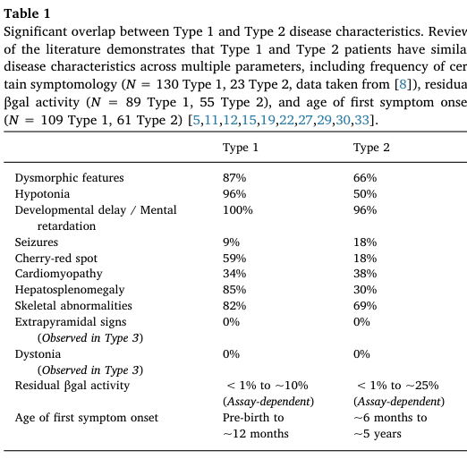
A large literature-based cohort (N=154 type 1) provides frequencies (Table 1). (lang2020thenaturalhistory pages 2-3, lang2020thenaturalhistory media 45298dc3)
Key high-frequency features (Type 1): * Developmental delay / intellectual disability (100%) (lang2020thenaturalhistory pages 2-3, lang2020thenaturalhistory media 45298dc3) * Hypotonia (96%) (lang2020thenaturalhistory pages 2-3, lang2020thenaturalhistory media 45298dc3) * Dysmorphic/coarse facial features (87%) (lang2020thenaturalhistory pages 2-3, lang2020thenaturalhistory media 45298dc3) * Hepatosplenomegaly (85%) (lang2020thenaturalhistory pages 2-3, lang2020thenaturalhistory media 45298dc3) * Skeletal abnormalities (dysostosis multiplex/skeletal dysplasia) (82%) (lang2020thenaturalhistory pages 2-3, lang2020thenaturalhistory media 45298dc3) * Cherry-red spot (59%) (lang2020thenaturalhistory pages 2-3, lang2020thenaturalhistory media 45298dc3) * Seizures (9%; described as later onset, ~10–12 months in the meta-analysis narrative) (lang2020thenaturalhistory pages 2-3, lang2020thenaturalhistory pages 3-4)
Additional phenotypes noted in summaries include Mongolian spots and cardiomyopathy in some infants. (lang2020thenaturalhistory pages 3-4, lang2020thenaturalhistory pages 1-2)
3.2 Age of onset, severity, progression
Type 1 is the most severe subtype, “traditionally… characterized by an age of first symptom onset between birth and 6 months” and death “often before 3 years.” (lang2020thenaturalhistory pages 1-2)
The meta-analysis reports that a “first symptom onset was often before 3 months of age” and identifies 6–18 months as a period of “significant neurodevelopmental regression and multi-organ system deterioration.” (lang2020thenaturalhistory pages 6-7)
3.3 Quality of life impact
Direct quantitative QoL metrics in type 1 were not found in the retrieved primary texts; however, GM1 type 1 is described as rapidly neurodegenerative with loss/failure to acquire major motor milestones (e.g., crawling/standing/walking), strongly implying profound impact on daily function and caregiving burden. (lang2020thenaturalhistory pages 6-7)
3.4 Suggested HPO terms (examples)
Based on the phenotypes above (frequency-supported where available): * Hypotonia — HP:0001252 (lang2020thenaturalhistory pages 2-3) * Global developmental delay — HP:0001263 (lang2020thenaturalhistory pages 2-3) * Hepatosplenomegaly — HP:0001433 (lang2020thenaturalhistory pages 2-3) * Coarse facial features — HP:0000280 (lang2020thenaturalhistory pages 2-3) * Cherry red spot of the macula — HP:0010729 (lang2020thenaturalhistory pages 2-3) * Seizures — HP:0001250 (lang2020thenaturalhistory pages 2-3) * Skeletal dysplasia / dysostosis multiplex — HP:0002652 (lang2020thenaturalhistory pages 2-3)
4. Genetic / molecular information
4.1 Causal gene(s)
- GLB1 (galactosidase beta 1; β-galactosidase) is the primary causal gene for GM1 gangliosidosis across subtypes. (lang2020thenaturalhistory pages 1-2, nicoli2021gm1gangliosidosis—aminireview pages 1-2, OpenTargets Search: GM1 gangliosidosis)
4.2 Pathogenic variants and variant spectrum
Lang et al. note “Over 165 mutations… reported in the GLB1 gene (ClinVar Database)” and provide type-associated examples: “Common pathogenic mutations associated with Type 1 disease, including p.R59H and c.1622-1627insG…” (lang2020thenaturalhistory pages 1-2)
Nicoli et al. report: “So far 261 pathogenic variants have been described, missense/nonsense mutations being the most prevalent,” with clustering “in exons 2, 6, 15, and 16.” (nicoli2021gm1gangliosidosis—aminireview pages 1-2)
4.3 Functional consequences
The dominant mechanism is loss of function (little or no residual β-gal activity), leading to lysosomal substrate accumulation and downstream neuronal dysfunction/neurodegeneration. (lang2020thenaturalhistory pages 1-2, rha2021gm1gangliosidosismechanisms pages 1-2)
4.4 Modifier genes / epigenetics / chromosomal abnormalities
No specific modifier genes, epigenetic signatures, or chromosomal abnormalities were supported by the retrieved evidence in this run.
4.5 Suggested ontology terms
- GO (biological process): lysosomal catabolic process (GO:0007040), sphingolipid metabolic process (GO:0006665), glycosaminoglycan catabolic process (GO:0006027)
- GO (cellular component): lysosome (GO:0005764)
(These are suggested based on the described lysosomal enzyme deficiency and substrate accumulation; the sources establish lysosomal dysfunction and substrate buildup but do not explicitly list GO terms.) (rha2021gm1gangliosidosismechanisms pages 1-2, lang2020thenaturalhistory pages 1-2)
5. Environmental information
No environmental, lifestyle, or infectious etiologies were supported by the retrieved evidence for this Mendelian disorder.
6. Mechanism / pathophysiology
6.1 Causal chain (upstream → downstream)
1) Biallelic GLB1 pathogenic variants → 2) β-galactosidase deficiency → 3) accumulation of GM1 ganglioside/GA1 and other β-galactose–terminal substrates in lysosomes (especially in neurons) → 4) impaired cellular physiology and neurodegeneration, with multisystem involvement (liver/spleen, skeleton, heart). (rha2021gm1gangliosidosismechanisms pages 1-2, lang2020thenaturalhistory pages 1-2)
6.2 Pathways / processes implicated
- Lysosomal glycosphingolipid degradation (GM1/GA1) and keratan sulfate catabolism are central biochemical pathways. (lang2020thenaturalhistory pages 1-2, NCT04713475 chunk 1)
- Pathophysiology is constrained by the blood–brain barrier for therapies (important for treatment design). (foster2024therapeuticdevelopmentsfor pages 2-4)
6.3 Suggested GO and CL terms (examples)
- GO processes: lysosomal lumen acid hydrolase activity–related pathways; neuronal cell death (GO:0070997) (suggested; neurodegeneration is supported but specific GO annotations not directly extracted). (lang2020thenaturalhistory pages 1-2)
- CL cell types (suggested): neuron (CL:0000540), astrocyte (CL:0000127), microglial cell (CL:0000129), based on CNS storage/neurodegeneration framing. (lang2020thenaturalhistory pages 1-2, rha2021gm1gangliosidosismechanisms pages 1-2)
7. Anatomical structures affected
7.1 Organ and system involvement
Type 1 involves: * Central nervous system (neuronopathic neurodegeneration) (lang2020thenaturalhistory pages 1-2) * Hepatosplenomegaly (liver/spleen involvement) (lang2020thenaturalhistory pages 2-3) * Skeletal system (skeletal dysplasia/dysostosis multiplex) (lang2020thenaturalhistory pages 2-3) * Eye (cherry-red maculae) (lang2020thenaturalhistory pages 2-3) * Heart (cardiomyopathy noted as frequent accompanying sign in infants) (lang2020thenaturalhistory pages 1-2)
7.2 Suggested UBERON terms (examples)
- Brain — UBERON:0000955 (lang2020thenaturalhistory pages 1-2)
- Liver — UBERON:0002107 (lang2020thenaturalhistory pages 2-3)
- Spleen — UBERON:0002106 (lang2020thenaturalhistory pages 2-3)
- Skeleton/bone — UBERON:0002481 (lang2020thenaturalhistory pages 2-3)
- Retina/macula — UBERON:0000966 (for macular cherry-red spot phenotype) (lang2020thenaturalhistory pages 2-3)
8. Temporal development
8.1 Onset
In a type 1 cohort (N=154), mean age of first symptom onset was 2.8 months (median 2.5; range 0–11). (lang2020thenaturalhistory pages 3-4)
8.2 Progression and milestones
The meta-analysis highlights a “predictable clinical course,” with marked regression/deterioration in 6–18 months, and failure to attain later milestones (crawling/standing/walking). (lang2020thenaturalhistory pages 6-7)
8.3 Duration
Mean age at death in the meta-analysis was 18.9 months (median 20.0; range 2–35). (lang2020thenaturalhistory pages 3-4)
9. Inheritance and population
9.1 Inheritance
Autosomal recessive inheritance is consistently described (biallelic GLB1 mutations). (lang2020thenaturalhistory pages 1-2, NCT04713475 chunk 1)
9.2 Epidemiology (statistics)
- Incidence estimates: “reported to be 1 in 100,000–200,000 live births,” with higher incidence in founder populations. (lang2020thenaturalhistory pages 1-2)
- A 2019 clinical series similarly states: “The overall incidence… is estimated to be 1:100 000–1:200 000 live births worldwide.” (Arash-Kaps et al., J Pediatr, Dec 2019; URL: https://doi.org/10.1016/j.jpeds.2019.08.016) (arashkaps2019theclinicaland pages 1-2)
9.3 Founder effects and high-incidence populations
A 2021 review reports markedly elevated incidence/carrier rates in specific populations (e.g., Malta; Roma; Cyprus/Pelendri). (rha2021gm1gangliosidosismechanisms pages 1-2)
10. Diagnostics
10.1 Clinical/biochemical tests
Enzyme assay: In a 22-patient cohort, “Residual β-galactosidase activity was measured in leucocytes… and in fibroblasts… using the artificial 4-methylumbelliferyl-β-galactopyranoside as substrate.” (Arash-Kaps et al., Dec 2019; URL: https://doi.org/10.1016/j.jpeds.2019.08.016) (arashkaps2019theclinicaland pages 1-2)
Supportive screening labs (reported abnormal proportions in that cohort): increased ASAT (13/20), chitotriosidase (12/15), pathologic urinary oligosaccharides (10/19). (arashkaps2019theclinicaland pages 1-2)
10.2 Genetic testing
Sanger sequencing of GLB1 was used in the 2019 cohort: “In 20 of 22 patients, molecular analysis of the GLB1 gene was performed… by Sanger sequencing.” (arashkaps2019theclinicaland pages 1-2)
Clinical trials specify diagnosis confirmation via genotyping (biallelic GLB1 mutations) and documented β-gal deficiency in a CLIA-certified laboratory for eligibility. (NCT03952637 chunk 1, NCT04713475 chunk 1)
10.3 Biomarkers (including recent 2023–2024 developments)
Pentasaccharide H3N2b (pharmacodynamic biomarker; 2023): * Abstract quote: “We identified two pentasaccharide biomarkers, H3N2a and H3N2b, that were elevated more than 18-fold in patient plasma, cerebrospinal fluid (CSF), and urine.” (Kell et al., eBioMedicine, published online 31 May 2023; Jun 2023; URL: https://doi.org/10.1016/j.ebiom.2023.104627) (kell2023apentasaccharidefor pages 1-2) * Abstract quote (treatment monitoring): “Following intravenous (IV) AAV9 gene therapy treatment, reduction of H3N2b was observed…” and “Reduction of H3N2b accurately reflected normalization of neuropathology… and improvement of clinical outcomes…” (kell2023apentasaccharidefor pages 1-2)
Trial-integrated biomarkers (gene therapy trials): ClinicalTrials.gov endpoints for PBGM01 include NfL in plasma/CSF, MRI measures, and ventilator-free survival (vs natural history). (NCT04713475 chunk 1)
10.4 Neuroimaging and functional testing (trial-based implementation)
The NIH AAV9-GLB1 trial protocol includes baseline and longitudinal testing: MRI/MRS/fMRI, EEG, skeletal survey, ophthalmology exam, modified barium swallow, echocardiogram/EKG, and others. (NCT03952637 chunk 1)
10.5 Newborn screening
The type 1 meta-analysis explicitly suggests improved diagnostic timing via “Implementation of newborn screening and increased use of whole exome sequencing.” (lang2020thenaturalhistory pages 6-7)
11. Outcome / prognosis
11.1 Natural history timing and survival (key statistics)
From the type 1 meta-analysis (N=154): * Mean age at diagnosis 8.7 months (lang2020thenaturalhistory pages 1-2) * Mean age at death 18.9 months (lang2020thenaturalhistory pages 1-2) * Survival distribution: “96% of patients are alive before 12 months” and “more than half of patients die between 12 and 24 months.” (lang2020thenaturalhistory pages 6-7)
From the large RETRIEVE natural history study (early-onset GM1; includes infantile presentations): * “In Group A, median (95% CI) survival was 19.0 (18.0, 22.0) months in patients with GM1.” (Héron et al., Orphanet J Rare Dis, Dec 2024; URL: https://doi.org/10.1186/s13023-024-03409-1) (heron2024anaturalhistory pages 1-2)
11.2 Prognostic factors
Earlier onset is associated with more rapid progression (concept supported in type I definition and natural history patterns), but specific quantitative prognostic models were not extracted from retrieved texts in this run. (lang2020thenaturalhistory pages 1-2, lang2020thenaturalhistory pages 6-7)
12. Treatment
12.1 Current standard of care (real-world implementation)
No approved disease-modifying therapy is documented in the retrieved sources; care is “limited to symptomatic supportive care.” (lang2020thenaturalhistory pages 1-2)
Supportive approaches referenced include seizure control and feeding/airway interventions. (rha2021gm1gangliosidosismechanisms pages 1-2)
Suggested MAXO terms (examples): * Supportive care — MAXO:0000147 (suggested) * Anticonvulsant therapy — MAXO term for antiseizure medication (suggested) * Gastrostomy tube feeding — MAXO:0000647 (suggested)
12.2 Advanced therapeutics / clinical trials (with NCT identifiers)
AAV9-GLB1 (intravenous) — NIH Phase 1/2
- ClinicalTrials.gov: “A Phase 1/2 Study of Intravenous Gene Transfer With an AAV9 Vector Expressing Human Beta-galactosidase in Type I and Type II GM1 Gangliosidosis” — NCT03952637. Start date 2019-08-19; status Recruiting (as of 2025-12 update). (NCT03952637 chunk 1)
- Route: intravenous infusion; vector: AAV9/GLB1. (NCT03952637 chunk 1)
PBGM01 (AAVhu68-GLB1; cisterna magna) — Imagine-1
- ClinicalTrials.gov: “Phase 1/2… PBGM01 delivered into the cisterna magna… Type 1 and Type 2a infantile GM1” — NCT04713475. Start date 2021-03-17; status Active not recruiting (as of 2025-05 update). (NCT04713475 chunk 1)
- Primary outcomes include treatment-related AEs/SAEs (CTCAE v5.0) up to 5 years and Bayley developmental milestones change through 2 years. (NCT04713475 chunk 1)
- Secondary outcomes include biomarkers of β-gal activity/substrates, NfL, MRI, QoL scales, and ventilator-free survival compared with natural history. (NCT04713475 chunk 1)
LYS-GM101 (AAVrh.10-GLB1; intracisternal)
- ClinicalTrials.gov: NCT04273269; status Terminated (closure not due to safety; cessation of activities). (NCT04273269 chunk 1)
- Intervention: AAVrh.10 carrying human β-galactosidase cDNA; single intracisternal injection; secondary outcomes included motor function, MRI, developmental scales, and β-gal/GM1 biomarkers in blood and CSF. (NCT04273269 chunk 1)
12.3 Substrate reduction therapy (SRT)
A 2024 review summarizes limited clinical experience with miglustat and notes a U.S. infantile GM1 trial (NCT02030015) was unsuccessful with high mortality; efficacy for type 1 remains unproven in the retrieved text. (foster2024therapeuticdevelopmentsfor pages 5-6)
A mini-review notes that a trial using a glucosylceramide synthase inhibitor venglustat is recruiting type II and III patients (not type I). (nicoli2021gm1gangliosidosis—aminireview pages 1-2)
12.4 Recent research developments (prioritizing 2023–2024)
Biomarker advancement (2023): H3N2b pentasaccharide validated as a pharmacodynamic biomarker for monitoring response to gene therapy, including evidence of reduction in treated cats and a treated patient. (kell2023apentasaccharidefor pages 1-2)
Natural history dataset expansion (2024): RETRIEVE provides one of the larger early-onset GM1 survival datasets with median survival and a multi-country cohort design, intended to support future trials and historical controls. (heron2024anaturalhistory pages 1-2)
13. Prevention
13.1 Primary prevention
For an autosomal recessive disorder, prevention primarily involves carrier screening, reproductive counseling, and prenatal/preimplantation testing, but specific guideline documents were not retrieved in this run.
13.2 Secondary prevention (early detection)
The type 1 natural history meta-analysis explicitly argues for improved public awareness/screening and mentions “Implementation of newborn screening.” (lang2020thenaturalhistory pages 6-7)
14. Other species / natural disease
Naturally occurring GM1 has been documented in multiple species, particularly as translational large-animal models: * Feline GM1 gangliosidosis (natural model used extensively for AAV gene therapy and biomarker work) (rha2021gm1gangliosidosismechanisms pages 21-21) * Ovine (sheep) GM1 (natural model) (rha2021gm1gangliosidosismechanisms pages 21-21) * Additional species reported in a model-focused source include dogs, calves, and sheep. (gross2020minimallyinvasiveroutes pages 16-20)
15. Model organisms
15.1 Mouse models
A mini-review states: “There are five mouse models of GM1-gangliosidosis… [that] share the major abnormalities and neurological symptoms that are characteristic of the most severe forms of GM1-gangliosidosis,” and have been used “to study pathogenic mechanisms, to identify biomarkers, and to evaluate therapeutic strategies.” (nicoli2021gm1gangliosidosis—aminireview pages 1-2)
15.2 Feline (cat) model
A 2021 review notes feline GM1 has been used for intracranial gene therapy with “sustained normalization of neurological disease” and for identifying “novel biomarkers… [that] reflect the clinical efficacy of gene therapy.” (rha2021gm1gangliosidosismechanisms pages 21-21)
15.3 Model limitations
A model-focused source argues murine models are invaluable but do not fully recapitulate human disease because of differences in lifespan, body/brain size, and cerebral complexity. (gross2020minimallyinvasiveroutes pages 16-20)
Evidence tables and compact reference
The following artifact consolidates key identifiers, natural history statistics, and phenotype frequencies.
| Item | Value | Evidence/source (with citation IDs) | URL (if in evidence) |
|---|---|---|---|
| Disease name | GM1 gangliosidosis type 1 | OpenTargets disease association lists “GM1 gangliosidosis type 1” with MONDO_0009260; clinical literature describes Type 1 as the infantile form (OpenTargets Search: GM1 gangliosidosis, nicoli2021gm1gangliosidosis—aminireview pages 1-2, lang2020thenaturalhistory pages 1-2) | https://platform.opentargets.org |
| MONDO ID | MONDO:0009260 | OpenTargets context for “GM1 gangliosidosis type 1” (OpenTargets Search: GM1 gangliosidosis) | https://platform.opentargets.org |
| OMIM / MIM | MIM #230500 | Lang 2020 introduction: “GM1 gangliosidosis (MIM# 230500)” (lang2020thenaturalhistory pages 1-2) | https://doi.org/10.1016/j.ymgme.2019.12.012 |
| Key causal gene | GLB1 (galactosidase beta 1) | GM1 is caused by biallelic GLB1 mutations causing β-galactosidase deficiency (nicoli2021gm1gangliosidosis—aminireview pages 1-2, lang2020thenaturalhistory pages 1-2, rha2021gm1gangliosidosismechanisms pages 1-2) | https://doi.org/10.3389/fgene.2021.734878 |
| Common synonyms | Infantile GM1 gangliosidosis; Type I GM1 gangliosidosis; early onset infantile GM1 gangliosidosis; GLB1 deficiency | Sources classify disease as Type I/infantile and note “GLB1 deficiency” as a condition label (NCT04713475 chunk 1, nicoli2021gm1gangliosidosis—aminireview pages 1-2, arashkaps2019theclinicaland pages 1-2) | https://clinicaltrials.gov/study/NCT04713475 |
| Short definition | Progressive neuronopathic lysosomal storage disorder caused by β-galactosidase deficiency, leading to accumulation of GM1 ganglioside and other β-linked galactose-containing substrates; infantile/type I is the most severe form | Review and mini-review definitions (nicoli2021gm1gangliosidosis—aminireview pages 1-2, lang2020thenaturalhistory pages 1-2, rha2021gm1gangliosidosismechanisms pages 1-2) | https://doi.org/10.2147/TACG.S206076 |
| Mean age at first symptom onset | 2.8 months (median 2.5; range 0–11) | Literature-based meta-analysis of 154 Type 1 cases (lang2020thenaturalhistory pages 3-4) | https://doi.org/10.1016/j.ymgme.2019.12.012 |
| Mean age at first hospital admission | 6.3 months (median 6.0; range 0–24) | Lang 2020 natural history meta-analysis (lang2020thenaturalhistory pages 3-4) | https://doi.org/10.1016/j.ymgme.2019.12.012 |
| Mean age at diagnosis | 8.7 months (median 8.0; range 0–30) | Lang 2020 reports “average age of diagnosis was 8.7 months” (lang2020thenaturalhistory pages 1-2, lang2020thenaturalhistory pages 3-4) | https://doi.org/10.1016/j.ymgme.2019.12.012 |
| Diagnostic delay from first symptoms | 5.9 months | Lang 2020 meta-analysis (lang2020thenaturalhistory pages 3-4) | https://doi.org/10.1016/j.ymgme.2019.12.012 |
| Mean age at death | 18.9 months (median 20.0; range 2–35) | Lang 2020 reports “average age of death was 18.9 months” (lang2020thenaturalhistory pages 1-2, lang2020thenaturalhistory pages 3-4) | https://doi.org/10.1016/j.ymgme.2019.12.012 |
| Survival pattern | 96% alive before 12 months; >50% die between 12 and 24 months; life expectancy <3 years | Lang 2020 and review summary (lang2020thenaturalhistory pages 6-7, lang2020thenaturalhistory pages 1-2) | https://doi.org/10.1016/j.ymgme.2019.12.012 |
| Median survival in early-onset GM1 cohort | 19.0 months (95% CI 18.0–22.0) | RETRIEVE natural history study, Group A, 60 GM1 patients (heron2024anaturalhistory pages 1-2) | https://doi.org/10.1186/s13023-024-03409-1 |
| Developmental delay / mental retardation frequency | 100% | Lang 2020 Table 1, Type 1 frequency summary (lang2020thenaturalhistory pages 2-3, lang2020thenaturalhistory media 45298dc3) | https://doi.org/10.1016/j.ymgme.2019.12.012 |
| Hypotonia frequency | 96% | Lang 2020 Table 1, Type 1 frequency summary (lang2020thenaturalhistory pages 2-3, lang2020thenaturalhistory media 45298dc3) | https://doi.org/10.1016/j.ymgme.2019.12.012 |
| Dysmorphic / coarse facial features frequency | 87% | Lang 2020 Table 1, Type 1 frequency summary (lang2020thenaturalhistory pages 2-3, lang2020thenaturalhistory media 45298dc3) | https://doi.org/10.1016/j.ymgme.2019.12.012 |
| Hepatosplenomegaly frequency | 85% | Lang 2020 Table 1, Type 1 frequency summary (lang2020thenaturalhistory pages 2-3, lang2020thenaturalhistory media 45298dc3) | https://doi.org/10.1016/j.ymgme.2019.12.012 |
| Skeletal abnormalities frequency | 82% | Lang 2020 Table 1, Type 1 frequency summary (lang2020thenaturalhistory pages 2-3, lang2020thenaturalhistory media 45298dc3) | https://doi.org/10.1016/j.ymgme.2019.12.012 |
| Cherry-red spot frequency | 59% | Lang 2020 Table 1, Type 1 frequency summary (lang2020thenaturalhistory pages 2-3, lang2020thenaturalhistory media 45298dc3) | https://doi.org/10.1016/j.ymgme.2019.12.012 |
| Seizure frequency | 9% | Lang 2020 Table 1, Type 1 frequency summary; later onset around 10–12 months noted in meta-analysis (lang2020thenaturalhistory pages 2-3, lang2020thenaturalhistory pages 3-4) | https://doi.org/10.1016/j.ymgme.2019.12.012 |
Table: This table condenses identifiers, nomenclature, core definition, key natural-history statistics, and major phenotype frequencies for GM1 gangliosidosis type 1 (infantile). It is useful as a compact evidence-backed reference for disease knowledge base population.
Notes on gaps vs requested template
- ICD-10/ICD-11, Orphanet IDs, and MeSH terms were not available in the retrieved evidence and thus are not reported.
- Some requested elements (e.g., formal diagnostic criteria, validated QoL instruments in type 1 cohorts, modifier genes, epigenetics) were not supported by tool-retrieved primary sources in this run.
References
-
(lang2020thenaturalhistory pages 1-2): Frederick M. Lang, Paul Korner, Mark Harnett, Ajith Karunakara, and Cynthia J. Tifft. The natural history of type 1 infantile gm1 gangliosidosis: a literature-based meta-analysis. Mar 2020. URL: https://doi.org/10.1016/j.ymgme.2019.12.012, doi:10.1016/j.ymgme.2019.12.012. This article has 54 citations and is from a peer-reviewed journal.
-
(lang2020thenaturalhistory pages 3-4): Frederick M. Lang, Paul Korner, Mark Harnett, Ajith Karunakara, and Cynthia J. Tifft. The natural history of type 1 infantile gm1 gangliosidosis: a literature-based meta-analysis. Mar 2020. URL: https://doi.org/10.1016/j.ymgme.2019.12.012, doi:10.1016/j.ymgme.2019.12.012. This article has 54 citations and is from a peer-reviewed journal.
-
(lang2020thenaturalhistory pages 6-7): Frederick M. Lang, Paul Korner, Mark Harnett, Ajith Karunakara, and Cynthia J. Tifft. The natural history of type 1 infantile gm1 gangliosidosis: a literature-based meta-analysis. Mar 2020. URL: https://doi.org/10.1016/j.ymgme.2019.12.012, doi:10.1016/j.ymgme.2019.12.012. This article has 54 citations and is from a peer-reviewed journal.
-
(rha2021gm1gangliosidosismechanisms pages 1-2): Allisandra K. Rha, Anne S. Maguire, and Douglas R. Martin. Gm1 gangliosidosis: mechanisms and management. The Application of Clinical Genetics, 14:209-233, Apr 2021. URL: https://doi.org/10.2147/tacg.s206076, doi:10.2147/tacg.s206076. This article has 96 citations.
-
(OpenTargets Search: GM1 gangliosidosis): Open Targets Query (GM1 gangliosidosis, 9 results). Buniello, A. et al. (2025). Open Targets Platform: facilitating therapeutic hypotheses building in drug discovery. Nucleic Acids Research.
-
(arashkaps2019theclinicaland pages 1-2): Laila Arash-Kaps, Katalin Komlosi, Marlene Seegräber, Stefan Diederich, Eduard Paschke, Yasmina Amraoui, Skadi Beblo, Andrea Dieckmann, Martin Smitka, and Julia B. Hennermann. The clinical and molecular spectrum of gm1 gangliosidosis. The Journal of Pediatrics, 215:152-157.e3, Dec 2019. URL: https://doi.org/10.1016/j.jpeds.2019.08.016, doi:10.1016/j.jpeds.2019.08.016. This article has 49 citations.
-
(NCT04713475 chunk 1): Study of Safety, Tolerability and Efficacy of PBGM01 in Pediatric Participants With GM1 Gangliosidosis. Gemma Biotherapeutics. 2021. ClinicalTrials.gov Identifier: NCT04713475
-
(heron2024anaturalhistory pages 1-2): Bénédicte Héron, Spyros Batzios, Eugen Mengel, Roberto Giugliani, Marc Patterson, Matthias Gautschi, Peter Cornelisse, Luba Trokan, Barbara Schwierin, and Marianne Rohrbach. A natural history study of pediatric patients with early onset of gm1 gangliosidosis, gm2 gangliosidoses, or gaucher disease type 2 (retrieve). Orphanet Journal of Rare Diseases, Dec 2024. URL: https://doi.org/10.1186/s13023-024-03409-1, doi:10.1186/s13023-024-03409-1. This article has 2 citations and is from a peer-reviewed journal.
-
(nicoli2021gm1gangliosidosis—aminireview pages 1-2): Elena-Raluca Nicoli, Ida Annunziata, Alessandra d’Azzo, Frances M. Platt, Cynthia J. Tifft, and Karolina M. Stepien. Gm1 gangliosidosis—a mini-review. Frontiers in Genetics, Sep 2021. URL: https://doi.org/10.3389/fgene.2021.734878, doi:10.3389/fgene.2021.734878. This article has 107 citations and is from a peer-reviewed journal.
-
(lang2020thenaturalhistory pages 2-3): Frederick M. Lang, Paul Korner, Mark Harnett, Ajith Karunakara, and Cynthia J. Tifft. The natural history of type 1 infantile gm1 gangliosidosis: a literature-based meta-analysis. Mar 2020. URL: https://doi.org/10.1016/j.ymgme.2019.12.012, doi:10.1016/j.ymgme.2019.12.012. This article has 54 citations and is from a peer-reviewed journal.
-
(lang2020thenaturalhistory media 45298dc3): Frederick M. Lang, Paul Korner, Mark Harnett, Ajith Karunakara, and Cynthia J. Tifft. The natural history of type 1 infantile gm1 gangliosidosis: a literature-based meta-analysis. Mar 2020. URL: https://doi.org/10.1016/j.ymgme.2019.12.012, doi:10.1016/j.ymgme.2019.12.012. This article has 54 citations and is from a peer-reviewed journal.
-
(foster2024therapeuticdevelopmentsfor pages 2-4): Dorian Foster, Lucian Williams, Noah Arnold, and Jessica Larsen. Therapeutic developments for neurodegenerative gm1 gangliosidosis. Frontiers in Neuroscience, Apr 2024. URL: https://doi.org/10.3389/fnins.2024.1392683, doi:10.3389/fnins.2024.1392683. This article has 21 citations and is from a peer-reviewed journal.
-
(NCT03952637 chunk 1): A Phase 1/2 Study of Intravenous Gene Transfer With an AAV9 Vector Expressing Human Beta-galactosidase in Type I and Type II GM1 Gangliosidosis. National Human Genome Research Institute (NHGRI). 2019. ClinicalTrials.gov Identifier: NCT03952637
-
(kell2023apentasaccharidefor pages 1-2): Pamela Kell, Rohini Sidhu, Mingxing Qian, Sonali Mishra, Elena-Raluca Nicoli, Precilla D'Souza, Cynthia J. Tifft, Amanda L. Gross, Heather L. Gray-Edwards, Douglas R. Martin, Miguel Sena- Esteves, Dennis J. Dietzen, Manmilan Singh, Jingqin Luo, Jean E. Schaffer, Daniel S. Ory, and Xuntian Jiang. A pentasaccharide for monitoring pharmacodynamic response to gene therapy in gm1 gangliosidosis. Jun 2023. URL: https://doi.org/10.1016/j.ebiom.2023.104627, doi:10.1016/j.ebiom.2023.104627. This article has 19 citations and is from a peer-reviewed journal.
-
(NCT04273269 chunk 1): A Safety and Efficacy Study of LYS-GM101 Gene Therapy in Patients With GM1 Gangliosidosis. LYSOGENE. 2021. ClinicalTrials.gov Identifier: NCT04273269
-
(foster2024therapeuticdevelopmentsfor pages 5-6): Dorian Foster, Lucian Williams, Noah Arnold, and Jessica Larsen. Therapeutic developments for neurodegenerative gm1 gangliosidosis. Frontiers in Neuroscience, Apr 2024. URL: https://doi.org/10.3389/fnins.2024.1392683, doi:10.3389/fnins.2024.1392683. This article has 21 citations and is from a peer-reviewed journal.
-
(rha2021gm1gangliosidosismechanisms pages 21-21): Allisandra K. Rha, Anne S. Maguire, and Douglas R. Martin. Gm1 gangliosidosis: mechanisms and management. The Application of Clinical Genetics, 14:209-233, Apr 2021. URL: https://doi.org/10.2147/tacg.s206076, doi:10.2147/tacg.s206076. This article has 96 citations.
-
(gross2020minimallyinvasiveroutes pages 16-20): AL Gross. Minimally invasive routes ok aav administration to treat gm1 gangliosidosis. Unknown journal, 2020.