Autosomal Recessive Spastic Ataxia 9 (SPAX9): Disease-Characteristics Research Report
Evidence cutoff: searches emphasized literature through 2024. Critical limitation: SPAX9 is exceptionally rare. The disease-specific human evidence retrieved consists of one 2018 report describing two affected siblings from one family. Consequently, phenotype frequencies such as “2/2” are descriptive of that family—not population estimates—and most natural-history, epidemiologic, and treatment fields remain unknown. No additional 2023–2024 SPAX9 clinical series or disease-specific trial was identified.
The following table provides a knowledge-base-oriented synopsis; ontology mappings marked “suggested” are annotations rather than assertions made by the source authors.
Table (click to expand)
| Domain | Summary | Ontology / Identifier Suggestions | Evidence |
|---|---|---|---|
| Identity / OMIM | Autosomal recessive spastic ataxia 9 (SPAX9); Mendelian, neurogenetic complex spastic ataxia. OMIM #618438. Do not infer MONDO/Orphanet/ICD identifiers from current evidence; unknown/not confirmed here. | OMIM: 618438; disease label: SPAX9; MONDO/Orphanet/ICD: unknown/not established in retrieved evidence | (mendozaferreira2018biallelicchp1mutation pages 1-2, mendozaferreira2018biallelicchp1mutation pages 2-4) |
| Causal gene and variant | Causal gene: CHP1 (calcineurin-like EF-hand protein 1). Founding human family carried homozygous NM_007236.4:c.52_54del, p.Lys19del (p.K19del). Variant segregated with disease in a consanguineous Moroccan pedigree; absent from public databases in the discovery study. | Gene: CHP1; variant class: in-frame 3-bp deletion; inheritance origin: germline | (mendozaferreira2018biallelicchp1mutation pages 2-4, mendozaferreira2018biallelicchp1mutation media 9ad7cc66) |
| Inheritance | Autosomal recessive; disease established in one consanguineous family with affected homozygous siblings and heterozygous parents. | Inheritance: AR | (mendozaferreira2018biallelicchp1mutation pages 1-2, mendozaferreira2018biallelicchp1mutation pages 2-4, mendozaferreira2018biallelicchp1mutation media 9ad7cc66) |
| Human evidence size | Extremely limited evidence base: 2 affected siblings in the index report; no additional pathogenic CHP1 variants found in screening cohorts (ARCA n=319; NeurOmics n=657), supporting rarity. | Evidence status: ultra-rare / sparse human evidence | (mendozaferreira2018biallelicchp1mutation pages 1-2, mendozaferreira2018biallelicchp1mutation pages 2-4) |
| Onset / course | Onset during the first decade of life; chronic progressive neurodegenerative course with gait instability, spastic ataxia, and cerebellar involvement. | HPO (inferred): Childhood onset HP:0011463; Progressive neurologic deterioration HP:0002344 | (mendozaferreira2018biallelicchp1mutation pages 2-4) |
| Core phenotypes | Core reported phenotype: gait instability / ataxia, spastic paraparesis, upper and lower motor neuron involvement, motor neuropathy, slow ocular saccades, intellectual disability, growth retardation; ovarian failure reported in the female proband, but likely not clearly attributable to CHP1 alone. | HPO (inferred): Ataxia HP:0001251; Spastic paraplegia / paraparesis HP:0001258 or HP:0002313; Peripheral neuropathy HP:0009830; Abnormal pyramidal signs HP:0002493; Slow saccadic eye movements HP:0001276; Intellectual disability HP:0001249; Short stature / growth delay HP:0004322; Primary ovarian insufficiency HP:0008209 (uncertain disease attribution) | (mendozaferreira2018biallelicchp1mutation pages 2-4, mendozaferreira2018biallelicchp1mutation pages 1-2, mendozaferreira2018biallelicchp1mutation pages 8-10) |
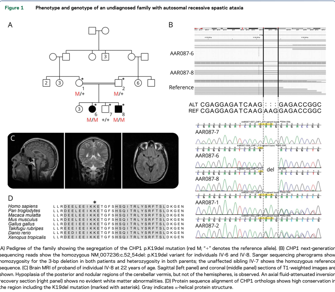
| MRI / anatomy | Brain MRI in one affected individual showed moderate cerebellar atrophy with hypoplasia of posterior and nodular regions of the cerebellar vermis, while cerebellar hemispheres were not hypoplastic; no evident white-matter abnormalities on the cited axial FLAIR image. | UBERON (inferred): cerebellum UBERON:0002037; cerebellar vermis UBERON:0002245; nervous system: UBERON:0001016 | (mendozaferreira2018biallelicchp1mutation pages 2-4, mendozaferreira2018biallelicchp1mutation media 9ad7cc66) |
| Molecular causal chain | Upstream: biallelic CHP1 p.Lys19del → reduced soluble CHP1, increased insoluble fraction, aggregation propensity, abnormal higher-molecular-weight complexes. Intermediate: impaired CHP1 support of NHE1/SLC9A1 maturation and membrane targeting. Downstream: reduced NHE1 membrane localization/function → disturbed intracellular pH/ion homeostasis → Purkinje-neuron and motor-system dysfunction → spastic ataxia phenotype. | GO (inferred): protein folding GO:0006457; protein complex assembly GO:0065003; protein localization to plasma membrane GO:1903076; sodium:hydrogen antiporter activity / regulation GO:0015385-related; intracellular pH reduction/homeostasis GO:0051453 / GO:0055078; neuron degeneration GO:0070997 | (mendozaferreira2018biallelicchp1mutation pages 1-2, mendozaferreira2018biallelicchp1mutation pages 2-2, mendozaferreira2018biallelicchp1mutation pages 4-5, mendozaferreira2018biallelicchp1mutation pages 5-8, mendozaferreira2018biallelicchp1mutation pages 8-10) |
| Affected cell types | Human phenotype and model data implicate Purkinje neurons and motor neurons / motor axons as key vulnerable populations; additional CNS regions may be sensitive to NHE1 depletion in animal models. | CL (inferred): Purkinje cell CL:0000121; motor neuron CL:0000100; broader: neuron CL:0000540 | (mendozaferreira2018biallelicchp1mutation pages 8-10, janzen2019pls3overexpressiondelays pages 5-7) |
| Diagnostic strategy | Recommended current approach: clinical recognition of childhood-onset progressive spastic ataxia plus MRI evidence of cerebellar involvement, followed by exome/genome sequencing or ataxia/spastic paraplegia gene panel including CHP1; confirm by segregation testing. No disease-specific biomarker is established. | Testing modalities: WES/WGS/panel sequencing; family segregation; MRI. Biomarker status: unknown/not established | (mendozaferreira2018biallelicchp1mutation pages 1-2, mendozaferreira2018biallelicchp1mutation pages 2-4) |
| Epidemiology | No prevalence or incidence estimates identified in retrieved evidence. Present evidence supports an ultra-rare disorder. Geographic signal from current human data: one consanguineous Moroccan family. | Epidemiology: unknown; founder effect: not established; sex ratio: unknown | (mendozaferreira2018biallelicchp1mutation pages 1-2, mendozaferreira2018biallelicchp1mutation pages 2-4) |
| Treatment / status | No SPAX9-specific approved disease-modifying therapy and no relevant registered clinical trial found in retrieved evidence. Current real-world management is expected to be supportive/multidisciplinary (rehabilitation, spasticity and mobility management, genetic counseling). PLS3 overexpression is a preclinical modifier only, not a human therapy. | NCIT (inferred supportive care): Physical Therapy C15329; Occupational Therapy C15231; Genetic Counseling C15709; disease-modifying therapy: none established | (janzen2019pls3overexpressiondelays pages 5-7, janzen2019pls3overexpressiondelays pages 1-2, janzen2019pls3overexpressiondelays pages 7-9) |
| Model organisms | Zebrafish: chp1 morphants show motor-axon defects, cerebellar hypoplasia, increased spontaneous contractions, and spastic-like trunk movements; rescued by WT but not mutant human CHP1 mRNA. Mouse: Chp1 vacillator mutants develop early balance deficits, progressive ataxic gait, Purkinje axon hypertrophy/swellings, later Purkinje-cell loss; PLS3 overexpression delays early but not late phenotype and trends toward improved NHE1 membrane localization. | Species/models: Danio rerio chp1 knockdown; Mus musculus Chp1 vacillator; evidence class: in vivo functional / modifier | (mendozaferreira2018biallelicchp1mutation pages 8-10, janzen2019pls3overexpressiondelays pages 5-7, janzen2019pls3overexpressiondelays pages 4-5) |
Table: This table condenses the currently retrievable evidence for autosomal recessive spastic ataxia 9, emphasizing the very small human evidence base, the CHP1→NHE1 mechanistic model, and practical knowledge-base fields with clearly marked inferred ontologies and unknowns.
1. Disease information
Definition
Autosomal recessive spastic ataxia 9 is a childhood-onset, complex hereditary spastic ataxia caused by biallelic pathogenic variation in CHP1, encoding calcineurin-like EF-hand protein 1. Its defining combination is cerebellar ataxia/atrophy, pyramidal involvement with spastic paraparesis, motor neuropathy, slow saccades, and neurodevelopmental impairment. It is a Mendelian neurogenetic disease rather than an acquired ataxia. The original investigators described it as “autosomal recessive spastic ataxia” and established CHP1 as an ataxia-causative gene. (mendozaferreira2018biallelicchp1mutation pages 1-2, mendozaferreira2018biallelicchp1mutation pages 2-4)
Identifiers and synonyms
- OMIM: 618438, Spastic ataxia 9, autosomal recessive.
- Common labels: SPAX9; autosomal recessive spastic ataxia type 9; CHP1-associated ataxia; CHP1-associated autosomal recessive ataxia.
- MONDO, Orphanet, MeSH: no disease-specific identifier was verified in the retrieved sources; these should remain unpopulated pending direct database validation rather than inferred.
- ICD-10/ICD-11: no unique SPAX9 code was identified. In practice, broader ataxia/hereditary spastic paraplegia codes may be used, but they are not disease-specific.
The evidence is primarily individual-patient/family-level research data, subsequently represented in aggregated disease resources such as OMIM. It is not an EHR-derived population cohort.
2. Etiology, risk, protection, and gene–environment interaction
Causal factor
The demonstrated cause is a germline, homozygous, in-frame three-base deletion in CHP1, transcript NM_007236.4:c.52_54del, protein p.Lys19del (p.K19del). It cosegregated with disease in two affected siblings; both consanguineous parents were heterozygous and an unaffected sibling carried the reference genotype. The variant was absent from the public population databases examined in 2018 and alters a highly conserved residue. (mendozaferreira2018biallelicchp1mutation media 9ad7cc66, mendozaferreira2018biallelicchp1mutation pages 2-4)
Risk factors
- Established genetic risk: two pathogenic CHP1 alleles. For the reported family, homozygosity for p.Lys19del was causal; parental consanguinity increased the probability of inheriting the same rare allele from both parents.
- Family history: an affected sibling or known carrier parents materially raises reproductive risk.
- Other variants: no validated susceptibility loci or severity-modifier alleles have been demonstrated in humans.
- Environmental, infectious, occupational, lifestyle, age, and sex risks: none established. Childhood age concerns expression/onset, not acquisition of disease.
Protective factors and gene–environment interaction
No human protective allele, diet, exercise exposure, toxin avoidance strategy, or gene–environment interaction has been established. PLS3 overexpression delayed early disease in Chp1-mutant mice, making PLS3 a candidate genetic modifier, but this has not been demonstrated in affected humans. (janzen2019pls3overexpressiondelays pages 5-7, janzen2019pls3overexpressiondelays pages 7-9)
The female proband’s ovarian failure should not automatically be considered part of SPAX9: the investigators found an additional homozygous BNC1 p.Gly258Glu candidate and noted normal reproductive anatomy in Chp1-mutant mice, supporting possible independent etiology. (mendozaferreira2018biallelicchp1mutation pages 4-5)
3. Phenotypes
Because only two affected siblings are documented, precise population frequencies and variability cannot be estimated.
- Gait instability/cerebellar ataxia: childhood onset, chronic and apparently progressive; reported in both siblings. Suggested HPO: Ataxia (HP:0001251), Gait ataxia (HP:0002066).
- Spastic paraparesis and pyramidal signs: upper-motor-neuron phenotype contributing to stiff, unstable gait; reported in the affected sibship. HPO: Spastic paraplegia (HP:0001258), Hyperreflexia (HP:0001347), Babinski sign (HP:0003487) where clinically documented.
- Motor neuropathy/lower-motor-neuron involvement: a clinical and electrophysiologic component of the complex phenotype. HPO: Motor axonal neuropathy (HP:0007002) or broader Peripheral neuropathy (HP:0009830).
- Cerebellar atrophy/hypoplasia: moderate cerebellar abnormality; MRI at age 22 showed selective hypoplasia of posterior and nodular vermian regions, sparing the hemispheres, with no evident white-matter abnormality. HPO: Cerebellar vermis hypoplasia (HP:0001320) and Cerebellar atrophy (HP:0001272). (mendozaferreira2018biallelicchp1mutation media 9ad7cc66, mendozaferreira2018biallelicchp1mutation pages 2-4)
- Slow ocular saccades: HPO Slow saccadic eye movements (HP:0000514; identifier should be ontology-validated before import).
- Intellectual disability: present in the reported siblings; severity was not robustly quantified. HPO: Intellectual disability (HP:0001249).
- Growth retardation: reported, without sufficient longitudinal anthropometry for frequency or severity estimates. HPO: Growth delay (HP:0001510) or Short stature (HP:0004322) as appropriate to measured data.
- Ovarian failure: reported in the female proband but attribution to CHP1 is uncertain because of the BNC1 finding. HPO: Primary ovarian insufficiency (HP:0008209), flagged as uncertain. (mendozaferreira2018biallelicchp1mutation pages 4-5, mendozaferreira2018biallelicchp1mutation pages 2-4)
The likely quality-of-life burden includes impaired ambulation, balance, education/cognition, independence, and participation. No SPAX9-specific EQ-5D, SF-36, PROMIS, SARA, or activities-of-daily-living dataset exists. Contemporary ataxia experts regard speech and other digital-motor measures as useful cross-stage endpoints, but that is general ataxia guidance, not SPAX9 validation.
4. Genetic and molecular information
Gene and variant
- Gene: CHP1 (calcineurin-like EF-hand protein 1); NCBI Gene 11261 was used in the experimental report.
- Disease mechanism: recessive, hypomorphic loss of normal protein function.
- Established human variant: NM_007236.4:c.52_54del; p.Lys19del, an in-frame deletion.
- Origin: constitutional/germline, not somatic.
- Population frequency: absent from databases available to the 2018 investigators; no reliable current gnomAD frequency was retrieved.
- Clinical classification: compelling disease-causal evidence comes from segregation, extreme rarity, conservation, cellular dysfunction, and animal rescue. A current ClinVar submitter-level ACMG classification was not independently retrieved and should be checked before importing a formal “pathogenic/likely pathogenic” label.
The study screened 319 ARCA cases plus 657 NeurOmics participants (976 total) and found no additional qualifying CHP1 variant; GeneMatcher also yielded none. This demonstrates extreme rarity, although it does not provide prevalence. (mendozaferreira2018biallelicchp1mutation pages 2-4)
Functional consequence
Relative to wild-type CHP1, p.Lys19del produced approximately 62% less protein in the soluble fraction and 68% more in the insoluble fraction in transfected cells. About 50% of mutant-expressing N2A cells contained aggregates versus approximately 20% of wild-type-expressing cells; large aggregates occurred in approximately 22% versus 4%, respectively. Mutant aggregates colocalized with ubiquitin and p62. The authors interpreted aggregation mainly as a readout of abnormal folding—not necessarily the primary toxic mechanism. (mendozaferreira2018biallelicchp1mutation pages 4-5, mendozaferreira2018biallelicchp1mutation pages 5-8, mendozaferreira2018biallelicchp1mutation pages 8-10)
No validated human modifier gene, disease-specific methylation signature, chromatin abnormality, structural variant, aneuploidy, or somatic mechanism is known. PLS3 is a preclinical candidate modifier only.
5. Environmental information
No toxin, radiation, pollution, occupation, smoking, alcohol, diet, exercise pattern, or infectious organism causes or triggers SPAX9. These factors may affect general health and disability but are not established etiologic modifiers. The disease is noninfectious and noncommunicable; zoonotic and pathogen-trigger concepts are not applicable.
6. Mechanism and pathophysiology
Causal chain
- Upstream genetic lesion: biallelic CHP1 p.Lys19del disrupts the conserved N-terminal α-helix.
- Protein-level defect: mutant CHP1 is unstable/poorly soluble, aggregates, and shifts into abnormal high-molecular-weight complexes.
- Transporter defect: CHP1 normally supports maturation, glycosylation, complex formation, and plasma-membrane targeting of NHE1, the SLC9A1 Na+/H+ exchanger. Mutant CHP1 reduces NHE1 membrane localization.
- Cellular consequence: impaired NHE1 compromises Na+/H+ exchange and intracellular pH/ion homeostasis.
- Selective neuronal injury: Purkinje-cell axons and motor axons are particularly vulnerable; mouse pathology supports a “dying-back” sequence in which axonal swelling/degeneration precedes Purkinje-cell loss.
- Clinical output: cerebellar dysfunction produces ataxia and vermian atrophy, while corticospinal and motor-neuron/axon involvement produces spastic paraparesis and neuropathy. (mendozaferreira2018biallelicchp1mutation pages 1-2, janzen2019pls3overexpressiondelays pages 5-7, mendozaferreira2018biallelicchp1mutation pages 5-8, mendozaferreira2018biallelicchp1mutation pages 8-10)
Suggested GO biological-process/function terms: protein folding (GO:0006457), protein-complex assembly (GO:0065003), protein localization to plasma membrane, intracellular pH homeostasis (GO:0030641/related ontology term), sodium:proton antiporter activity (GO:0015385), axon degeneration (GO:0061564), and neuron death (GO:0070997). Exact term versions should be ontology-validated.
Suggested cell types: Purkinje cell CL:0000121, motor neuron CL:0000100, and neuron CL:0000540. Compartments: plasma membrane (GO:0005886), cytosol (GO:0005829), axon (GO:0030424), and protein-containing complex (GO:0032991).
Molecular profiling and advanced technologies
No SPAX9 patient-derived transcriptomic, proteomic, metabolomic, lipidomic, single-cell, spatial-transcriptomic, organoid, multi-omic, CRISPR-screen, or disease-specific epigenomic dataset was identified. A 2023 study clarified epigenetic regulation of PLS3, but it was not a SPAX9 patient study and does not establish an SPAX9 epigenetic mechanism. (strathmann2023epigeneticregulationof pages 1-7)
7. Anatomical structures affected
- Primary system: nervous system—cerebellar and motor systems.
- Primary site: cerebellum, particularly the posterior/nodular cerebellar vermis; suggested UBERON: cerebellum UBERON:0002037, cerebellar vermis UBERON:0002245.
- Cells/tissues: Purkinje neurons and their axons; corticospinal/upper-motor pathways; peripheral motor neurons/axons.
- Secondary/uncertain: growth and cognitive systems. Ovarian involvement is not securely attributable to CHP1.
- Subcellular sites: CHP1-containing complexes, cytosol/insoluble aggregates, and NHE1 at the plasma membrane.
- Lateralization: no unilateral or asymmetric pattern reported; the syndrome is systemic/bilateral in clinical expression.
The pedigree, variant segregation, residue conservation, and selective vermian MRI abnormality are directly visualized in the discovery report’s Figure 1. (mendozaferreira2018biallelicchp1mutation media 9ad7cc66)
8. Temporal development
Onset occurred in the first decade, apparently insidiously rather than acutely. Available human observations support a chronic, lifelong, progressive course, but no formal stages or annual progression rate exist. A practical—not validated—staging concept is: early gait imbalance; intermediate combined ataxia/spasticity with neuropathy; and advanced mobility dependence. There is no documented spontaneous remission, relapsing-remitting behavior, or treatment-induced remission. (mendozaferreira2018biallelicchp1mutation pages 2-4)
Animal evidence suggests an early therapeutic window before irreversible Purkinje-cell death: vacillator mice have balance abnormalities by three weeks, progressive gait ataxia from approximately six weeks, and marked Purkinje loss by four months. PLS3 helped at three to four weeks but not at six weeks, supporting early axonal dysfunction as a potentially more modifiable phase. This remains a model-based inference, not a human critical period. (janzen2019pls3overexpressiondelays pages 5-7, janzen2019pls3overexpressiondelays pages 4-5)
9. Inheritance and population
Inheritance is autosomal recessive. For two heterozygous carrier parents, each conception has the standard Mendelian probabilities of 25% affected, 50% carrier, and 25% unaffected/noncarrier, assuming full penetrance of the familial pathogenic genotype. Penetrance cannot be independently estimated from one family; the observed homozygotes were affected. Expressivity, anticipation, and germline mosaicism are unknown. Anticipation is not expected for an in-frame deletion disorder but has not been formally studied.
The index family was consanguineous and Moroccan. This demonstrates neither a Moroccan founder allele nor elevated regional prevalence. There are no prevalence, incidence, carrier-frequency, sex-ratio, or geographic-distribution estimates. The negative screen of 976 additional individuals supports extreme rarity. (mendozaferreira2018biallelicchp1mutation pages 1-2, mendozaferreira2018biallelicchp1mutation pages 2-4)
10. Diagnostics
Clinical work-up
Suspect SPAX9 in childhood-onset complex spastic ataxia combining cerebellar signs, spastic paraparesis/pyramidal signs, motor neuropathy, slow saccades, intellectual disability, and cerebellar vermian atrophy. Recommended evaluations, extrapolated from hereditary ataxia practice, include neurologic examination, developmental/cognitive assessment, brain MRI, nerve-conduction studies/EMG, ophthalmologic examination, mobility and swallowing/speech evaluation, and endocrine assessment only when clinically indicated.
There is no validated blood, CSF, enzyme, metabolite, proteomic, or pharmacodynamic biomarker. MRI is supportive but not specific. Biopsy is not routinely indicated.
Genetic testing
- Use a hereditary ataxia/hereditary spastic paraplegia panel that includes CHP1, or preferably trio/affected-sibling WES/WGS for an unsolved complex phenotype.
- Confirm candidate CHP1 variants by an orthogonal method and test segregation.
- Assess copy-number and splice-altering variants when sequence analysis is negative; WGS/RNA studies may help unresolved cases, although disease-specific diagnostic yield is unknown.
- Single-variant testing is appropriate for relatives of a family with known p.Lys19del.
- CMA/karyotype/FISH, mitochondrial sequencing, and repeat-expansion testing do not directly diagnose known SPAX9, but may be useful in the broader differential.
The discovery used linkage plus WES and Sanger segregation. The abstract states: “We identified a biallelic 3-bp deletion (p.K19del) in CHP1 that cosegregates with the disease.” (mendozaferreira2018biallelicchp1mutation pages 1-2)
Differential diagnosis
Important alternatives include other recessive spastic ataxias and complicated HSPs; SLC9A1-related Lichtenstein–Knorr syndrome is mechanistically close but classically includes sensorineural deafness. Other differentials include NKX6-2-, KIF1C-, MAG-, CAPN1-, PNPLA6-, KIAA0415/SPG48-, CYP2U1-, and mitochondrial/repeat-expansion ataxias. Absence of deafness or epilepsy in the reported CHP1 family may reflect residual NHE1 function but is not a definitive discriminator. (mendozaferreira2018biallelicchp1mutation pages 2-2, mendozaferreira2018biallelicchp1mutation pages 8-10)
No population or newborn-screening program exists. Cascade testing of relatives is appropriate after a molecular diagnosis.
11. Outcome and prognosis
No survival curve, mortality rate, life-expectancy estimate, hospitalization rate, or validated quality-of-life measure exists. The known morbidity is progressive neurologic disability affecting gait, balance, cognition, and motor function. Recovery of lost neurons is not demonstrated; rehabilitation may preserve safety, conditioning, and function but is not known to alter neurodegeneration.
Potential complications—based on the phenotype and general neurologic care, not measured SPAX9 rates—include falls, contractures, loss of ambulation, scoliosis, pain, dysarthria/dysphagia, and caregiver burden. No prognostic biomarker or genotype–phenotype model exists. Earlier onset, severe motor neuropathy, and extent of cerebellar involvement are plausible clinical indicators but are unvalidated.
12. Treatment
No approved disease-modifying, gene, cell, RNA, editing, targeted, immunologic, or surgical therapy exists for SPAX9, and no relevant SPAX9-specific ClinicalTrials.gov study was found.
Current care is supportive and individualized:
- physical therapy, balance and gait training, stretching, strengthening, fall prevention, orthoses, walking aids, and wheelchair assessment;
- occupational therapy and accessibility/adaptive equipment;
- speech-language therapy for dysarthria, communication, and swallowing if affected;
- conventional symptomatic treatment of spasticity (e.g., oral antispastic agents or focal botulinum toxin) under specialist supervision, with caution because weakness can worsen function;
- educational/neuropsychological support, nutrition, ophthalmology, and psychosocial care;
- surveillance for orthopedic and swallowing complications.
Suggested NCIT annotations include Physical Therapy (C15329), Occupational Therapy (C15231), Speech Therapy, Assistive Device, Genetic Counseling (C15709), and Supportive Care; identifiers should be checked against the current NCIT release.
Experimental direction
In Chp1-vacillator mice, transgenic PLS3 expression was 27-fold higher in cerebellum and improved three-week beam crossing from 11.00 seconds in mutants to 8.50 seconds, versus 5.86 seconds in wild type. It reduced Purkinje axonal hypertrophy/swellings and increased NHE1 in membrane fractions, but benefits disappeared by six weeks and late gait measures were not improved. Thus PLS3/NHE1 stabilization is a mechanistic lead, not a therapy ready for clinical use. (janzen2019pls3overexpressiondelays pages 4-5, janzen2019pls3overexpressiondelays pages 5-7)
The mouse-study abstract’s appropriately limited conclusion was: “PLS3 overexpression (OE) delays the ataxic phenotype of the vacillator mice at an early but not later disease stage.” (janzen2019pls3overexpressiondelays pages 1-2)
13. Prevention
- Primary prevention: acquired-disease prevention is not applicable. Reproductive options after identifying familial variants include genetic counseling, partner/family testing, prenatal diagnosis, and preimplantation genetic testing where legally and ethically available.
- Secondary prevention: cascade testing can identify carriers and presymptomatic biallelic relatives; whether presymptomatic rehabilitation changes disease course is unknown.
- Tertiary prevention: fall prevention, contracture management, vaccinations/general preventive care, exercise adapted to ability, swallowing surveillance, and assistive devices may reduce complications.
- Vaccines, antimicrobial prophylaxis, environmental remediation, and public-health infection control: not disease-specific and not etiologically relevant.
14. Other species and natural disease
No naturally occurring veterinary counterpart or breed-associated CHP1 disease was identified. There is no zoonotic transmission. Orthologous CHP1/Chp1 genes are strongly conserved: the discovery study reported approximately 99% amino-acid identity in mouse and 92% in zebrafish, supporting comparative modeling. (mendozaferreira2018biallelicchp1mutation pages 8-10)
Relevant taxa are Homo sapiens (NCBI Taxon 9606), Mus musculus (10090), and Danio rerio (7955). Mouse Chp1 disease is a spontaneous/experimental genetic model rather than evidence of a recognized natural livestock or companion-animal syndrome.
15. Model organisms
Zebrafish
Morpholino-mediated chp1 depletion caused caudal-primary-motor-neuron truncation/absence in approximately 23% of analyzed axons, increased terminal branching in approximately 35%, and severe cerebellar hypoplasia in approximately 70% of morphants. It also produced increased spontaneous contractions and spastic-like trunk movements. Wild-type human CHP1 mRNA improved axonal defects by approximately 13–20% and cerebellar hypoplasia by 25%; p.Lys19del mRNA failed to rescue. This is strong in-vivo functional evidence, although transient morpholino models have known off-target and developmental-dose limitations. (mendozaferreira2018biallelicchp1mutation pages 5-8, mendozaferreira2018biallelicchp1mutation pages 8-10)
The discovery abstract states: “Chp1 deficiency in zebrafish, resembling the affected individuals, led to movement defects, cerebellar hypoplasia, and motor axon abnormalities, which were ameliorated by coinjection with wild-type, but not mutant, human CHP1 messenger RNA.” (mendozaferreira2018biallelicchp1mutation pages 1-2)
Mouse
The vacillator (Chp1vac/vac) mouse carries biallelic splice-altering Chp1 variants that markedly reduce CHP1. It develops early balance impairment, progressive ataxia, Purkinje-axon hypertrophy and PKCγ-positive spheroids, followed by marked axonal loss and Purkinje-cell death. The model recapitulates cerebellar degeneration and NHE1 mislocalization but does not reproduce every human feature, including intellectual disability, growth phenotype, or ovarian failure. (janzen2019pls3overexpressiondelays pages 2-4, janzen2019pls3overexpressiondelays pages 5-7)
Cellular models
HEK293T, N2A, PC12, and HeLa expression systems demonstrated mutant instability, insolubility, ubiquitin/p62-positive aggregation, abnormal complex assembly, and reduced NHE1 membrane targeting. These clarify mechanism but cannot reproduce circuit-level disease or human natural history. (mendozaferreira2018biallelicchp1mutation pages 4-5, mendozaferreira2018biallelicchp1mutation pages 5-8)
Recent developments and expert interpretation
The principal disease-defining work remains the 2018 human/genetic-functional study; the major translational extension is the 2019 PLS3-modifier mouse study. A 2023 investigation of PLS3 epigenetic regulation may inform future manipulation of this modifier, but it did not study SPAX9 patients and should not be interpreted as a clinical advance for SPAX9. No 2023–2024 expansion of the human allelic spectrum, natural-history cohort, biomarker, or therapeutic trial was found. (janzen2019pls3overexpressiondelays pages 5-7, strathmann2023epigeneticregulationof pages 1-7)
The most defensible current expert interpretation is therefore: CHP1–NHE1 dysfunction is well supported mechanistically, but disease characterization remains preliminary because the human phenotype rests on two siblings and one allele. Immediate real-world value lies in adding CHP1 to genomic evaluation of unsolved childhood complex spastic ataxia and providing molecularly informed counseling; treatment translation remains preclinical.
Key references
- Mendoza-Ferreira N, et al. Biallelic CHP1 mutation causes human autosomal recessive ataxia by impairing NHE1 function. Neurology: Genetics. Published February 2018;4(1):e209. DOI: 10.1212/NXG.0000000000000209. URL: https://doi.org/10.1212/NXG.0000000000000209. Primary human genetics, cellular assays, and zebrafish rescue. (mendozaferreira2018biallelicchp1mutation pages 1-2, mendozaferreira2018biallelicchp1mutation pages 8-10)
- Janzen E, et al. PLS3 Overexpression Delays Ataxia in Chp1 Mutant Mice. Frontiers in Neuroscience. Published September 2019;13:993. DOI: 10.3389/fnins.2019.00993. URL: https://doi.org/10.3389/fnins.2019.00993. Primary mouse modifier study. (janzen2019pls3overexpressiondelays pages 5-7)
- Strathmann EA, et al. Epigenetic regulation of plastin 3 expression by the macrosatellite DXZ4 and the transcriptional regulator CHD4. American Journal of Human Genetics. Published March 2023;110:442–459. DOI: 10.1016/j.ajhg.2023.02.004. URL: https://doi.org/10.1016/j.ajhg.2023.02.004. Relevant to regulation of the candidate modifier PLS3, not direct SPAX9 clinical evidence. (strathmann2023epigeneticregulationof pages 1-7)
Knowledge-base caution: do not infer prevalence, complete penetrance, a founder effect, ovarian involvement, treatment efficacy, or additional pathogenic CHP1 alleles from the present evidence. These remain high-priority gaps for international case matching, ClinVar reassessment, longitudinal phenotyping, patient-derived neuronal models, and natural-history study.
References
-
(mendozaferreira2018biallelicchp1mutation pages 1-2): Natalia Mendoza-Ferreira, Marie Coutelier, Eva Janzen, Seyyedmohsen Hosseinibarkooie, Heiko Löhr, Svenja Schneider, Janine Milbradt, Mert Karakaya, Markus Riessland, Christian Pichlo, Laura Torres-Benito, Andrew Singleton, Stephan Zuchner, Alexis Brice, Alexandra Durr, Matthias Hammerschmidt, Giovanni Stevanin, and Brunhilde Wirth. Biallelic chp1 mutation causes human autosomal recessive ataxia by impairing nhe1 function. Neurology Genetics, Feb 2018. URL: https://doi.org/10.1212/nxg.0000000000000209, doi:10.1212/nxg.0000000000000209. This article has 34 citations.
-
(mendozaferreira2018biallelicchp1mutation pages 2-4): Natalia Mendoza-Ferreira, Marie Coutelier, Eva Janzen, Seyyedmohsen Hosseinibarkooie, Heiko Löhr, Svenja Schneider, Janine Milbradt, Mert Karakaya, Markus Riessland, Christian Pichlo, Laura Torres-Benito, Andrew Singleton, Stephan Zuchner, Alexis Brice, Alexandra Durr, Matthias Hammerschmidt, Giovanni Stevanin, and Brunhilde Wirth. Biallelic chp1 mutation causes human autosomal recessive ataxia by impairing nhe1 function. Neurology Genetics, Feb 2018. URL: https://doi.org/10.1212/nxg.0000000000000209, doi:10.1212/nxg.0000000000000209. This article has 34 citations.
-
(mendozaferreira2018biallelicchp1mutation media 9ad7cc66): Natalia Mendoza-Ferreira, Marie Coutelier, Eva Janzen, Seyyedmohsen Hosseinibarkooie, Heiko Löhr, Svenja Schneider, Janine Milbradt, Mert Karakaya, Markus Riessland, Christian Pichlo, Laura Torres-Benito, Andrew Singleton, Stephan Zuchner, Alexis Brice, Alexandra Durr, Matthias Hammerschmidt, Giovanni Stevanin, and Brunhilde Wirth. Biallelic chp1 mutation causes human autosomal recessive ataxia by impairing nhe1 function. Neurology Genetics, Feb 2018. URL: https://doi.org/10.1212/nxg.0000000000000209, doi:10.1212/nxg.0000000000000209. This article has 34 citations.
-
(mendozaferreira2018biallelicchp1mutation pages 8-10): Natalia Mendoza-Ferreira, Marie Coutelier, Eva Janzen, Seyyedmohsen Hosseinibarkooie, Heiko Löhr, Svenja Schneider, Janine Milbradt, Mert Karakaya, Markus Riessland, Christian Pichlo, Laura Torres-Benito, Andrew Singleton, Stephan Zuchner, Alexis Brice, Alexandra Durr, Matthias Hammerschmidt, Giovanni Stevanin, and Brunhilde Wirth. Biallelic chp1 mutation causes human autosomal recessive ataxia by impairing nhe1 function. Neurology Genetics, Feb 2018. URL: https://doi.org/10.1212/nxg.0000000000000209, doi:10.1212/nxg.0000000000000209. This article has 34 citations.
-
(mendozaferreira2018biallelicchp1mutation pages 2-2): Natalia Mendoza-Ferreira, Marie Coutelier, Eva Janzen, Seyyedmohsen Hosseinibarkooie, Heiko Löhr, Svenja Schneider, Janine Milbradt, Mert Karakaya, Markus Riessland, Christian Pichlo, Laura Torres-Benito, Andrew Singleton, Stephan Zuchner, Alexis Brice, Alexandra Durr, Matthias Hammerschmidt, Giovanni Stevanin, and Brunhilde Wirth. Biallelic chp1 mutation causes human autosomal recessive ataxia by impairing nhe1 function. Neurology Genetics, Feb 2018. URL: https://doi.org/10.1212/nxg.0000000000000209, doi:10.1212/nxg.0000000000000209. This article has 34 citations.
-
(mendozaferreira2018biallelicchp1mutation pages 4-5): Natalia Mendoza-Ferreira, Marie Coutelier, Eva Janzen, Seyyedmohsen Hosseinibarkooie, Heiko Löhr, Svenja Schneider, Janine Milbradt, Mert Karakaya, Markus Riessland, Christian Pichlo, Laura Torres-Benito, Andrew Singleton, Stephan Zuchner, Alexis Brice, Alexandra Durr, Matthias Hammerschmidt, Giovanni Stevanin, and Brunhilde Wirth. Biallelic chp1 mutation causes human autosomal recessive ataxia by impairing nhe1 function. Neurology Genetics, Feb 2018. URL: https://doi.org/10.1212/nxg.0000000000000209, doi:10.1212/nxg.0000000000000209. This article has 34 citations.
-
(mendozaferreira2018biallelicchp1mutation pages 5-8): Natalia Mendoza-Ferreira, Marie Coutelier, Eva Janzen, Seyyedmohsen Hosseinibarkooie, Heiko Löhr, Svenja Schneider, Janine Milbradt, Mert Karakaya, Markus Riessland, Christian Pichlo, Laura Torres-Benito, Andrew Singleton, Stephan Zuchner, Alexis Brice, Alexandra Durr, Matthias Hammerschmidt, Giovanni Stevanin, and Brunhilde Wirth. Biallelic chp1 mutation causes human autosomal recessive ataxia by impairing nhe1 function. Neurology Genetics, Feb 2018. URL: https://doi.org/10.1212/nxg.0000000000000209, doi:10.1212/nxg.0000000000000209. This article has 34 citations.
-
(janzen2019pls3overexpressiondelays pages 5-7): Eva Janzen, Lisa Wolff, Natalia Mendoza-Ferreira, Kristina Hupperich, Andrea Delle Vedove, Seyyedmohsen Hosseinibarkooie, Min Jeong Kye, and Brunhilde Wirth. Pls3 overexpression delays ataxia in chp1 mutant mice. Frontiers in Neuroscience, Sep 2019. URL: https://doi.org/10.3389/fnins.2019.00993, doi:10.3389/fnins.2019.00993. This article has 11 citations and is from a peer-reviewed journal.
-
(janzen2019pls3overexpressiondelays pages 1-2): Eva Janzen, Lisa Wolff, Natalia Mendoza-Ferreira, Kristina Hupperich, Andrea Delle Vedove, Seyyedmohsen Hosseinibarkooie, Min Jeong Kye, and Brunhilde Wirth. Pls3 overexpression delays ataxia in chp1 mutant mice. Frontiers in Neuroscience, Sep 2019. URL: https://doi.org/10.3389/fnins.2019.00993, doi:10.3389/fnins.2019.00993. This article has 11 citations and is from a peer-reviewed journal.
-
(janzen2019pls3overexpressiondelays pages 7-9): Eva Janzen, Lisa Wolff, Natalia Mendoza-Ferreira, Kristina Hupperich, Andrea Delle Vedove, Seyyedmohsen Hosseinibarkooie, Min Jeong Kye, and Brunhilde Wirth. Pls3 overexpression delays ataxia in chp1 mutant mice. Frontiers in Neuroscience, Sep 2019. URL: https://doi.org/10.3389/fnins.2019.00993, doi:10.3389/fnins.2019.00993. This article has 11 citations and is from a peer-reviewed journal.
-
(janzen2019pls3overexpressiondelays pages 4-5): Eva Janzen, Lisa Wolff, Natalia Mendoza-Ferreira, Kristina Hupperich, Andrea Delle Vedove, Seyyedmohsen Hosseinibarkooie, Min Jeong Kye, and Brunhilde Wirth. Pls3 overexpression delays ataxia in chp1 mutant mice. Frontiers in Neuroscience, Sep 2019. URL: https://doi.org/10.3389/fnins.2019.00993, doi:10.3389/fnins.2019.00993. This article has 11 citations and is from a peer-reviewed journal.
-
(strathmann2023epigeneticregulationof pages 1-7): Eike A. Strathmann, Irmgard Hölker, Nikolai Tschernoster, Seyyedmohsen Hosseinibarkooie, Julien Come, Cecile Martinat, Janine Altmüller, and Brunhilde Wirth. Epigenetic regulation of plastin 3 expression by the macrosatellite dxz4 and the transcriptional regulator chd4. The American Journal of Human Genetics, 110:442-459, Mar 2023. URL: https://doi.org/10.1016/j.ajhg.2023.02.004, doi:10.1016/j.ajhg.2023.02.004. This article has 15 citations.
-
(janzen2019pls3overexpressiondelays pages 2-4): Eva Janzen, Lisa Wolff, Natalia Mendoza-Ferreira, Kristina Hupperich, Andrea Delle Vedove, Seyyedmohsen Hosseinibarkooie, Min Jeong Kye, and Brunhilde Wirth. Pls3 overexpression delays ataxia in chp1 mutant mice. Frontiers in Neuroscience, Sep 2019. URL: https://doi.org/10.3389/fnins.2019.00993, doi:10.3389/fnins.2019.00993. This article has 11 citations and is from a peer-reviewed journal.
Artifacts
Reference Validation
Checked with linkml-reference-validator 0.2.1.
Table (click to expand)
| Outcome | Count |
|---|---|
| References checked | 4 |
| Resolved | 4 |
| Unresolved (possible confabulation) | 0 |
| Unverifiable | 0 |
| References weighed for topical relevance | 4 |
| On topic | 0 |
| Off topic | 0 |
All extracted references resolved successfully.