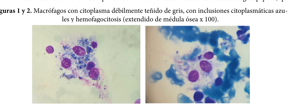
Sea-Blue Histiocyte Syndrome
Sea-blue histiocyte syndrome (sea-blue histiocytosis) is a lipid-storage histiocytosis defined by the accumulation of histiocytes (tissue macrophages) laden with deep blue/blue-green cytoplasmic granules on Wright-Giemsa (May-Grunwald-Giemsa) staining, classically identified in the bone marrow and also in the spleen, liver, lymph nodes, and occasionally lung. The granules represent lysosomal accumulation of indigestible, oxidized lipid and lipoprotein material that polymerizes into ceroid/lipofuscin pigment. The entity is heterogeneous. A rare primary (idiopathic/genetic) form presents as a clinical syndrome of splenomegaly, thrombocytopenia, and hypertriglyceridemia; the best molecularly characterized primary kindreds carry an autosomal dominant APOE mutation (p.Leu149del) in the receptor-binding region, and historically a primary sphingomyelinase-related form was grouped with Niemann-Pick disease (the now-disfavored "type F"). Much more commonly, sea-blue histiocytes are a secondary/reactive marrow finding accompanying lysosomal storage diseases (Niemann-Pick, Gaucher), states of high marrow-precursor turnover (myeloproliferative neoplasms, myelodysplasia, ineffective erythropoiesis), severe dyslipidemias, prolonged total parenteral nutrition, and certain drugs. Because the finding itself often carries little independent prognostic weight, its discovery should prompt a search for an underlying disorder. The course of primary disease is generally chronic and benign, although a minority develop progressive hepatic disease.
Ask OpenScientist
Ask a research question about Sea-Blue Histiocyte Syndrome. OpenScientist will conduct autonomous deep research using the Disorder Mechanisms Knowledge Base and PubMed literature (typically 10-30 minutes).
Do not include personal health information in your question. Questions and results are cached in your browser's local storage.
Mappings
Definitions
1Show evidence (1 reference)
Inheritance
1Show evidence (1 reference)
Subtypes
2Discussions and Knowledge Gaps
1Pathophysiology
3Show evidence (1 reference)
Show evidence (1 reference)
Show evidence (1 reference)
Pathograph
Phenotypes
6Blood 2
Show evidence (2 references)
Show evidence (1 reference)
Cardiovascular 2
Show evidence (1 reference)
Show evidence (1 reference)
Digestive 1
Show evidence (1 reference)
Metabolism 1
Show evidence (1 reference)
Genetic Associations
1Show evidence (2 references)
Medical Actions
2Show evidence (1 reference)
Show evidence (1 reference)
Biochemical Markers
1Show evidence (1 reference)
Diagnosis
3Show evidence (1 reference)
Show evidence (1 reference)
Show evidence (1 reference)
Progression
1Show evidence (1 reference)
Differential Diagnoses
3Conditions with similar clinical presentations that must be differentiated from Sea-Blue Histiocyte Syndrome:
- Confirmed by deficient acid (lysosomal) sphingomyelinase activity on enzyme assay and SMPD1 genotyping.
- Disease-modifying enzyme replacement therapy (olipudase alfa) is available, unlike idiopathic sea-blue histiocyte syndrome.
Show evidence (2 references)
- Caused by NPC1/NPC2 defects of intracellular cholesterol trafficking, with prominent neurological involvement.
- Confirmed by molecular testing and biochemical markers (e.g., plasma oxysterols); substrate-reduction therapy with miglustat is available.
Show evidence (1 reference)
- Gaucher cells have a characteristic crumpled-tissue-paper cytoplasm rather than sea-blue granules.
- Confirmed by deficient glucocerebrosidase activity and GBA genotyping.
Show evidence (1 reference)
Source YAML
click to showname: Sea-Blue Histiocyte Syndrome
creation_date: "2026-06-13T00:00:00Z"
description: >-
Sea-blue histiocyte syndrome (sea-blue histiocytosis) is a lipid-storage histiocytosis
defined by the accumulation of histiocytes (tissue macrophages) laden with deep
blue/blue-green cytoplasmic granules on Wright-Giemsa (May-Grunwald-Giemsa) staining,
classically identified in the bone marrow and also in the spleen, liver, lymph nodes,
and occasionally lung. The granules represent lysosomal accumulation of indigestible,
oxidized lipid and lipoprotein material that polymerizes into ceroid/lipofuscin pigment.
The entity is heterogeneous. A rare primary (idiopathic/genetic) form presents as a
clinical syndrome of splenomegaly, thrombocytopenia, and hypertriglyceridemia; the
best molecularly characterized primary kindreds carry an autosomal dominant APOE
mutation (p.Leu149del) in the receptor-binding region, and historically a primary
sphingomyelinase-related form was grouped with Niemann-Pick disease (the now-disfavored
"type F"). Much more commonly, sea-blue histiocytes are a secondary/reactive marrow
finding accompanying lysosomal storage diseases (Niemann-Pick, Gaucher), states of
high marrow-precursor turnover (myeloproliferative neoplasms, myelodysplasia,
ineffective erythropoiesis), severe dyslipidemias, prolonged total parenteral
nutrition, and certain drugs. Because the finding itself often carries little
independent prognostic weight, its discovery should prompt a search for an underlying
disorder. The course of primary disease is generally chronic and benign, although a
minority develop progressive hepatic disease.
category: Mendelian
disease_term:
preferred_term: sea-blue histiocyte syndrome
term:
id: MONDO:0010017
label: sea-blue histiocyte syndrome
mappings:
mondo_mappings:
- term:
id: MONDO:0010017
label: sea-blue histiocyte syndrome
mapping_predicate: skos:exactMatch
mapping_source: MONDO
mapping_justification: Primary MONDO disease identifier for this sea-blue histiocyte syndrome entry.
synonyms:
- Sea-blue histiocytosis
- Primary sea-blue histiocytosis
- Inherited lipemic splenomegaly
- Niemann-Pick disease type F
parents:
- non-Langerhans cell histiocytosis
- sphingolipidosis
has_subtypes:
- name: Primary
display_name: Primary (idiopathic/genetic) sea-blue histiocyte syndrome
description: >-
A rare primary form presenting as a clinical syndrome of splenomegaly,
thrombocytopenia, and hypertriglyceridemia in the absence of an identifiable
underlying disorder. The best molecularly defined primary kindreds carry an autosomal
dominant APOE p.Leu149del mutation; a historical sphingomyelinase-deficient primary
form was grouped with Niemann-Pick disease.
- name: Secondary
display_name: Secondary (reactive) sea-blue histiocytosis
description: >-
The more common situation, in which sea-blue histiocytes are a reactive marrow finding
secondary to another disorder: lysosomal storage diseases (Niemann-Pick, Gaucher),
high marrow-precursor turnover (myeloproliferative neoplasms, myelodysplasia,
ineffective erythropoiesis, immune thrombocytopenia), severe dyslipidemia, prolonged
total parenteral nutrition, or drug exposure.
pathophysiology:
- name: Defective Macrophage Lipid and Lipoprotein Handling
description: >-
The initiating defect is impaired macrophage handling of lipids and lipoproteins. In
the best-characterized primary form, an autosomal dominant APOE mutation (p.Leu149del)
in the LDL-receptor-binding region deranges lipoprotein metabolism and drives
macrophage hypercatabolism of lipoproteins. In secondary forms the upstream cause is a
lysosomal hydrolase deficiency (e.g., acid sphingomyelinase) or an excess substrate
load (high cell turnover, dyslipidemia, intravenous lipid emulsion).
gene:
preferred_term: APOE
term:
id: hgnc:613
label: APOE
cell_types:
- preferred_term: macrophage
term:
id: CL:0000235
label: macrophage
biological_processes:
- preferred_term: lipoprotein metabolic process
modifier: ABNORMAL
term:
id: GO:0042157
label: lipoprotein metabolic process
evidence:
- reference: PMID:11095479
reference_title: "Familial splenomegaly: macrophage hypercatabolism of lipoproteins associated with apolipoprotein E mutation [apolipoprotein E (delta149 Leu)]."
supports: SUPPORT
evidence_source: HUMAN_CLINICAL
snippet: "we provide evidence linking the syndrome to an inherited dominant mutation in the apoE gene, a 3-bp deletion on the background of an apoE 3 allele that causes a derangement in lipid metabolism and leads to splenomegaly"
explanation: "Links primary sea-blue histiocyte syndrome to a dominant APOE mutation deranging lipid metabolism in two kindreds."
downstream:
- target: Lysosomal Ceroid Accumulation and Sea-Blue Pigment Formation
description: Defective lipid handling overloads the macrophage lysosome with indigestible lipid.
- name: Lysosomal Ceroid Accumulation and Sea-Blue Pigment Formation
description: >-
Indigestible, oxidized lipid and lipoprotein material accumulates within macrophage
lysosomes and polymerizes into ceroid/lipofuscin pigment. The resulting lipid-laden
macrophages display the characteristic deep blue cytoplasmic granules on Wright-Giemsa
staining that define the sea-blue histiocyte.
cell_types:
- preferred_term: macrophage
term:
id: CL:0000235
label: macrophage
cellular_components:
- preferred_term: lysosome
term:
id: GO:0005764
label: lysosome
biological_processes:
- preferred_term: lipid storage
modifier: INCREASED
term:
id: GO:0019915
label: lipid storage
evidence:
- reference: PMID:33062516
reference_title: "Sea Blue Histiocytosis Concordant With Immune Thrombocytopenic Purpura."
supports: SUPPORT
evidence_source: HUMAN_CLINICAL
snippet: "lipid-laden blue-stained macrophages"
explanation: "The defining cell is a lipid-laden macrophage that stains blue, reflecting stored lipid pigment."
downstream:
- target: Sea-blue histiocytosis
description: >-
Ceroid/lipofuscin accumulation in macrophage lysosomes produces the
lipid-laden blue-staining histiocytes that define sea-blue histiocytosis.
- target: Storage Histiocyte Infiltration of Reticuloendothelial Organs
description: Pigment-laden histiocytes accumulate in and infiltrate reticuloendothelial tissues.
- name: Storage Histiocyte Infiltration of Reticuloendothelial Organs
description: >-
Sea-blue histiocytes accumulate in the bone marrow, spleen, and liver (and at times
lymph nodes and lung), producing organomegaly. Splenic involvement with sequestration
and marrow infiltration underlies the thrombocytopenia and other cytopenias.
cell_types:
- preferred_term: macrophage
term:
id: CL:0000235
label: macrophage
locations:
- preferred_term: bone marrow
term:
id: UBERON:0002371
label: bone marrow
- preferred_term: spleen
term:
id: UBERON:0002106
label: spleen
- preferred_term: liver
term:
id: UBERON:0002107
label: liver
evidence:
- reference: PMID:37865572
reference_title: "Sea-blue histiocytosis."
supports: SUPPORT
evidence_source: HUMAN_CLINICAL
snippet: "hepatomegaly and splenomegaly were identified, with PAS and Giemsa positive intracellular ceroid granular deposits"
explanation: "Autopsy shows hepatosplenomegaly with ceroid-laden histiocytes, reflecting storage-histiocyte infiltration of reticuloendothelial organs."
downstream:
- target: Splenomegaly
description: Splenic histiocyte accumulation produces splenomegaly.
- target: Hepatomegaly
description: Hepatic histiocyte accumulation produces hepatomegaly.
- target: Thrombocytopenia
description: Splenic sequestration and marrow infiltration drive thrombocytopenia.
- target: Lymphadenopathy
description: Histiocyte infiltration of lymphoid tissue can produce lymphadenopathy.
phenotypes:
- category: Cellular
name: Sea-blue histiocytosis
description: >-
The pathognomonic finding is lipid-laden macrophages with deep blue/blue-green
granular cytoplasm on Wright/May-Grunwald-Giemsa staining in the bone marrow; the
ceroid granules are also PAS positive. Its discovery should prompt evaluation for an
underlying disorder.
diagnostic: true
phenotype_term:
preferred_term: Sea-blue histiocytosis
term:
id: HP:0001982
label: Sea-blue histiocytosis
evidence:
- reference: PMID:33062516
reference_title: "Sea Blue Histiocytosis Concordant With Immune Thrombocytopenic Purpura."
supports: SUPPORT
evidence_source: HUMAN_CLINICAL
snippet: "Sea blue histiocytosis is an unusual bone marrow finding in many haematological conditions or lipid metabolic diseases that by itself may not carry any prognostic value."
explanation: "Sea-blue histiocytes are an unusual bone marrow morphologic finding that defines the entity and prompts a search for an underlying cause."
- reference: PMID:37865572
reference_title: "Sea-blue histiocytosis."
supports: SUPPORT
evidence_source: HUMAN_CLINICAL
snippet: "PAS and Giemsa positive intracellular ceroid granular deposits"
explanation: "The histiocytes contain PAS- and Giemsa-positive ceroid granules."
- name: Splenomegaly
description: >-
Splenomegaly is the most consistent clinical finding, reflecting accumulation of
storage histiocytes in the spleen.
phenotype_term:
preferred_term: Splenomegaly
term:
id: HP:0001744
label: Splenomegaly
evidence:
- reference: PMID:33062516
reference_title: "Sea Blue Histiocytosis Concordant With Immune Thrombocytopenic Purpura."
supports: SUPPORT
evidence_source: HUMAN_CLINICAL
snippet: "It may occur rarely as a primary genetic clinical syndrome characterized by splenomegaly, hypertriglyceridemia and thrombocytopenia."
explanation: "Splenomegaly is a defining feature of the primary genetic syndrome."
- name: Hepatomegaly
description: Hepatomegaly frequently accompanies splenomegaly.
phenotype_term:
preferred_term: Hepatomegaly
term:
id: HP:0002240
label: Hepatomegaly
evidence:
- reference: PMID:37865572
reference_title: "Sea-blue histiocytosis."
supports: SUPPORT
evidence_source: HUMAN_CLINICAL
snippet: "hepatomegaly and splenomegaly were identified"
explanation: "Hepatomegaly is documented together with splenomegaly."
- name: Thrombocytopenia
description: >-
Thrombocytopenia, often with a hemorrhagic diathesis, is part of the classic primary
syndrome and is commonly aggravated by hypersplenism.
phenotype_term:
preferred_term: Thrombocytopenia
term:
id: HP:0001873
label: Thrombocytopenia
evidence:
- reference: PMID:33062516
reference_title: "Sea Blue Histiocytosis Concordant With Immune Thrombocytopenic Purpura."
supports: SUPPORT
evidence_source: HUMAN_CLINICAL
snippet: "It may occur rarely as a primary genetic clinical syndrome characterized by splenomegaly, hypertriglyceridemia and thrombocytopenia."
explanation: "Thrombocytopenia is a defining feature of the primary genetic syndrome."
- name: Hypertriglyceridemia
description: >-
Elevated serum triglycerides are characteristic of the primary APOE-associated
syndrome and may worsen markedly after splenectomy.
phenotype_term:
preferred_term: Hypertriglyceridemia
term:
id: HP:0002155
label: Hypertriglyceridemia
evidence:
- reference: PMID:16094309
reference_title: "Variable expressivity of the clinical and biochemical phenotype associated with the apolipoprotein E p.Leu149del mutation."
supports: SUPPORT
evidence_source: HUMAN_CLINICAL
snippet: "Splenomegaly with sea-blue histiocytes, thrombocytopenia and hypertriglyceridemia is a very rare association"
explanation: "Hypertriglyceridemia is part of the characteristic primary triad with splenomegaly and thrombocytopenia."
- name: Lymphadenopathy
description: Lymph node involvement can occur as histiocytes infiltrate lymphoid tissue.
phenotype_term:
preferred_term: Lymphadenopathy
term:
id: HP:0002716
label: Lymphadenopathy
evidence:
- reference: PMID:37865572
reference_title: "Sea-blue histiocytosis."
supports: SUPPORT
evidence_source: HUMAN_CLINICAL
snippet: "It is characterized by hepatosplenomegaly, thrombocytopenia and lymphadenopathy."
explanation: "Lymphadenopathy is listed among the characteristic features of sea-blue histiocytosis."
biochemical:
- name: Hypertriglyceridemia
presence: INCREASED
context: >-
Elevated serum triglycerides are a biochemical hallmark of the primary
APOE-associated syndrome and characteristically rise further after splenectomy.
biomarker_term:
preferred_term: triglyceride
term:
id: CHEBI:17855
label: triglyceride
evidence:
- reference: PMID:33062516
reference_title: "Sea Blue Histiocytosis Concordant With Immune Thrombocytopenic Purpura."
supports: SUPPORT
evidence_source: HUMAN_CLINICAL
snippet: "It may occur rarely as a primary genetic clinical syndrome characterized by splenomegaly, hypertriglyceridemia and thrombocytopenia."
explanation: "Elevated triglycerides (hypertriglyceridemia) are a defining biochemical feature of the primary syndrome."
genetic:
- name: APOE
association: Autosomal dominant APOE p.Leu149del in the receptor-binding region (primary form)
relationship_type: CAUSATIVE
variant_origin: GERMLINE
gene_term:
preferred_term: APOE
term:
id: hgnc:613
label: APOE
inheritance:
- name: Autosomal dominant
evidence:
- reference: PMID:11095479
reference_title: "Familial splenomegaly: macrophage hypercatabolism of lipoproteins associated with apolipoprotein E mutation [apolipoprotein E (delta149 Leu)]."
supports: SUPPORT
evidence_source: HUMAN_CLINICAL
snippet: "we provide evidence linking the syndrome to an inherited dominant mutation in the apoE gene"
explanation: "Primary sea-blue histiocyte syndrome in these kindreds follows dominant inheritance of an APOE mutation."
notes: >-
Sea-blue histiocyte syndrome is genetically heterogeneous. The molecularly defined
primary kindreds carry a 3-bp deletion removing leucine 149 of APOE (a French-Canadian
founder allele); a historical sphingomyelinase-deficient primary form overlaps with
Niemann-Pick disease.
evidence:
- reference: PMID:11095479
reference_title: "Familial splenomegaly: macrophage hypercatabolism of lipoproteins associated with apolipoprotein E mutation [apolipoprotein E (delta149 Leu)]."
supports: SUPPORT
evidence_source: HUMAN_CLINICAL
snippet: "A mutation of apoE was demonstrated, with a 3-bp deletion resulting in the loss of a leucine at position 149 in the receptor-binding region of the apoE molecule"
explanation: "Identifies the APOE p.Leu149del receptor-binding-region variant in primary sea-blue histiocyte syndrome."
- reference: PMID:16094309
reference_title: "Variable expressivity of the clinical and biochemical phenotype associated with the apolipoprotein E p.Leu149del mutation."
supports: SUPPORT
evidence_source: HUMAN_CLINICAL
snippet: "An apoE p.Leu149del mutation was found in both subjects."
explanation: "Independent family confirms the APOE p.Leu149del mutation in the primary syndrome."
inheritance:
- name: Autosomal dominant
inheritance_term:
preferred_term: Autosomal dominant inheritance
term:
id: HP:0000006
label: Autosomal dominant inheritance
evidence:
- reference: PMID:11095479
reference_title: "Familial splenomegaly: macrophage hypercatabolism of lipoproteins associated with apolipoprotein E mutation [apolipoprotein E (delta149 Leu)]."
supports: SUPPORT
evidence_source: HUMAN_CLINICAL
snippet: "we provide evidence linking the syndrome to an inherited dominant mutation in the apoE gene"
explanation: "The molecularly defined primary APOE-associated form is inherited as an autosomal dominant trait; a historical sphingomyelinase-deficient primary form is recessive and overlaps Niemann-Pick disease."
progression:
- phase: Chronic benign course
subtype: Primary
notes: >-
Primary sea-blue histiocyte syndrome generally follows a chronic, benign course,
although a minority of patients develop progressive hepatic disease.
evidence:
- reference: PMID:37865572
reference_title: "Sea-blue histiocytosis."
supports: SUPPORT
evidence_source: HUMAN_CLINICAL
snippet: "Sea-blue histiocytosis is an extremely rare, chronic and benign deposit disease."
explanation: "Characterizes the typical chronic, benign natural history."
diagnosis:
- name: Bone marrow examination
diagnosis_term:
preferred_term: clinical cytological testing
term:
id: NCIT:C16490
label: Cytological Procedure
description: >-
Wright/May-Grunwald-Giemsa-stained bone marrow aspirate or biopsy demonstrating
lipid-laden macrophages with deep blue (sea-blue) cytoplasmic granules. This
morphologic finding establishes the diagnosis and should prompt a search for an
underlying disorder.
markers: Sea-blue histiocytes with PAS- and Giemsa-positive ceroid granules on marrow smear.
evidence:
- reference: PMID:33062516
reference_title: "Sea Blue Histiocytosis Concordant With Immune Thrombocytopenic Purpura."
supports: SUPPORT
evidence_source: HUMAN_CLINICAL
snippet: "Sea blue histiocytosis is an unusual bone marrow finding in many haematological conditions or lipid metabolic diseases that by itself may not carry any prognostic value."
explanation: "The diagnosis rests on identifying sea-blue histiocytes on bone marrow examination."
- name: Acid sphingomyelinase enzyme assay
diagnosis_term:
preferred_term: clinical laboratory procedure
term:
id: NCIT:C25294
label: Laboratory Procedure
description: >-
Leukocyte acid (lysosomal) sphingomyelinase activity assay to evaluate for underlying
Niemann-Pick disease (acid sphingomyelinase deficiency) when sea-blue histiocytes are
found.
markers: Reduced leukocyte acid sphingomyelinase activity in acid sphingomyelinase deficiency.
evidence:
- reference: PMID:11732877
reference_title: "Sea-blue histiocytosis secondary to Niemann-Pick disease type B: a case report."
supports: SUPPORT
evidence_source: HUMAN_CLINICAL
snippet: "confirmed by the measurement of acid lysosomal sphingomyelinase activity below normal values"
explanation: "A sphingomyelinase enzyme assay confirms or excludes underlying Niemann-Pick disease in patients with sea-blue histiocytes."
- name: Molecular genetic testing
diagnosis_term:
preferred_term: genetic testing
term:
id: NCIT:C15709
label: Genetic Testing
description: >-
Molecular testing (e.g., APOE, SMPD1, NPC1/NPC2) is increasingly the preferred
confirmatory approach over biopsy for the lysosomal storage-disease differentials.
evidence:
- reference: PMID:38592278
reference_title: "Hepatomegaly and Splenomegaly: An Approach to the Diagnosis of Lysosomal Storage Diseases."
supports: SUPPORT
evidence_source: OTHER
snippet: "preferred confirmatory test (over biopsy), accompanied by enzymatic testing"
explanation: "For the storage-disease differentials, molecular testing is now the preferred confirmatory modality."
differential_diagnoses:
- name: Niemann-Pick disease type B (acid sphingomyelinase deficiency)
description: >-
Sea-blue histiocytes occur secondarily in acid sphingomyelinase deficiency (SMPD1).
Historically, the benign non-neuronopathic sphingomyelinase-deficient form was even
designated Niemann-Pick "type F" / sea-blue histiocyte disease.
disease_term:
preferred_term: Niemann-Pick disease type B
term:
id: MONDO:0011871
label: Niemann-Pick disease type B
distinguishing_features:
- Confirmed by deficient acid (lysosomal) sphingomyelinase activity on enzyme assay and SMPD1 genotyping.
- Disease-modifying enzyme replacement therapy (olipudase alfa) is available, unlike idiopathic sea-blue histiocyte syndrome.
evidence:
- reference: PMID:11732877
reference_title: "Sea-blue histiocytosis secondary to Niemann-Pick disease type B: a case report."
supports: SUPPORT
evidence_source: HUMAN_CLINICAL
snippet: "We report a rare case of sea-blue histiocytosis associated with a mild phenotype of Niemann-Pick disease (NPD) type B in a 44-year-old man who presented with splenomegaly and mild thrombocytopenia."
explanation: "Sea-blue histiocytosis can be the presenting marrow clue to underlying Niemann-Pick disease type B."
- reference: PMID:38397448
reference_title: "The Genetic Basis, Lung Involvement, and Therapeutic Options in Niemann-Pick Disease: A Comprehensive Review."
supports: SUPPORT
evidence_source: OTHER
snippet: "NPD type A and B are caused by mutations in the gene SMPD1 coding for sphingomyelin phosphodiesterase 1, with a consequent lack of acid sphingomyelinase activity."
explanation: "Defines the SMPD1/acid sphingomyelinase basis distinguishing NPD-B from idiopathic sea-blue histiocyte syndrome."
- name: Niemann-Pick disease type C
description: >-
Foam cells and sea-blue histiocytes can be seen in NP-C, but they are not specific;
NP-C is a neurodegenerative cholesterol-trafficking disorder.
disease_term:
preferred_term: Niemann-Pick disease type C
term:
id: MONDO:0018982
label: Niemann-Pick disease type C
distinguishing_features:
- Caused by NPC1/NPC2 defects of intracellular cholesterol trafficking, with prominent neurological involvement.
- Confirmed by molecular testing and biochemical markers (e.g., plasma oxysterols); substrate-reduction therapy with miglustat is available.
evidence:
- reference: PMID:38397448
reference_title: "The Genetic Basis, Lung Involvement, and Therapeutic Options in Niemann-Pick Disease: A Comprehensive Review."
supports: SUPPORT
evidence_source: OTHER
snippet: "NPD type C is a neurologic disorder due to mutations in the genes NPC1 or NPC2, causing a defect of cholesterol trafficking and esterification."
explanation: "Defines NP-C as a distinct NPC1/NPC2 cholesterol-trafficking neurodegenerative disorder."
- name: Gaucher disease
description: >-
Gaucher disease produces lipid-laden storage macrophages (Gaucher cells) that enter
the differential of marrow storage histiocytes.
disease_term:
preferred_term: Gaucher disease
term:
id: MONDO:0018150
label: Gaucher disease
distinguishing_features:
- Gaucher cells have a characteristic crumpled-tissue-paper cytoplasm rather than sea-blue granules.
- Confirmed by deficient glucocerebrosidase activity and GBA genotyping.
evidence:
- reference: PMID:28218669
reference_title: "A Review of Gaucher Disease Pathophysiology, Clinical Presentation and Treatments."
supports: SUPPORT
evidence_source: OTHER
snippet: "deficiency of the lysosomal enzyme, glucocerebrosidase, which leads to an accumulation of its substrate, glucosylceramide, in macrophages"
explanation: "Gaucher disease stores glucosylceramide in macrophages from glucocerebrosidase deficiency, a distinct enzymatic basis from sea-blue histiocyte syndrome."
treatments:
- name: Management of the Underlying Disorder
description: >-
Sea-blue histiocytosis itself has no specific therapy; management is directed at any
identified underlying disorder. For lysosomal storage differentials, disease-modifying
therapy exists (enzyme replacement with olipudase alfa for acid sphingomyelinase
deficiency; substrate reduction with miglustat for Niemann-Pick disease type C).
treatment_term:
preferred_term: supportive care
term:
id: NCIT:C15747
label: Supportive Care
evidence:
- reference: PMID:38397448
reference_title: "The Genetic Basis, Lung Involvement, and Therapeutic Options in Niemann-Pick Disease: A Comprehensive Review."
supports: SUPPORT
evidence_source: OTHER
snippet: "Enzyme replacement therapy with Olipudase α is the first and only approved disease-modifying therapy for patients with ASMD."
explanation: "When a treatable underlying lysosomal storage disorder is identified, disease-modifying therapy is directed at it."
- name: Splenectomy
description: >-
Splenectomy may be considered for symptomatic hypersplenism or severe thrombocytopenia,
but in the primary APOE-associated form it can be followed by a marked rise in serum
triglycerides and should be approached cautiously.
therapeutic_modality: SURGERY
treatment_term:
preferred_term: surgical procedure
term:
id: NCIT:C15329
label: Surgical Procedure
evidence:
- reference: PMID:11095479
reference_title: "Familial splenomegaly: macrophage hypercatabolism of lipoproteins associated with apolipoprotein E mutation [apolipoprotein E (delta149 Leu)]."
supports: SUPPORT
evidence_source: HUMAN_CLINICAL
snippet: "After splenectomy both patients developed severe hypertriglyceridemia."
explanation: "Documents worsening hypertriglyceridemia after splenectomy in primary APOE-associated disease, a caution for this intervention."
definitions:
- name: Morphologic and clinical case definition of sea-blue histiocyte syndrome
definition_type: CASE_DEFINITION
description: >-
Sea-blue histiocyte syndrome is defined by the presence of sea-blue histiocytes
(lipid-laden macrophages with deep blue Wright-Giemsa cytoplasmic granules) in the
bone marrow. The primary syndrome additionally comprises splenomegaly,
thrombocytopenia, and hypertriglyceridemia in the absence of an identifiable cause;
secondary sea-blue histiocytosis is the same morphologic finding accompanying another
disorder.
scope: Disease-level morphologic plus clinical case definition spanning primary and secondary forms.
evidence:
- reference: PMID:33062516
reference_title: "Sea Blue Histiocytosis Concordant With Immune Thrombocytopenic Purpura."
supports: SUPPORT
evidence_source: HUMAN_CLINICAL
snippet: "It may occur rarely as a primary genetic clinical syndrome characterized by splenomegaly, hypertriglyceridemia and thrombocytopenia."
explanation: "Anchors the primary-syndrome case definition (splenomegaly, hypertriglyceridemia, thrombocytopenia with sea-blue histiocytes)."
discussions:
- discussion_id: gap_sbhs_distinct_entity_vs_pattern
prompt: >-
Is "primary sea-blue histiocyte syndrome" a single distinct disease entity, or a
heterogeneous group of disorders (e.g., APOE p.Leu149del-associated dyslipidemic
splenomegaly versus a sphingomyelinase-deficient Niemann-Pick "type F") that share
only the non-specific sea-blue histiocyte morphology?
kind: KNOWLEDGE_GAP
status: OPEN
attaches_to:
- pathophysiology#Defective Macrophage Lipid and Lipoprotein Handling
rationale: >-
The literature applies "primary sea-blue histiocyte syndrome" to genetically distinct
conditions and repeatedly frames the morphologic finding as often non-specific and of
little independent prognostic value. Whether MONDO:0010017 denotes one disease or a
shared pattern affects gene-disease assertions, the boundary with Niemann-Pick disease,
and how secondary cases should be curated.
References & Deep Research
Deep Research
1Deep research results are used as seeds for research; they do not undergo the same validation as the main records and may contain errors. How we use deep research.
1. Disease Information
1.1 What is the disease? (overview; current understanding)
SBHS refers to the presence of “sea-blue histiocytes”—lipid-laden macrophages with deep blue/blue-green cytoplasmic granules on Romanowsky/May–Grünwald–Giemsa stains—typically identified in bone marrow and sometimes in liver/spleen/other tissues. (caetano2016anemiainvestigationreveals pages 3-4, caetano2016anemiainvestigationreveals pages 1-2, fernandez2023síndromedelhistiocito pages 1-3)
A key modern framing is that sea-blue histiocytosis often “may not carry any prognostic value” by itself and should be treated as a morphologic clue prompting evaluation for an underlying disorder (hematologic high-turnover states, lipid storage diseases, dyslipidemias, nutrition/drug exposures). (bhardwaj2020seabluehistiocytosis pages 3-4, bhardwaj2020seabluehistiocytosis pages 1-3)
1.2 Key identifiers (OMIM/Orphanet/ICD/MeSH/MONDO)
The retrieved primary texts and reviews did not provide OMIM/Orphanet/ICD/MeSH/MONDO identifiers specific to SBHS. Accordingly, these identifiers are not asserted here. (caetano2016anemiainvestigationreveals pages 3-4, caetano2016anemiainvestigationreveals pages 1-2)
Related, well-defined differential diagnoses (examples) referenced in the retrieved sources include Niemann–Pick disease spectrum disorders and Niemann–Pick disease type C (NP-C), which have established genetic/diagnostic frameworks in the literature. (tirelli2024thegeneticbasis pages 1-3, ferreira2017lysosomalstoragediseases pages 13-16, ferreira2017lysosomalstoragediseases pages 11-13)
1.3 Synonyms / alternative names
Common terms used across reports/reviews include: - Sea-blue histiocyte syndrome / sea-blue histiocytosis (euch2013seabluehistiocytessyndrome pages 1-2, caetano2016anemiainvestigationreveals pages 1-2) - Primary/idiopathic sea-blue histiocytosis (bhardwaj2020seabluehistiocytosis pages 3-4, caetano2016anemiainvestigationreveals pages 3-4) - Inherited lipemic splenomegaly (used as an alternative descriptor for a rare primary genetic phenotype in a subset of literature) (bhardwaj2020seabluehistiocytosis pages 3-4)
1.4 Evidence source type
Most SBHS knowledge in the retrieved corpus is derived from: - Case reports and case-based reviews (human clinical pathology) (gunay2012pulmonaryinvolvementin pages 1-3, euch2013seabluehistiocytessyndrome pages 1-2, caetano2016anemiainvestigationreveals pages 1-2, fernandez2023síndromedelhistiocito pages 1-3) - Broader lysosomal storage disease reviews (expert synthesis) that contextualize sea-blue histiocytes as a histologic clue (ferreira2017lysosomalstoragediseases pages 13-16, ferreira2017lysosomalstoragediseases pages 11-13)
2. Etiology
2.1 Disease causal factors (primary vs secondary)
Primary SBHS (rare/idiopathic): - Defined operationally when no associated cause is found despite a thorough evaluation. (caetano2016anemiainvestigationreveals pages 3-4, caetano2016anemiainvestigationreveals pages 5-6)
Secondary sea-blue histiocytosis (more common): documented associations include: - Lysosomal storage diseases (e.g., Gaucher disease, Niemann–Pick disorders) (bhardwaj2020seabluehistiocytosis pages 3-4, caetano2016anemiainvestigationreveals pages 3-4, euch2013seabluehistiocytessyndrome pages 1-2, ferreira2017lysosomalstoragediseases pages 13-16) - Hematologic disorders with increased marrow precursor turnover (e.g., myeloproliferative disease, myelodysplasia, ineffective erythropoiesis; even ITP as an incidental finding via rapid megakaryocytic turnover) (bhardwaj2020seabluehistiocytosis pages 3-4, bhardwaj2020seabluehistiocytosis pages 1-3) - Severe dyslipidemias / hypertriglyceridemia (bhardwaj2020seabluehistiocytosis pages 3-4, euch2013seabluehistiocytessyndrome pages 1-2) - Prolonged total parenteral nutrition (TPN)—documented as a real-world acquired cause (fernandez2023síndromedelhistiocito pages 1-3) - Drug/toxin exposures reported in the literature (e.g., liposomal amphotericin B mentioned in reviews) (bhardwaj2020seabluehistiocytosis pages 3-4, caetano2016anemiainvestigationreveals pages 4-5)
2.2 Genetic risk factors / Mendelian context (key genes)
The SBHS label itself is not tied to a single causal gene in the retrieved evidence; rather, sea-blue histiocytes appear in multiple Mendelian disorders.
Key Mendelian differentials and genes explicitly described in retrieved authoritative reviews: - Acid sphingomyelinase deficiency (ASMD; Niemann–Pick A/B): caused by SMPD1 mutations with lack of acid sphingomyelinase activity. (tirelli2024thegeneticbasis pages 1-3) - Niemann–Pick disease type C (NP-C): caused by NPC1 (majority) or NPC2 mutations, affecting intracellular cholesterol trafficking/esterification. (tirelli2024thegeneticbasis pages 1-3, ferreira2017lysosomalstoragediseases pages 13-16)
Sea-blue histiocytes specifically in Niemann–Pick context: a comprehensive lysosomal storage disease review captions a marrow image: “A sea-blue histiocyte is present in the marrow. These cells are predominantly seen in types C and F.” (ferreira2017lysosomalstoragediseases pages 11-13)
2.3 Environmental and iatrogenic contributors
A 2023 case report (Argentina) describes sea-blue histiocyte syndrome in bone marrow secondary to TPN, emphasizing lysosomal accumulation of oxidized lipids/lipoprotein material and reticuloendothelial deposition during prolonged lipid emulsion exposure. (fernandez2023síndromedelhistiocito pages 1-3)
3. Phenotypes (clinical features) and Ontology Suggestions
3.1 Core phenotypes (SBHS as a syndrome; primary cases)
From case-based reviews: - Splenomegaly is reported as the most consistent clinical finding in primary SBHS. (caetano2016anemiainvestigationreveals pages 4-5) - Hepatomegaly accompanies splenomegaly in ~60% of primary cases (reviewed in Caetano et al.). (caetano2016anemiainvestigationreveals pages 4-5) - Thrombocytopenia with hemorrhagic diathesis/purpura is part of the classic primary phenotype described in reviews. (bhardwaj2020seabluehistiocytosis pages 3-4, caetano2016anemiainvestigationreveals pages 4-5) - Hypertriglyceridemia is highlighted in the “primary genetic clinical syndrome” framing. (bhardwaj2020seabluehistiocytosis pages 3-4) - Cytopenias (including pancytopenia) can occur (reported across cases). (euch2013seabluehistiocytessyndrome pages 1-2, fernandez2023síndromedelhistiocito pages 1-3)
3.2 Secondary/associated phenotypes (depend on underlying cause)
- Hematologic-disease associated: cytopenias, marrow hypercellularity/turnover patterns. (bhardwaj2020seabluehistiocytosis pages 1-3)
- Niemann–Pick-related: hepatosplenomegaly, possible pulmonary involvement depending on subtype and disease burden. (gunay2012pulmonaryinvolvementin pages 1-3, tirelli2024thegeneticbasis pages 1-3)
3.3 Suggested HPO terms (examples; for knowledge base structuring)
(Provided as ontology suggestions consistent with described findings; not asserted as exhaustive) - Splenomegaly (HP:0001744) - Hepatomegaly (HP:0002240) - Thrombocytopenia (HP:0001873) - Pancytopenia (HP:0001876) - Anemia (HP:0001903) - Hypertriglyceridemia (HP:0002155) - Hepatic failure (HP:0001410) / Cirrhosis (HP:0001394) - Pulmonary infiltrates / interstitial lung disease (context-dependent; e.g., HP:0006530)
3.4 Quality-of-life impact
No SBHS-specific validated QoL instruments or quantitative QoL data were present in the retrieved texts. Clinical impact is inferred through organomegaly, cytopenias, bleeding risk, and (in severe cases) liver failure/multiorgan dysfunction. (caetano2016anemiainvestigationreveals pages 3-4, fernandez2023síndromedelhistiocito pages 1-3)
4. Genetic/Molecular Information
4.1 Causal genes for SBHS
Not established as a single-gene disorder in the retrieved evidence. SBHS is best treated as a pattern that can arise from diverse causes. (bhardwaj2020seabluehistiocytosis pages 3-4, caetano2016anemiainvestigationreveals pages 3-4)
4.2 Mendelian disorders where sea-blue histiocytes are a diagnostic clue
- NP-C: pathology “includes the presence of foam cells or sea-blue histiocytes in many tissues,” but these cells are not specific and can be absent without visceromegaly. (ferreira2017lysosomalstoragediseases pages 13-16, ferreira2017lysosomalstoragediseases pages 16-19)
- Niemann–Pick context: marrow sea-blue histiocytes noted as “predominantly seen in types C and F” in a figure caption from a major LSD review. (ferreira2017lysosomalstoragediseases pages 11-13)
4.3 Variant-level data, allele frequencies, modifier genes
No SBHS-specific variant cataloging (ClinVar-style) or allele frequency data were present in retrieved SBHS-focused case reports/reviews. For the Niemann–Pick disorders, gene-level causation is stated in a 2024 review (SMPD1; NPC1/NPC2), but specific variant spectra were not extracted from the retrieved pages. (tirelli2024thegeneticbasis pages 1-3)
5. Environmental Information
5.1 Environmental/lifestyle factors
SBHS is not presented as a lifestyle-mediated disease entity in the retrieved evidence. However, iatrogenic lipid exposure via prolonged TPN is a documented acquired contributor. (fernandez2023síndromedelhistiocito pages 1-3)
5.2 Infectious triggers
Not a primary theme for SBHS in the retrieved literature; however, secondary sea-blue histiocytes may be encountered in broad hematologic/inflammatory contexts depending on underlying disease. (bhardwaj2020seabluehistiocytosis pages 3-4)
6. Mechanism / Pathophysiology
6.1 Key concept: what are sea-blue granules?
Multiple sources converge on the concept that sea-blue histiocytes are macrophages with lysosomal accumulation of lipid-derived material, including oxidized, indigestible lipid/lipoprotein material with ceroid-like features. - Fernández et al. (2023) abstract: sea-blue histiocytes are “macrophages laden with phospholipid granules … granules result from lysosomal accumulation of indigestible oxidized lipid or lipoprotein material.” (fernandez2023síndromedelhistiocito pages 1-3) - Bhardwaj et al. (2020) describes the blue material as ceroid derived from oxidation/polymerization of unsaturated lipids. (bhardwaj2020seabluehistiocytosis pages 3-4)
6.2 Causal chain (high-level)
- Upstream trigger (examples):
- Increased intramedullary cell death/turnover (myeloproliferation, ineffective erythropoiesis) (bhardwaj2020seabluehistiocytosis pages 3-4)
- Primary lipid trafficking/lysosomal dysfunction (e.g., NP-C; ASMD) (tirelli2024thegeneticbasis pages 1-3, ferreira2017lysosomalstoragediseases pages 13-16)
- Prolonged exposure to intravenous lipid emulsions (TPN-associated acquired form) (fernandez2023síndromedelhistiocito pages 1-3)
- Macrophage processing overload / lysosomal lipid accumulation → formation of pigmented granules (ceroid/lipofuscin-like) (caetano2016anemiainvestigationreveals pages 4-5, fernandez2023síndromedelhistiocito pages 1-3)
- Tissue infiltration (marrow ± liver/spleen/lymphoid organs/lung) → clinical manifestations: cytopenias (marrow involvement/hypersplenism), organomegaly, hypertriglyceridemia in some primary phenotypes, and liver dysfunction in severe cases. (caetano2016anemiainvestigationreveals pages 4-5, caetano2016anemiainvestigationreveals pages 3-4, fernandez2023síndromedelhistiocito pages 1-3)
6.3 Suggested GO terms (examples)
- GO:0006629 lipid metabolic process
- GO:0006897 endocytosis
- GO:0005764 lysosome (cellular component)
- GO:0030198 extracellular matrix organization (if fibrosis/cirrhosis mechanisms are being captured downstream)
- GO:0006954 inflammatory response (when associated with immune/hematologic conditions)
6.4 Suggested Cell Ontology (CL) terms
- Macrophage (CL:0000235)
- Monocyte (CL:0000576) (as precursor/related circulating compartment)
7. Anatomical Structures Affected
7.1 Organ/tissue distribution
- Bone marrow (primary diagnostic site in many cases) (caetano2016anemiainvestigationreveals pages 3-4, bhardwaj2020seabluehistiocytosis pages 1-3, fernandez2023síndromedelhistiocito pages 1-3)
- Spleen and liver (common sites; organomegaly and infiltration) (caetano2016anemiainvestigationreveals pages 4-5, fernandez2023síndromedelhistiocito pages 1-3)
- Lung (less frequent in classic primary descriptions; may be involved in Niemann–Pick spectrum or in reported cases) (gunay2012pulmonaryinvolvementin pages 1-3, tirelli2024thegeneticbasis pages 1-3)
- Lymphoid organs/lymph nodes (occasionally) (fernandez2023síndromedelhistiocito pages 1-3)
7.2 Suggested UBERON terms (examples)
- Bone marrow (UBERON:0002371)
- Spleen (UBERON:0002106)
- Liver (UBERON:0002107)
- Lung (UBERON:0002048)
7.3 Subcellular localization
- Lysosome (GO:0005764) is central to the granule concept (lysosomal storage/accumulation). (fernandez2023síndromedelhistiocito pages 1-3, ferreira2017lysosomalstoragediseases pages 13-16)
8. Temporal Development (onset and progression)
- Onset: highly variable and depends on etiology. Primary cases are often described in childhood/young adulthood in case literature; secondary forms may present at any age as a manifestation of the underlying condition. (caetano2016anemiainvestigationreveals pages 4-5, gunay2012pulmonaryinvolvementin pages 1-3)
- Course: generally described as benign/chronic in many primary cases, but severe organ involvement can occur. (caetano2016anemiainvestigationreveals pages 3-4, euch2013seabluehistiocytessyndrome pages 1-2)
- Severe progression: older case-based literature cited in SBHS reviews reports a minority progressing to fatal liver failure/cirrhosis. (caetano2016anemiainvestigationreveals pages 3-4, fernandez2023síndromedelhistiocito pages 1-3)
9. Inheritance and Population
9.1 Inheritance
- Some literature describes primary SBHS as an autosomal recessive congenital disorder (historical framing). (fernandez2023síndromedelhistiocito pages 1-3)
- However, the more consistent contemporary message across reports is that most sea-blue histiocytosis is secondary, and the primary form is rare and poorly defined genetically. (bhardwaj2020seabluehistiocytosis pages 3-4, caetano2016anemiainvestigationreveals pages 3-4)
9.2 Epidemiology
Robust SBHS-specific incidence/prevalence estimates were not present in the retrieved SBHS-focused papers.
Useful contextual statistics from authoritative reviews: - Lysosomal storage diseases (as a group): “collective incidence of 1 in 5000 live births.” (Serrano et al., published 2 Mar 2024; https://doi.org/10.3390/jcm13051465) (serrano2024hepatomegalyandsplenomegaly pages 1-2) - NP-C prevalence: approximately 1 in 150,000 (Ferreira & Gahl 2017; https://doi.org/10.3233/TRD-160005). (ferreira2017lysosomalstoragediseases pages 13-16)
10. Diagnostics (current practice and real-world implementation)
10.1 Pathology / morphology (core diagnostic clue)
- Key marrow finding: lipid-laden macrophages with deep blue granular cytoplasm on Wright/Giemsa-type stains. (bhardwaj2020seabluehistiocytosis pages 1-3, caetano2016anemiainvestigationreveals pages 1-2)
- Histochemical context: PAS positivity and lipid stains may help characterize granules; CD68 supports histiocytic lineage in some reports. (gunay2012pulmonaryinvolvementin pages 1-3, caetano2016anemiainvestigationreveals pages 2-3)
Visual evidence (bone marrow sea-blue histiocytes): Fernández et al. (2023) includes marrow image panels showing macrophages with blue cytoplasmic granules consistent with sea-blue histiocytes in TPN-associated acquired syndrome. (fernandez2023síndromedelhistiocito media 38826773, fernandez2023síndromedelhistiocito media 3a59548e)
10.2 Differential diagnosis (must be pursued)
Finding sea-blue histiocytes should trigger evaluation for: - Hematologic malignancy / myelodysplasia / myeloproliferation (bhardwaj2020seabluehistiocytosis pages 3-4) - Lysosomal storage diseases (e.g., Niemann–Pick, Gaucher; NP-C) (caetano2016anemiainvestigationreveals pages 3-4, ferreira2017lysosomalstoragediseases pages 13-16) - Dyslipidemias (e.g., severe hypertriglyceridemia) (bhardwaj2020seabluehistiocytosis pages 3-4, euch2013seabluehistiocytessyndrome pages 1-2) - TPN exposure and medications associated with acquired lipidosis (bhardwaj2020seabluehistiocytosis pages 3-4, fernandez2023síndromedelhistiocito pages 1-3)
10.3 Laboratory and molecular testing (modern approach)
A major shift in the broader storage-disease diagnostic paradigm is toward molecular confirmation: - Serrano et al. (2024) abstract: “molecular testing, arguably the preferred confirmatory test (over biopsy), accompanied by enzymatic testing when feasible.” (serrano2024hepatomegalyandsplenomegaly pages 1-2)
For NP-C specifically (as a key Mendelian differential): - Ferreira & Gahl (2017) highlights biochemical markers such as plasma oxysterols (cholestane-3,5,6-triol; 7-ketocholesterol) and describes fibroblast-based filipin testing as available in some labs, with increasing reliance on molecular testing. (ferreira2017lysosomalstoragediseases pages 13-16)
For Niemann–Pick/ASMD and NP-C (2024 expert review): - Tirelli et al. (published 11 Feb 2024; https://doi.org/10.3390/biom14020211) explicitly states genetic bases (SMPD1; NPC1/NPC2) and indicates bronchoscopy/BAL/biopsy and HRCT as key tools for pulmonary assessment when relevant. (tirelli2024thegeneticbasis pages 1-3)
11. Outcome / Prognosis
- SBHS as a morphologic finding is often not prognostic by itself; prognosis depends on the underlying disorder. (bhardwaj2020seabluehistiocytosis pages 3-4)
- In primary SBHS case-based literature, the course is often described as benign, but a minority progress to severe liver disease. (caetano2016anemiainvestigationreveals pages 3-4, euch2013seabluehistiocytessyndrome pages 1-2)
- Statistic: SBHS case-based reviews report that ~15% of cases may develop fatal liver failure/cirrhosis (stated in SBHS-focused review/case report literature). (caetano2016anemiainvestigationreveals pages 3-4, fernandez2023síndromedelhistiocito pages 1-3)
12. Treatment
12.1 SBHS-directed therapy
SBHS itself generally has no specific treatment, other than management of the underlying disorder when identified. (caetano2016anemiainvestigationreveals pages 3-4)
12.2 Disease-specific therapies relevant to key Mendelian differentials
From a 2024 comprehensive Niemann–Pick review: - ASMD (SMPD1-related): “Enzyme replacement therapy with Olipudase α is the first and only approved disease-modifying therapy for patients with ASMD.” (tirelli2024thegeneticbasis pages 1-3) - NP-C (NPC1/NPC2-related): “The only approved disease-modifying therapy in NPD type C is miglustat, a substrate-reduction treatment.” (tirelli2024thegeneticbasis pages 1-3)
12.3 Supportive care / complication management
- Cytopenia management, bleeding risk mitigation, and management of organ complications (e.g., liver disease; pulmonary involvement when present). (gunay2012pulmonaryinvolvementin pages 1-3, fernandez2023síndromedelhistiocito pages 1-3)
12.4 Suggested MAXO terms (examples)
- Enzyme replacement therapy (MAXO term; for ASMD/olipudase alfa context)
- Substrate reduction therapy (MAXO term; for miglustat in NP-C)
- Genetic counseling (MAXO)
- Lipid-lowering therapy (MAXO; for dyslipidemia-associated forms)
- Parenteral nutrition modification (MAXO; for TPN-associated acquired form)
13. Prevention
- Primary prevention is not defined for SBHS as a heterogeneous pattern.
- Secondary/tertiary prevention focuses on early recognition of the marrow finding, prompt etiologic workup, and early initiation of disease-modifying therapy where available for underlying Mendelian disorders (e.g., ASMD, NP-C), consistent with the broader LSD literature emphasizing early diagnosis and targeted management. (serrano2024hepatomegalyandsplenomegaly pages 1-2, tirelli2024thegeneticbasis pages 1-3)
- For iatrogenic acquired forms (e.g., TPN-associated), prevention/mitigation may include lipid formulation/dose adjustments and monitoring for hepatic and hematologic complications. (fernandez2023síndromedelhistiocito pages 1-3)
14. Other Species / Natural Disease
No non-human natural disease evidence for “sea-blue histiocyte syndrome” was present in the retrieved texts.
15. Model Organisms
No SBHS-specific model organism systems were described in the retrieved texts. Mechanistic and model-organism work is more developed for underlying Mendelian lysosomal storage diseases (e.g., NP-C), but those details were not extracted in the retrieved evidence set.
Recent Developments (prioritizing 2023–2024)
- Real-world acquired SBHS linked to parenteral nutrition (case report, 2023): provides a contemporary, clinically actionable example of secondary sea-blue histiocytosis and includes marrow pathology images. (Fernández et al., received 29 Mar 2023; approved 30 May 2023; published 2023; https://doi.org/10.48057/hematologa.v27i2.519) (fernandez2023síndromedelhistiocito pages 1-3, fernandez2023síndromedelhistiocito media 38826773, fernandez2023síndromedelhistiocito media 3a59548e)
- Modern genetic/therapeutic framing in Niemann–Pick disorders (review, published Feb 2024): clarifies gene causation (SMPD1; NPC1/NPC2) and states approved disease-modifying therapies (olipudase alfa for ASMD; miglustat for NP-C), providing an up-to-date expert synthesis relevant to SBHS differential diagnosis. (Tirelli et al., published 11 Feb 2024; https://doi.org/10.3390/biom14020211) (tirelli2024thegeneticbasis pages 1-3)
- Contemporary diagnostic philosophy for hepatosplenomegaly/LSDs (review, published Mar 2024): emphasizes that molecular testing is now commonly preferred over biopsy for confirmation in suspected LSDs, which directly impacts how a sea-blue histiocyte finding should be followed up. (Serrano et al., published 2 Mar 2024; https://doi.org/10.3390/jcm13051465) (serrano2024hepatomegalyandsplenomegaly pages 1-2)
Expert opinions & analysis (authoritative synthesis)
- SBHS as a marker rather than a stand-alone disease: The Cureus case-based discussion stresses sea-blue histiocytosis is often an “unusual bone marrow finding” and should prompt evaluation for underlying disorders (myelodysplasia, infiltrative disease, storage disease, high turnover states). (bhardwaj2020seabluehistiocytosis pages 3-4)
- Niemann–Pick/NP-C diagnostic caution: A highly cited lysosomal storage disease review notes sea-blue histiocytes can be present in NP-C but are not specific, and may be absent without visceromegaly—supporting the need for biochemical/molecular confirmation. (ferreira2017lysosomalstoragediseases pages 13-16, ferreira2017lysosomalstoragediseases pages 16-19)
Evidence Table (cross-walk summary)
| Topic | Summary of finding | Key genes/disorders in context | Diagnostic tests / pathology clues | Key statistics | Source (year) | DOI / URL | Evidence |
|---|---|---|---|---|---|---|---|
| Histologic definition of sea-blue histiocytes | Sea-blue histiocytes are lipid-laden macrophages with deep blue/blue-green cytoplasmic granules on Romanovsky or May-Grünwald-Giemsa stain; granules reflect lysosomal accumulation of oxidized indigestible lipid/lipoprotein material. | Morphologic finding rather than a single-gene disease entity | Bone marrow aspirate/biopsy; Giemsa stain; PAS and Sudan stains may be positive; autofluorescence and birefringence reported in reviews/case reports | No disease-specific prevalence available | Caetano 2016; Fernández 2023 | https://doi.org/10.23937/2469-5807/1510019 ; https://doi.org/10.48057/hematologa.v27i2.519 | (caetano2016anemiainvestigationreveals pages 1-2, caetano2016anemiainvestigationreveals pages 4-5, fernandez2023síndromedelhistiocito pages 1-3) |
| Primary vs secondary concept | Primary/idiopathic SBHS is rare; secondary sea-blue histiocytosis is more common and occurs with hematologic disorders, lipid storage diseases, hypertriglyceridemia, parenteral nutrition, and drug/toxic exposures. Bhardwaj emphasizes it often “does not signify a distinct entity” but a morphologic marker of underlying disease/high marrow turnover. | Primary SBHS; secondary forms linked to Niemann-Pick, Gaucher, LCAT deficiency, hyperlipoproteinemia, myeloproliferative disorders, ITP | Requires exclusion workup for storage disease, hematologic neoplasm, ineffective erythropoiesis, nutrition/drug causes | Primary form is rare; no robust prevalence estimate found | Bhardwaj 2020; Caetano 2016; El Euch 2013 | https://doi.org/10.7759/cureus.10396 ; https://doi.org/10.23937/2469-5807/1510019 ; https://doi.org/10.4236/ojim.2013.31005 | (bhardwaj2020seabluehistiocytosis pages 3-4, bhardwaj2020seabluehistiocytosis pages 1-3, caetano2016anemiainvestigationreveals pages 3-4, euch2013seabluehistiocytessyndrome pages 1-2) |
| Typical clinical phenotype in reported primary SBHS | Most consistent reported features are splenomegaly, often hepatomegaly, thrombocytopenia/hemorrhagic diathesis; pancytopenia can occur. Hypertriglyceridemia is part of the classic primary syndrome description. | Primary SBHS phenotype; inherited lipemic splenomegaly concept | Clinical exam plus CBC, lipid profile, liver studies; marrow biopsy often prompted by cytopenias/splenomegaly | Hepatomegaly reported in ~60% of primary cases in review literature | Caetano 2016; Bhardwaj 2020 | https://doi.org/10.23937/2469-5807/1510019 ; https://doi.org/10.7759/cureus.10396 | (caetano2016anemiainvestigationreveals pages 4-5, bhardwaj2020seabluehistiocytosis pages 3-4) |
| Reported prognosis in primary SBHS literature | Course is usually benign/chronic, but severe organ infiltration can occur; fatal liver failure/cirrhosis is the major feared complication in older primary SBHS literature. | Primary SBHS | Monitor liver involvement, cytopenias, organomegaly | Roughly 15% of cases reported to develop fatal liver failure/cirrhosis | Caetano 2016; Fernández 2023; El Euch 2013 | https://doi.org/10.23937/2469-5807/1510019 ; https://doi.org/10.48057/hematologa.v27i2.519 ; https://doi.org/10.4236/ojim.2013.31005 | (caetano2016anemiainvestigationreveals pages 3-4, fernandez2023síndromedelhistiocito pages 1-3, euch2013seabluehistiocytessyndrome pages 1-2) |
| Niemann-Pick / ASMD context | Sea-blue histiocytes are strongly associated with Niemann-Pick spectrum disorders in pathology practice. Older literature proposed “type F”/sea-blue histiocytosis for a relatively benign form without neurologic involvement, but this designation has fallen out of favor. | SMPD1-related acid sphingomyelinase deficiency (ASMD/NPD A/B); historical NPD “type F” | Bone marrow, liver, spleen, lung pathology may show foamy histiocytes/sea-blue histiocytes | NPD types A/B combined prevalence in review excerpt ~1 in 250,000; historical type F label no longer favored | Ferreira & Gahl 2017; Günay 2012; Tirelli 2024 | https://doi.org/10.3233/trd-160005 ; https://doi.org/10.5578/tt.2215 ; https://doi.org/10.3390/biom14020211 | (ferreira2017lysosomalstoragediseases pages 11-13, ferreira2017lysosomalstoragediseases pages 9-11, gunay2012pulmonaryinvolvementin pages 1-3, tirelli2024thegeneticbasis pages 1-3) |
| Niemann-Pick type C context | In NP-C, pathology may include foam cells or sea-blue histiocytes in many tissues, but these are not specific and may be absent if visceromegaly is lacking; modern diagnosis relies on molecular and biochemical testing. | NPC1, NPC2 | Marrow/spleen biopsy can support suspicion; filipin-cholesterol staining in cultured fibroblasts; plasma oxysterols by LC-MS/MS; molecular testing | NP-C prevalence ~1 in 150,000 | Ferreira & Gahl 2017 | https://doi.org/10.3233/trd-160005 | (ferreira2017lysosomalstoragediseases pages 13-16, ferreira2017lysosomalstoragediseases pages 16-19) |
| ASMD genetics and modern therapy context | ASMD/NPD A and B are autosomal recessive disorders due to SMPD1 mutations causing acid sphingomyelinase deficiency; olipudase alfa is the first approved disease-modifying therapy. This is relevant because sea-blue histiocytes can be a marrow/tissue clue to underlying ASMD. | SMPD1 | Enzyme assay for acid sphingomyelinase; molecular confirmation; HRCT/BAL/biopsy for lung disease when indicated | No SBHS-specific treatment; ASMD has disease-modifying therapy | Tirelli 2024 | https://doi.org/10.3390/biom14020211 | (tirelli2024thegeneticbasis pages 1-3) |
| Other associated disorders | Reviews/case reports list Gaucher disease, LCAT deficiency, cholesterol ester storage disease, Tangier disease, severe hypertriglyceridemia/hyperlipoproteinemia, chronic myelogenous leukemia, myelodysplastic syndromes, thalassemia, ITP, and prolonged total parenteral nutrition as associations. | Gaucher (GBA context not detailed in gathered evidence), LCAT deficiency, lipid disorders, hematologic disorders | Directed enzyme assays, lipid studies, hematology workup, marrow review, medication/nutrition history | No unified frequency across causes | Caetano 2016; El Euch 2013; Bhardwaj 2020; Fernández 2023 | https://doi.org/10.23937/2469-5807/1510019 ; https://doi.org/10.4236/ojim.2013.31005 ; https://doi.org/10.7759/cureus.10396 ; https://doi.org/10.48057/hematologa.v27i2.519 | (caetano2016anemiainvestigationreveals pages 3-4, euch2013seabluehistiocytessyndrome pages 1-2, bhardwaj2020seabluehistiocytosis pages 3-4, fernandez2023síndromedelhistiocito pages 1-3) |
| Parenteral nutrition–associated acquired sea-blue histiocytosis | 2023 case report documents marrow sea-blue histiocytes in a patient on prolonged parenteral nutrition, supporting a real-world acquired form due to abnormal long-chain fatty acid/lipid handling and reticuloendothelial deposition. | Acquired/non-Mendelian mimic of SBHS | Bone marrow aspirate/biopsy showed macrophages with blue intracytoplasmic granules; clinical correlation with liver failure, hypertriglyceridemia, pancytopenia | Hypertriglyceridemia in case: 332 mg/dL; ferritin >1500 ng/mL | Fernández 2023 | https://doi.org/10.48057/hematologa.v27i2.519 | (fernandez2023síndromedelhistiocito pages 1-3, fernandez2023síndromedelhistiocito media 38826773, fernandez2023síndromedelhistiocito media 3a59548e) |
| Practical diagnostic approach | Current reviews on hepatosplenomegaly/LSDs recommend molecular testing as the preferred confirmatory test over biopsy when LSD is suspected, usually accompanied by enzymatic testing when feasible; biopsy remains useful as a clue when sea-blue histiocytes are discovered unexpectedly. | LSD differential includes NPC, ASMD, Gaucher, CESD | Imaging for hepatosplenomegaly; CBC/liver tests; molecular testing; enzyme testing; biopsy as supportive rather than definitive in many LSDs | LSD collective incidence ~1 in 5000 live births | Serrano 2024; Ferreira & Gahl 2017 | https://doi.org/10.3390/jcm13051465 ; https://doi.org/10.3233/trd-160005 | (serrano2024hepatomegalyandsplenomegaly pages 1-2, ferreira2017lysosomalstoragediseases pages 13-16, ferreira2017lysosomalstoragediseases pages 1-3) |
| Pulmonary/visceral involvement | Sea-blue histiocytes/foamy macrophages can infiltrate liver, spleen, marrow, lung, and lymphoid organs; pulmonary involvement is uncommon in primary sea-blue histiocytosis but recognized in Niemann-Pick/ASMD. | Particularly ASMD/Niemann-Pick; historical primary SBHS reports | BAL, lung biopsy, HRCT, pulmonary function testing in symptomatic patients | No general percentage reported; lung disease more frequent in NPD type B than C | Günay 2012; Tirelli 2024; Fernández 2023 | https://doi.org/10.5578/tt.2215 ; https://doi.org/10.3390/biom14020211 ; https://doi.org/10.48057/hematologa.v27i2.519 | (gunay2012pulmonaryinvolvementin pages 1-3, tirelli2024thegeneticbasis pages 1-3, fernandez2023síndromedelhistiocito pages 1-3) |
Table: This table consolidates the gathered evidence on Sea-Blue Histiocyte Syndrome/sea-blue histiocytosis, emphasizing that it is primarily a histopathologic pattern with a rare primary syndrome and many secondary causes. It also links the finding to modern lysosomal disease diagnostics and therapies relevant to the differential diagnosis.
Direct abstract quotes (selected)
-
Fernández et al. 2023 (TPN-associated acquired SBHS): “They are macrophages laden with phospholipid granules that stain dark blue with the May-Grünwald-Giemsa stain. These granules result from lysosomal accumulation of indigestible oxidized lipid or lipoprotein material.” (https://doi.org/10.48057/hematologa.v27i2.519; 2023) (fernandez2023síndromedelhistiocito pages 1-3)
-
Tirelli et al. 2024 (Niemann–Pick genetics/therapy): “NPD type A and B are caused by mutations in the gene SMPD1 … NPD type C is a neurologic disorder due to mutations in the genes NPC1 or NPC2 … Enzyme replacement therapy with Olipudase α is the first and only approved disease-modifying therapy for patients with ASMD … The only approved disease-modifying therapy in NPD type C is miglustat.” (https://doi.org/10.3390/biom14020211; published 11 Feb 2024) (tirelli2024thegeneticbasis pages 1-3)
-
Serrano et al. 2024 (HSM/LSD diagnostic approach): “lysosomal storage diseases (LSDs) are a group of rare and ultrarare conditions with a collective incidence of 1 in 5000 live births … we discuss molecular testing, arguably the preferred confirmatory test (over biopsy), accompanied by enzymatic testing when feasible.” (https://doi.org/10.3390/jcm13051465; published 2 Mar 2024) (serrano2024hepatomegalyandsplenomegaly pages 1-2)
Key data points (recent and/or quantitative)
- Primary SBHS: hepatomegaly reported with splenomegaly in ~60% of cases in a case-based review; fatal liver failure/cirrhosis in ~15% in older series cited in SBHS-focused literature. (caetano2016anemiainvestigationreveals pages 4-5, caetano2016anemiainvestigationreveals pages 3-4)
- NP-C prevalence: ~1 in 150,000 (contextual differential diagnosis statistic from a major LSD review). (ferreira2017lysosomalstoragediseases pages 13-16)
- LSD collective incidence: 1 in 5000 live births (2024 review). (serrano2024hepatomegalyandsplenomegaly pages 1-2)
Limitations of this evidence set
- SBHS-specific standardized disease identifiers (MONDO/Orphanet/MeSH/ICD) and SBHS-specific causal genes/variant spectra were not present in the retrieved full-text excerpts, so they are not inferred here.
- Much of SBHS literature is case-based; therefore, quantitative penetrance/expressivity and high-quality epidemiology are limited in the accessible sources.
References
-
(bhardwaj2020seabluehistiocytosis pages 3-4): Arshia Bhardwaj, Monica Gupta, Anita Tahlan, Sanjay D'Cruz, and Saurabh Gaba. Sea blue histiocytosis concordant with immune thrombocytopenic purpura. Cureus, Sep 2020. URL: https://doi.org/10.7759/cureus.10396, doi:10.7759/cureus.10396. This article has 3 citations.
-
(caetano2016anemiainvestigationreveals pages 3-4): AP Caetano, I de Figueiredo, and F Tortosa. Anemia investigation reveals a primary sea-blue histiocyte syndrome. ArXiv, Mar 2016. URL: https://doi.org/10.23937/2469-5807/1510019, doi:10.23937/2469-5807/1510019. This article has 2 citations.
-
(euch2013seabluehistiocytessyndrome pages 1-2): Mounira El Euch, Fatma Ben Fredj Ismail, Amel Rezgui, Monia Karmani, Belgacen Mrad, Hassen Hosi Mhiri, and Chedia Laouani Kechrid. Sea-blue histiocytes syndrome: case report and review of literature. Open Journal of Internal Medicine, 03:19-21, Mar 2013. URL: https://doi.org/10.4236/ojim.2013.31005, doi:10.4236/ojim.2013.31005. This article has 3 citations.
-
(fernandez2023síndromedelhistiocito pages 1-3): D. Fernández, D. Maymó, and G. Alfonso. Síndrome del histiocito azul marino en médula ósea secundario a nutrición parenteral total. reporte de un caso. Revista Hematología, 27:44-48, Sep 2023. URL: https://doi.org/10.48057/hematologa.v27i2.519, doi:10.48057/hematologa.v27i2.519. This article has 0 citations.
-
(caetano2016anemiainvestigationreveals pages 1-2): AP Caetano, I de Figueiredo, and F Tortosa. Anemia investigation reveals a primary sea-blue histiocyte syndrome. ArXiv, Mar 2016. URL: https://doi.org/10.23937/2469-5807/1510019, doi:10.23937/2469-5807/1510019. This article has 2 citations.
-
(bhardwaj2020seabluehistiocytosis pages 1-3): Arshia Bhardwaj, Monica Gupta, Anita Tahlan, Sanjay D'Cruz, and Saurabh Gaba. Sea blue histiocytosis concordant with immune thrombocytopenic purpura. Cureus, Sep 2020. URL: https://doi.org/10.7759/cureus.10396, doi:10.7759/cureus.10396. This article has 3 citations.
-
(tirelli2024thegeneticbasis pages 1-3): Claudio Tirelli, Ornella Rondinone, Marta Italia, Sabrina Mira, Luca Alessandro Belmonte, Mauro De Grassi, Gabriele Guido, Sara Maggioni, Michele Mondoni, Monica Rosa Miozzo, and Stefano Centanni. The genetic basis, lung involvement, and therapeutic options in niemann–pick disease: a comprehensive review. Biomolecules, 14:211, Feb 2024. URL: https://doi.org/10.3390/biom14020211, doi:10.3390/biom14020211. This article has 35 citations.
-
(ferreira2017lysosomalstoragediseases pages 13-16): Carlos R. Ferreira and William A. Gahl. Lysosomal storage diseases. Translational Science of Rare Diseases, 2:1-71, Sep 2017. URL: https://doi.org/10.3233/trd-160005, doi:10.3233/trd-160005. This article has 435 citations.
-
(ferreira2017lysosomalstoragediseases pages 11-13): Carlos R. Ferreira and William A. Gahl. Lysosomal storage diseases. Translational Science of Rare Diseases, 2:1-71, Sep 2017. URL: https://doi.org/10.3233/trd-160005, doi:10.3233/trd-160005. This article has 435 citations.
-
(gunay2012pulmonaryinvolvementin pages 1-3): Ersin GÜNAY, Selma FIRAT GÜVEN, Zafer AKTAŞ, Tuğrul ŞİPİT, Yetkin AĞAÇKIRAN, and Hakan ERTÜRK. Pulmonary involvement in sea-blue histiocytosis. Tuberkuloz ve toraks, 60 2:176-9, Jun 2012. URL: https://doi.org/10.5578/tt.2215, doi:10.5578/tt.2215. This article has 10 citations.
-
(caetano2016anemiainvestigationreveals pages 5-6): AP Caetano, I de Figueiredo, and F Tortosa. Anemia investigation reveals a primary sea-blue histiocyte syndrome. ArXiv, Mar 2016. URL: https://doi.org/10.23937/2469-5807/1510019, doi:10.23937/2469-5807/1510019. This article has 2 citations.
-
(caetano2016anemiainvestigationreveals pages 4-5): AP Caetano, I de Figueiredo, and F Tortosa. Anemia investigation reveals a primary sea-blue histiocyte syndrome. ArXiv, Mar 2016. URL: https://doi.org/10.23937/2469-5807/1510019, doi:10.23937/2469-5807/1510019. This article has 2 citations.
-
(ferreira2017lysosomalstoragediseases pages 16-19): Carlos R. Ferreira and William A. Gahl. Lysosomal storage diseases. Translational Science of Rare Diseases, 2:1-71, Sep 2017. URL: https://doi.org/10.3233/trd-160005, doi:10.3233/trd-160005. This article has 435 citations.
-
(serrano2024hepatomegalyandsplenomegaly pages 1-2): Teodoro Jerves Serrano, Jessica Gold, James A. Cooper, Heather J. Church, Karen L. Tylee, Hoi Yee Wu, Sun Young Kim, and Karolina M. Stepien. Hepatomegaly and splenomegaly: an approach to the diagnosis of lysosomal storage diseases. Journal of Clinical Medicine, 13:1465, Mar 2024. URL: https://doi.org/10.3390/jcm13051465, doi:10.3390/jcm13051465. This article has 17 citations.
-
(caetano2016anemiainvestigationreveals pages 2-3): AP Caetano, I de Figueiredo, and F Tortosa. Anemia investigation reveals a primary sea-blue histiocyte syndrome. ArXiv, Mar 2016. URL: https://doi.org/10.23937/2469-5807/1510019, doi:10.23937/2469-5807/1510019. This article has 2 citations.
-
(fernandez2023síndromedelhistiocito media 38826773): D. Fernández, D. Maymó, and G. Alfonso. Síndrome del histiocito azul marino en médula ósea secundario a nutrición parenteral total. reporte de un caso. Revista Hematología, 27:44-48, Sep 2023. URL: https://doi.org/10.48057/hematologa.v27i2.519, doi:10.48057/hematologa.v27i2.519. This article has 0 citations.
-
(fernandez2023síndromedelhistiocito media 3a59548e): D. Fernández, D. Maymó, and G. Alfonso. Síndrome del histiocito azul marino en médula ósea secundario a nutrición parenteral total. reporte de un caso. Revista Hematología, 27:44-48, Sep 2023. URL: https://doi.org/10.48057/hematologa.v27i2.519, doi:10.48057/hematologa.v27i2.519. This article has 0 citations.
-
(ferreira2017lysosomalstoragediseases pages 9-11): Carlos R. Ferreira and William A. Gahl. Lysosomal storage diseases. Translational Science of Rare Diseases, 2:1-71, Sep 2017. URL: https://doi.org/10.3233/trd-160005, doi:10.3233/trd-160005. This article has 435 citations.
-
(ferreira2017lysosomalstoragediseases pages 1-3): Carlos R. Ferreira and William A. Gahl. Lysosomal storage diseases. Translational Science of Rare Diseases, 2:1-71, Sep 2017. URL: https://doi.org/10.3233/trd-160005, doi:10.3233/trd-160005. This article has 435 citations.