Schindler Disease
Schindler disease (alpha-N-acetylgalactosaminidase deficiency type 1) is the severe, infantile-onset neuroaxonal-dystrophy form of NAGA deficiency, an autosomal recessive lysosomal storage disorder caused by biallelic NAGA variants that abolish lysosomal alpha-N-acetylgalactosaminidase. Enzyme deficiency causes lysosomal accumulation of glycopeptides and glycoconjugates bearing terminal alpha-N-acetylgalactosamine, producing a rapidly progressive infantile neurodegeneration with developmental regression, hypotonia, spasticity, blindness, and characteristic CNS axonal spheroids, typically fatal in early childhood, in contrast to the mild adult Kanzaki (type II) form.
Ask OpenScientist
Ask a research question about Schindler Disease. OpenScientist will conduct autonomous deep research using the Disorder Mechanisms Knowledge Base and PubMed literature (typically 10-30 minutes).
Do not include personal health information in your question. Questions and results are cached in your browser's local storage.
Mappings
Inheritance
1Show evidence (1 reference)
Pathophysiology
3Show evidence (1 reference)
Show evidence (1 reference)
Show evidence (1 reference)
Pathograph
Phenotypes
7Ear 1
Show evidence (1 reference)
Eye 1
Show evidence (1 reference)
Musculoskeletal 2
Show evidence (1 reference)
Show evidence (1 reference)
Nervous System 2
Show evidence (1 reference)
Show evidence (1 reference)
Other 1
Show evidence (1 reference)
Genetic Associations
1Show evidence (1 reference)
Medical Actions
1Show evidence (1 reference)
Biochemical Markers
1Show evidence (1 reference)
Differential Diagnoses
1Conditions with similar clinical presentations that must be differentiated from Schindler Disease:
Show evidence (1 reference)
Source YAML
click to showname: Schindler Disease
creation_date: "2026-06-13T00:00:00Z"
description: >-
Schindler disease (alpha-N-acetylgalactosaminidase deficiency type 1) is the severe,
infantile-onset neuroaxonal-dystrophy form of NAGA deficiency, an autosomal recessive
lysosomal storage disorder caused by biallelic NAGA variants that abolish lysosomal
alpha-N-acetylgalactosaminidase. Enzyme deficiency causes lysosomal accumulation of
glycopeptides and glycoconjugates bearing terminal alpha-N-acetylgalactosamine, producing a
rapidly progressive infantile neurodegeneration with developmental regression, hypotonia,
spasticity, blindness, and characteristic CNS axonal spheroids, typically fatal in early
childhood, in contrast to the mild adult Kanzaki (type II) form.
synonyms:
- alpha-N-acetylgalactosaminidase deficiency type 1
- Schindler disease type I
- infantile neuroaxonal dystrophy due to alpha-NAGA deficiency
- NAGA deficiency, infantile
category: Mendelian
disease_term:
preferred_term: alpha-N-acetylgalactosaminidase deficiency type 1
term:
id: MONDO:0012221
label: alpha-N-acetylgalactosaminidase deficiency type 1
mappings:
mondo_mappings:
- term:
id: MONDO:0012221
label: alpha-N-acetylgalactosaminidase deficiency type 1
mapping_predicate: skos:exactMatch
mapping_source: MONDO
parents:
- Lysosomal Storage Disorder
inheritance:
- name: Autosomal recessive
inheritance_term:
preferred_term: Autosomal recessive inheritance
term:
id: HP:0000007
label: Autosomal recessive inheritance
evidence:
- reference: PMID:2122121
reference_title: "Schindler disease: an inherited neuroaxonal dystrophy due to alpha-N-acetylgalactosaminidase deficiency."
supports: SUPPORT
evidence_source: HUMAN_CLINICAL
snippet: "consistent with the autosomal recessive transmission of this disease"
explanation: Intermediate parental enzyme activity established autosomal recessive inheritance.
pathophysiology:
- name: Alpha-N-Acetylgalactosaminidase Deficiency
conforms_to: "lysosomal_substrate_accumulation#Lysosomal Hydrolase or Cofactor Deficiency"
description: >-
Biallelic NAGA variants (e.g., the E325K substitution) abolish lysosomal
alpha-N-acetylgalactosaminidase activity, the hydrolase that removes terminal
alpha-N-acetylgalactosamine residues from glycopeptides and glycolipids.
gene:
preferred_term: NAGA
term:
id: hgnc:7631
label: NAGA
biological_processes:
- preferred_term: glycoprotein catabolic process
term:
id: GO:0006516
label: glycoprotein catabolic process
modifier: DECREASED
evidence:
- reference: PMID:2243144
reference_title: "Schindler disease: the molecular lesion in the alpha-N-acetylgalactosaminidase gene that causes an infantile neuroaxonal dystrophy."
supports: SUPPORT
evidence_source: HUMAN_CLINICAL
snippet: "Schindler disease is a recently recognized infantile neuroaxonal dystrophy resulting from the deficient activity of the lysosomal hydrolase"
explanation: Establishes alpha-NAGA (lysosomal hydrolase) deficiency as the primary defect in Schindler disease.
downstream:
- target: Lysosomal Glycopeptide Accumulation
description: Enzyme deficiency causes lysosomal accumulation of undegraded glycopeptides.
- name: Lysosomal Glycopeptide Accumulation
conforms_to: "lysosomal_substrate_accumulation#Lysosomal Substrate Accumulation"
description: >-
Failure to cleave terminal alpha-N-acetylgalactosamine causes accumulation of O-linked
glycopeptides and blood-group-A glycoconjugates, with marked urinary glycopeptide
excretion.
cell_types:
- preferred_term: neuron
term:
id: CL:0000540
label: neuron
evidence:
- reference: PMID:14685826
reference_title: "Structural and immunocytochemical studies on alpha-N-acetylgalactosaminidase deficiency (Schindler/Kanzaki disease)."
supports: SUPPORT
evidence_source: HUMAN_CLINICAL
snippet: "a wide spectrum including an early onset neuroaxonal dystrophy (Schindler disease) and late onset angiokeratoma corporis diffusum (Kanzaki disease)"
explanation: Schindler disease is the early-onset neuroaxonal-dystrophy end of the alpha-NAGA deficiency spectrum.
downstream:
- target: Neuroaxonal Dystrophy and Neurodegeneration
description: Substrate accumulation drives progressive infantile neurodegeneration.
- name: Neuroaxonal Dystrophy and Neurodegeneration
conforms_to: "lysosomal_substrate_accumulation#Progressive Multisystem and Neurodegenerative Disease"
description: >-
Progressive neuronal dysfunction produces an infantile neuroaxonal dystrophy with axonal
spheroids in grey matter and rapidly progressive neurodegeneration.
cell_types:
- preferred_term: neuron
term:
id: CL:0000540
label: neuron
evidence:
- reference: PMID:2122121
reference_title: "Schindler disease: an inherited neuroaxonal dystrophy due to alpha-N-acetylgalactosaminidase deficiency."
supports: SUPPORT
evidence_source: HUMAN_CLINICAL
snippet: "Axonal 'spheroids' were observed histologically in the grey matter"
explanation: Axonal spheroids are the histopathological hallmark of the neuroaxonal dystrophy in Schindler disease.
phenotypes:
- name: Developmental regression
description: Loss of acquired milestones with mental deterioration in infancy.
phenotype_term:
preferred_term: Developmental regression
term:
id: HP:0002376
label: Developmental regression
evidence:
- reference: PMID:31890708
reference_title: "A New Case of Schindler Disease."
supports: SUPPORT
evidence_source: HUMAN_CLINICAL
snippet: "Schindler disease type I generally develops around the second year of life, presenting with hypotonia, areflexia, rigidity, spasticity, deafness, blindness and mental deterioration"
explanation: Mental deterioration / developmental regression is a core feature of infantile Schindler disease.
- name: Hypotonia
description: Early hypotonia in the infantile presentation.
phenotype_term:
preferred_term: Hypotonia
term:
id: HP:0001252
label: Hypotonia
evidence:
- reference: PMID:31890708
reference_title: "A New Case of Schindler Disease."
supports: SUPPORT
evidence_source: HUMAN_CLINICAL
snippet: "presenting with hypotonia, areflexia, rigidity, spasticity, deafness, blindness and mental deterioration"
explanation: Hypotonia is part of the infantile Schindler disease phenotype.
- name: Spasticity
description: Spasticity and rigidity develop as the disease progresses.
phenotype_term:
preferred_term: Spasticity
term:
id: HP:0001257
label: Spasticity
evidence:
- reference: PMID:31890708
reference_title: "A New Case of Schindler Disease."
supports: SUPPORT
evidence_source: HUMAN_CLINICAL
snippet: "presenting with hypotonia, areflexia, rigidity, spasticity, deafness, blindness and mental deterioration"
explanation: Spasticity is a recognized feature of infantile Schindler disease.
- name: Areflexia
description: Loss of deep tendon reflexes.
phenotype_term:
preferred_term: Areflexia
term:
id: HP:0001284
label: Areflexia
evidence:
- reference: PMID:31890708
reference_title: "A New Case of Schindler Disease."
supports: SUPPORT
evidence_source: HUMAN_CLINICAL
snippet: "presenting with hypotonia, areflexia, rigidity, spasticity, deafness, blindness and mental deterioration"
explanation: Areflexia is part of the infantile Schindler disease phenotype.
- name: Blindness
description: Cortical visual impairment progressing to blindness.
phenotype_term:
preferred_term: Blindness
term:
id: HP:0000618
label: Blindness
evidence:
- reference: PMID:31890708
reference_title: "A New Case of Schindler Disease."
supports: SUPPORT
evidence_source: HUMAN_CLINICAL
snippet: "presenting with hypotonia, areflexia, rigidity, spasticity, deafness, blindness and mental deterioration"
explanation: Blindness develops in infantile Schindler disease.
- name: Hearing impairment
description: Deafness as part of the neurodegenerative phenotype.
phenotype_term:
preferred_term: Hearing impairment
term:
id: HP:0000365
label: Hearing impairment
evidence:
- reference: PMID:31890708
reference_title: "A New Case of Schindler Disease."
supports: SUPPORT
evidence_source: HUMAN_CLINICAL
snippet: "presenting with hypotonia, areflexia, rigidity, spasticity, deafness, blindness and mental deterioration"
explanation: Deafness is part of the infantile Schindler disease phenotype.
- name: Central nervous system axonal spheroids
category: Histopathology
description: Axonal spheroids in grey matter, the neuropathological hallmark of neuroaxonal dystrophy.
phenotype_term:
preferred_term: Central nervous system axonal spheroid
term:
id: HP:0034381
label: Central nervous system axonal spheroid
evidence:
- reference: PMID:2122121
reference_title: "Schindler disease: an inherited neuroaxonal dystrophy due to alpha-N-acetylgalactosaminidase deficiency."
supports: SUPPORT
evidence_source: HUMAN_CLINICAL
snippet: "Axonal 'spheroids' were observed histologically in the grey matter"
explanation: Axonal spheroids are the defining histopathological lesion of Schindler disease.
biochemical:
- name: Urinary glycopeptide excretion
presence: INCREASED
context: >-
Markedly increased urinary glycopeptides, including blood-group-A trisaccharide and
O-linked glycopeptides, reflecting impaired alpha-NAGA-mediated catabolism.
evidence:
- reference: PMID:2122121
reference_title: "Schindler disease: an inherited neuroaxonal dystrophy due to alpha-N-acetylgalactosaminidase deficiency."
supports: SUPPORT
evidence_source: HUMAN_CLINICAL
snippet: "Abnormally increased amounts of urinary glycopeptides were observed"
explanation: Urinary glycopeptide excretion is the characteristic biochemical abnormality.
genetic:
- name: NAGA pathogenic variants
gene_term:
preferred_term: NAGA
term:
id: hgnc:7631
label: NAGA
association: Causative
notes: >-
Biallelic loss-of-function NAGA variants cause Schindler disease; the original family D
brothers were homozygous for the E325K (p.Glu325Lys) substitution, which abolishes
enzyme activity.
evidence:
- reference: PMID:2243144
reference_title: "Schindler disease: the molecular lesion in the alpha-N-acetylgalactosaminidase gene that causes an infantile neuroaxonal dystrophy."
supports: SUPPORT
evidence_source: HUMAN_CLINICAL
snippet: "a glutamic acid to lysine substitution in residue 325 (E325K)"
explanation: Identifies the founding pathogenic NAGA variant (E325K) in Schindler disease.
diagnosis:
- name: Enzyme assay and molecular testing
description: >-
Diagnosis rests on demonstrating deficient alpha-NAGA activity, supportive urinary
glycopeptide excretion, and confirmatory NAGA molecular testing as the gold standard.
diagnosis_term:
preferred_term: clinical laboratory procedure
term:
id: MAXO:0000006
label: clinical laboratory procedure
evidence:
- reference: PMID:31890708
reference_title: "A New Case of Schindler Disease."
supports: SUPPORT
evidence_source: HUMAN_CLINICAL
snippet: "Enzymatic analysis through a blood test shows diminished α-NAGA activity"
explanation: Reduced alpha-NAGA enzyme activity is the key biochemical diagnostic test.
- reference: PMID:31890708
reference_title: "A New Case of Schindler Disease."
supports: SUPPORT
evidence_source: HUMAN_CLINICAL
snippet: "genetics testing (PCR) is the gold-standard method for diagnosis"
explanation: Molecular NAGA testing is the diagnostic gold standard.
differential_diagnoses:
- name: PLA2G6-associated infantile neuroaxonal dystrophy (Seitelberger disease)
description: >-
Classic infantile neuroaxonal dystrophy (Seitelberger disease, usually PLA2G6-related)
shares axonal spheroids and infantile neurodegeneration with Schindler disease but has
normal alpha-NAGA activity, distinguishing it biochemically.
disease_term:
preferred_term: PLA2G6-associated neurodegeneration
term:
id: MONDO:0017998
label: PLA2G6-associated neurodegeneration
evidence:
- reference: PMID:2122121
reference_title: "Schindler disease: an inherited neuroaxonal dystrophy due to alpha-N-acetylgalactosaminidase deficiency."
supports: SUPPORT
evidence_source: HUMAN_CLINICAL
snippet: "Of eight unrelated patients diagnosed as having infantile neuroaxonal dystrophy by pathological studies, none had deficient alpha-N-acetylgalactosaminidase activity"
explanation: Normal alpha-NAGA activity in classic INAD distinguishes it from Schindler disease.
treatments:
- name: Supportive and Symptomatic Care
description: >-
No disease-modifying therapy exists; management is supportive and symptomatic.
treatment_term:
preferred_term: supportive care
term:
id: MAXO:0000950
label: supportive care
evidence:
- reference: PMID:31890708
reference_title: "A New Case of Schindler Disease."
supports: SUPPORT
evidence_source: HUMAN_CLINICAL
snippet: "The only treatment for this disease consists of support measures and symptomatic treatment"
explanation: Supportive and symptomatic care is the only established management.
references:
- reference: PMID:39426251
title: "Assessment of genes involved in lysosomal diseases using the ClinGen clinical validity framework."
- reference: PMID:19683538
title: "The 1.9 A structure of human alpha-N-acetylgalactosaminidase: the molecular basis of Schindler and Kanzaki diseases."
- reference: PMID:14685826
title: "Structural and immunocytochemical studies on alpha-N-acetylgalactosaminidase deficiency (Schindler/Kanzaki disease)."
References & Deep Research
References
3Deep Research
1Schindler Disease (alpha-N-acetylgalactosaminidase deficiency) — Disease Characteristics Research Report
Executive summary
Schindler disease is an ultra-rare, autosomal recessive lysosomal storage disorder caused by biallelic pathogenic variants in NAGA (alpha-N-acetylgalactosaminidase), leading to α‑NAGA enzymatic deficiency and abnormal lysosomal glycan degradation. The clinical spectrum is classically divided into type I (infantile neuroaxonal dystrophy), type II (adult-onset “Kanzaki disease”), and type III (intermediate), but curated evidence emphasizes marked variable expressivity and uncertain penetrance, including clinically unaffected individuals with biochemical deficiency. (wang1990schindlerdiseasethe pages 1-2, castro2019anewcase pages 1-3, groopman2024assessmentofgenes pages 8-11)
Recent authoritative curation (ClinGen, 2024) classifies the gene–disease relationship as Definitive (MONDO:0017779), while still noting that the clinical impact of α‑NAGA deficiency can be unclear (variable phenotype). (groopman2024assessmentofgenes pages 8-11)
A key mechanistic feature in Kanzaki disease is lysosomal accumulation of Tn‑antigen (GalNAcα1‑O‑Ser/Thr) that co-localizes with the lysosomal marker LAMP‑1 in patient fibroblasts, supporting lysosome-localized substrate storage. (sakuraba2004structuralandimmunocytochemical pages 3-5, sakuraba2004structuralandimmunocytochemical media f06dc2b6)
Target disease and classification
- Disease name: Schindler disease / alpha-N-acetylgalactosaminidase deficiency
- Category: Mendelian (lysosomal storage disorder; glycoproteinosis/oligosaccharidosis)
- MONDO ID: MONDO:0017779 (alpha-N-acetylgalactosaminidase deficiency) (groopman2024assessmentofgenes pages 8-11, OpenTargets Search: Schindler disease,Kanzaki disease-NAGA)
1. Disease information (definition, identifiers, synonyms)
Concise overview. Schindler disease is a lysosomal enzyme deficiency syndrome due to absent or markedly reduced α‑N‑acetylgalactosaminidase (α‑NAGA) activity, with tissue storage and urinary excretion of α‑GalNAc–containing glycopeptides/oligosaccharides; severe infantile cases manifest as neuroaxonal dystrophy. (wang1990schindlerdiseasethe pages 1-2)
Synonyms / alternative names. Schindler disease, Schindler/Kanzaki disease, α‑NAGA deficiency, alpha‑N‑acetylgalactosaminidase deficiency; type II is historically termed Kanzaki disease. (sakuraba2004structuralandimmunocytochemical pages 1-2, castro2019anewcase pages 1-3)
Subtype concept. The spectrum is commonly divided into: - Type I: infantile neuroaxonal dystrophy (severe neurodegeneration). (wang1990schindlerdiseasethe pages 1-2, castro2019anewcase pages 1-3) - Type II: adult-onset angiokeratoma/lymphoedema ± neuropathy (Kanzaki disease). (castro2019anewcase pages 1-3, castro2019anewcase pages 3-3) - Type III: intermediate. (castro2019anewcase pages 1-3)
Key identifiers available in retrieved evidence. - MONDO:0017779 (alpha-N-acetylgalactosaminidase deficiency); MONDO subtypes also appear in OpenTargets (type 1/2/3 MONDO terms). (OpenTargets Search: Schindler disease,Kanzaki disease-NAGA, groopman2024assessmentofgenes pages 8-11) - MIM identifiers are present in Tajima et al. 2009 for Schindler disease (MIM 609241) and Kanzaki disease (MIM 609242). (tajima2009useofa pages 6-7)
Identifiers not found in retrieved full text: OMIM entry pages, Orphanet IDs, ICD-10/ICD-11 codes, and MeSH IDs were not present in the retrieved evidence set and therefore cannot be asserted here.
Evidence provenance (individual vs aggregated). The clinical characterization in the retrieved evidence largely derives from case reports/series and early biochemical genetics studies (e.g., Wang 1990; Keulemans 1996; Castro 2019). (wang1990schindlerdiseasethe pages 1-2, keulemans1996humanalphanacetylgalactosaminidase(alphanaga) pages 1-2, castro2019anewcase pages 1-3) Aggregated/curated disease-level evidence is represented by the ClinGen 2024 clinical validity curation and OpenTargets disease–target mapping. (groopman2024assessmentofgenes pages 8-11, OpenTargets Search: Schindler disease,Kanzaki disease-NAGA)
| Concept (disease/gene) | Preferred name | Synonyms | MONDO ID(s) | OMIM/MIM (if present in evidence) | Evidence notes |
|---|---|---|---|---|---|
| Disease | alpha-N-acetylgalactosaminidase deficiency | Schindler disease; Schindler/Kanzaki disease; α-NAGA deficiency (castro2019anewcase pages 1-3, sakuraba2004structuralandimmunocytochemical pages 1-2, wang1990schindlerdiseasethe pages 1-2) | MONDO:0017779 (alpha-N-acetylgalactosaminidase deficiency) (groopman2024assessmentofgenes pages 8-11, OpenTargets Search: Schindler disease,Kanzaki disease-NAGA) | Not found in retrieved full-text | ClinGen LD GCEP classified NAGA–alpha-N-acetylgalactosaminidase deficiency as Definitive; autosomal recessive in primary literature (groopman2024assessmentofgenes pages 8-11, wang1990schindlerdiseasethe pages 1-2) |
| Disease subtype | alpha-N-acetylgalactosaminidase deficiency type 1 | Schindler disease type I; infantile Schindler disease; infantile neuroaxonal dystrophy phenotype (castro2019anewcase pages 1-3, sakuraba2004structuralandimmunocytochemical pages 1-2, wang1990schindlerdiseasethe pages 1-2) | MONDO:0012221 (OpenTargets Search: Schindler disease,Kanzaki disease-NAGA) | Not found in retrieved full-text | Severe infantile, neuroaxonal dystrophy phenotype; associated with NAGA variants (wang1990schindlerdiseasethe pages 1-2, groopman2024assessmentofgenes pages 8-11) |
| Disease subtype | alpha-N-acetylgalactosaminidase deficiency type 2 | Schindler disease type II; Kanzaki disease (castro2019anewcase pages 1-3, sakuraba2004structuralandimmunocytochemical pages 1-2, castro2019anewcase pages 3-3) | MONDO:0012222 (OpenTargets Search: Schindler disease,Kanzaki disease-NAGA) | MIM 609242 (tajima2009useofa pages 6-7) | Adult-onset angiokeratoma/neuropathy phenotype in retrieved evidence; included in ClinGen curation under NAGA deficiency spectrum (castro2019anewcase pages 1-3, groopman2024assessmentofgenes pages 8-11, tajima2009useofa pages 6-7) |
| Disease subtype | alpha-N-acetylgalactosaminidase deficiency type 3 | Schindler disease type III; intermediate Schindler disease (castro2019anewcase pages 1-3, sakuraba2004structuralandimmunocytochemical pages 1-2) | MONDO:0019264 (OpenTargets Search: Schindler disease,Kanzaki disease-NAGA) | Not found in retrieved full-text | Intermediate phenotype; recognized in ClinGen/OpenTargets disease spectrum (castro2019anewcase pages 1-3, groopman2024assessmentofgenes pages 8-11, OpenTargets Search: Schindler disease,Kanzaki disease-NAGA) |
| Gene | NAGA | alpha-N-acetylgalactosaminidase; α-NAGA; alpha-galactosidase B (historical enzyme synonym) (keulemans1996humanalphanacetylgalactosaminidase(alphanaga) pages 1-2, castro2019anewcase pages 1-3, tajima2009useofa pages 6-7) | Not applicable / not provided for gene in retrieved evidence | Not found in retrieved full-text | Causal gene for Schindler/Kanzaki disease; all reported affected individuals in ClinGen curation had α-NAGA enzyme deficiency; variable expressivity noted (wang1990schindlerdiseasethe pages 1-2, groopman2024assessmentofgenes pages 8-11) |
| Disease (eponymic adult presentation) | Kanzaki disease | Schindler disease type II; adult-onset α-NAGA deficiency (castro2019anewcase pages 1-3, sakuraba2004structuralandimmunocytochemical pages 1-2, castro2019anewcase pages 3-3) | MONDO:0012222 when treated as type 2 in retrieved evidence (OpenTargets Search: Schindler disease,Kanzaki disease-NAGA) | MIM 609242 (tajima2009useofa pages 6-7) | Adult-onset, milder phenotype with angiokeratoma corporis diffusum and related systemic features; historically named separately but now part of the α-NAGA deficiency spectrum (castro2019anewcase pages 1-3, sakuraba2004structuralandimmunocytochemical pages 1-2, tajima2009useofa pages 6-7) |
| Disease (eponymic infantile presentation) | Schindler disease | Schindler disease type I; infantile α-NAGA deficiency (castro2019anewcase pages 1-3, sakuraba2004structuralandimmunocytochemical pages 1-2, wang1990schindlerdiseasethe pages 1-2) | MONDO:0012221 when treated as type 1 in retrieved evidence; broader disease MONDO:0017779 (OpenTargets Search: Schindler disease,Kanzaki disease-NAGA, groopman2024assessmentofgenes pages 8-11) | MIM 609241 (tajima2009useofa pages 6-7) | Original infantile neuroaxonal dystrophy presentation; primary report demonstrated autosomal recessive inheritance and E325K-associated enzyme deficiency (wang1990schindlerdiseasethe pages 1-2, tajima2009useofa pages 6-7) |
Table: This table summarizes the disease and gene nomenclature for Schindler disease/alpha-N-acetylgalactosaminidase deficiency using only identifiers explicitly supported in the retrieved evidence. It is useful for harmonizing preferred names, synonyms, subtype MONDO terms, and evidence-backed notes on inheritance and ClinGen validity.
2. Etiology
Disease causal factors
- Genetic cause: biallelic pathogenic variants in NAGA, encoding the lysosomal hydrolase α‑N‑acetylgalactosaminidase. (wang1990schindlerdiseasethe pages 1-2, groopman2024assessmentofgenes pages 8-11)
- Inheritance: autosomal recessive demonstrated in the original molecular report by segregation in a consanguineous family and intermediate parental enzyme activity. (wang1990schindlerdiseasethe pages 1-2)
Primary abstract-supported statement (direct quote-like from excerpt content). The 1990 JCI report describes Schindler disease as an infantile neuroaxonal dystrophy “resulting from the deficient activity of the lysosomal hydrolase, aN-acetylgalactosaminidase” and inherited “as an autosomal recessive trait,” with the causal molecular lesion producing p.E325K. (wang1990schindlerdiseasethe pages 1-2)
Risk factors
- Genetic risk factors: biallelic pathogenic NAGA variants (examples: p.E325K; p.E193*; p.R329W; p.R329Q; p.S160C). (wang1990schindlerdiseasethe pages 1-2, sakuraba2004structuralandimmunocytochemical pages 3-5, keulemans1996humanalphanacetylgalactosaminidase(alphanaga) pages 1-2, castro2019anewcase pages 1-3)
- Consanguinity: reported in early severe infantile families (consanguineous parents). (wang1990schindlerdiseasethe pages 1-2, keulemans1996humanalphanacetylgalactosaminidase(alphanaga) pages 1-2)
Non-genetic risk/protective factors. No environmental or lifestyle risk factors were identified in the retrieved evidence; disease is primarily Mendelian.
Protective factors / modifiers
Strong evidence for specific genetic modifiers is not present in the retrieved full texts; however, multiple reports emphasize genotype–phenotype discordance (including severe vs mild presentations not explained by residual enzyme alone), implying modifiers beyond NAGA may contribute. (keulemans1996humanalphanacetylgalactosaminidase(alphanaga) pages 1-2, sakuraba2004structuralandimmunocytochemical pages 3-5, groopman2024assessmentofgenes pages 8-11)
Gene–environment interaction
No gene–environment interaction evidence was found in the retrieved set.
3. Phenotypes
The phenotype spectrum is dominated by neurologic disease in type I and dermatologic/neuropathy/systemic manifestations in type II, with substantial heterogeneity.
Type I (infantile)
Onset and progression. Affected infants “appeared normal for the first 9 to 12 mo,” then in “the second year of life” developed delay with rapid regression; by “3–4 yr of age,” severe neurologic phenotype including “cortical blindness” and “myoclonic seizures” is described. (wang1990schindlerdiseasethe pages 1-2)
Key clinical features (HPO suggestions). - Developmental regression; psychomotor regression; seizures (myoclonic/convulsions); spasticity; cortical blindness; hypotonia/areflexia/rigidity (as summarized in case literature); childhood death. (wang1990schindlerdiseasethe pages 1-2, castro2019anewcase pages 1-3, naumchik2020theroleof pages 7-9)
Type II (Kanzaki/adult-onset)
Core features. Angiokeratoma corporis diffusum, lymphoedema, peripheral neuropathy, sensorineural hearing loss, recurrent vertigo; carpal tunnel syndrome and possible cardiomegaly/septal hypertrophy have been reported. (castro2019anewcase pages 1-3, castro2019anewcase pages 3-3)
Example case (real-world clinical phenotype). A 68-year-old man with “axonal and demyelinating polyneuropathy… sensorineural hearing loss, chronic lymphoedema, angiokeratoma corporis diffusum and bilateral carpal tunnel syndrome” with NAGA c.577G>T (p.Glu193*) illustrates the adult presentation. (castro2019anewcase pages 1-3, castro2019anewcase pages 3-3)
Type III (intermediate)
The intermediate form is recognized, but detailed phenotypic characterization and frequencies were not present in the retrieved evidence set. (castro2019anewcase pages 1-3)
Phenotype frequencies and QoL measures. No robust frequency estimates, patient-reported outcomes, EQ‑5D/SF‑36/PROMIS metrics, or standardized QoL studies were found in the retrieved evidence.
| Subtype | Typical onset | Core neurologic features | Core dermatologic/systemic features | Key lab/biomarker findings | Example features from case reports | Suggested HPO terms |
|---|---|---|---|---|---|---|
| Type I (infantile Schindler disease) | Normal development for first 9-12 months; developmental delay/regression begins in the second year of life; severe progression by age 3-4 years (wang1990schindlerdiseasethe pages 1-2, naumchik2020theroleof pages 7-9, castro2019anewcase pages 1-3) | Rapid psychomotor regression; developmental regression; cortical blindness; myoclonic seizures/convulsions; spasticity; decorticate posturing; profound psychomotor retardation; neuroaxonal dystrophy with axonal spheroids (wang1990schindlerdiseasethe pages 1-2, naumchik2020theroleof pages 7-9, sakuraba2004structuralandimmunocytochemical pages 1-2, castro2019anewcase pages 1-3) | Childhood death, typically before age 6 years; urinary glycopeptide/oligosaccharide excretion; no prominent dermal vacuolization in some severe infantile cases (castro2019anewcase pages 1-3, keulemans1996humanalphanacetylgalactosaminidase(alphanaga) pages 1-2, wang1990schindlerdiseasethe pages 1-2) | Markedly reduced α-NAGA activity, usually <1% of normal; urinary glycopeptides/oligosaccharides with α-GalNAc moieties; glycopeptiduria/abnormal urinary oligosaccharides (wang1990schindlerdiseasethe pages 1-2, keulemans1996humanalphanacetylgalactosaminidase(alphanaga) pages 1-2) | Two affected German brothers with infantile neuroaxonal dystrophy and E325K homozygosity; one additional severe patient died during convulsions (wang1990schindlerdiseasethe pages 1-2, keulemans1996humanalphanacetylgalactosaminidase(alphanaga) pages 1-2) | Developmental regression; Psychomotor regression; Cortical blindness; Myoclonic seizures; Spasticity; Decorticate posturing; Neuroaxonal dystrophy; Profound intellectual disability; Childhood death; Abnormal urinary oligosaccharide excretion |
| Type II (Kanzaki disease; adult-onset) | Adult/late-onset; exact age variable; adult presentations reported including age 68 years; frequency not reported (castro2019anewcase pages 1-3, keulemans1996humanalphanacetylgalactosaminidase(alphanaga) pages 1-2, makridou2025mappinglysosomalstorage pages 10-12) | Peripheral neuropathy/polyneuropathy; sensorineural hearing loss; recurrent vertigo; mild cognitive impairment or mild intellectual disability; some reports note no overt neurologic manifestations, reflecting variable expressivity (castro2019anewcase pages 1-3, castro2019anewcase pages 3-3, sakuraba2004structuralandimmunocytochemical pages 1-2, makridou2025mappinglysosomalstorage pages 10-12) | Angiokeratoma corporis diffusum; chronic lymphedema/lymphoedema; vacuolization in vascular and lymphatic endothelial cells, eccrine sweat gland cells, fibroblasts, dermal neural cells; possible cardiomegaly/interventricular septal hypertrophy; bilateral carpal tunnel syndrome; lymphadenopathy reported in review literature (castro2019anewcase pages 1-3, keulemans1996humanalphanacetylgalactosaminidase(alphanaga) pages 1-2, castro2019anewcase pages 3-3, makridou2025mappinglysosomalstorage pages 10-12) | Diminished α-NAGA activity in blood; urine glycopeptiduria/abnormal urinary oligosaccharides; lysosomal Tn-antigen accumulation in fibroblasts; genetic confirmation by NAGA testing (castro2019anewcase pages 3-3, sakuraba2004structuralandimmunocytochemical pages 1-2) | 68-year-old man with axonal and demyelinating polyneuropathy, sensorineural hearing loss, chronic lymphedema, angiokeratoma corporis diffusum, and bilateral carpal tunnel syndrome; homozygous c.577G>T (p.Glu193*) (castro2019anewcase pages 1-3, castro2019anewcase pages 3-3) | Angiokeratoma corporis diffusum; Peripheral neuropathy; Polyneuropathy; Sensorineural hearing impairment; Vertigo; Lymphedema; Mild intellectual disability; Cardiomegaly; Ventricular septal hypertrophy; Carpal tunnel syndrome; Glycopeptiduria |
| Type III (intermediate) | Intermediate between infantile and adult forms; detailed age range not reported (castro2019anewcase pages 1-3, naumchik2020theroleof pages 7-9) | Intermediate severity; specific neurologic phenotype not reported in retrieved evidence (castro2019anewcase pages 1-3, naumchik2020theroleof pages 7-9) | Intermediate phenotype between severe infantile neurodegeneration and milder adult angiokeratoma/lymphoedema spectrum; specific systemic findings not reported (castro2019anewcase pages 1-3, naumchik2020theroleof pages 7-9) | Presumed α-NAGA deficiency and urinary glycopeptide/oligosaccharide abnormalities as part of Schindler spectrum, but subtype-specific biomarker pattern not reported in retrieved evidence (castro2019anewcase pages 1-3, groopman2024assessmentofgenes pages 8-11) | No detailed individual type III case description found in retrieved evidence; frequency not reported (castro2019anewcase pages 1-3, naumchik2020theroleof pages 7-9) | Abnormality of the nervous system; Abnormality of the skin; Abnormal urinary oligosaccharide excretion; Phenotypic variability |
Table: This table summarizes the phenotype spectrum of Schindler disease across type I, type II/Kanzaki, and type III presentations using only supported evidence snippets. It is useful for subtype-aware curation of onset, neurologic and systemic manifestations, biomarkers, and candidate HPO mappings.
4. Genetic / molecular information
Causal gene
- NAGA (alpha-N-acetylgalactosaminidase). (groopman2024assessmentofgenes pages 8-11, wang1990schindlerdiseasethe pages 1-2)
Pathogenic variants (examples from primary literature)
- c.973G>A (p.E325K) identified in the original severe infantile family; <1% residual enzyme activity in affected individuals. (wang1990schindlerdiseasethe pages 1-2)
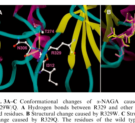
- p.R329W / p.R329Q associated with Kanzaki disease; structural modeling suggests destabilization at the domain I–II interface. (sakuraba2004structuralandimmunocytochemical pages 3-5)
- c.577G>T (p.Glu193*) / p.E193X observed in adult/mild spectrum; described as a null mutation in Spanish patients; paradoxically can be associated with milder phenotype despite very low activity. (sakuraba2004structuralandimmunocytochemical pages 3-5, castro2019anewcase pages 1-3)
- p.S160C reported as an additional mutation in α‑NAGA deficiency. (keulemans1996humanalphanacetylgalactosaminidase(alphanaga) pages 1-2)
Functional consequences
Evidence supports loss of function via absent enzyme protein (CRIM-negative) and/or conformational destabilization, resulting in lysosomal substrate accumulation. (wang1990schindlerdiseasethe pages 1-2, sakuraba2004structuralandimmunocytochemical pages 3-5)
Population allele frequency
Population allele frequency data (gnomAD/ExAC/TOPMed) were not available in retrieved full texts and are not reported here.
Modifier genes / epigenetics / chromosomal abnormalities
No specific modifier genes, epigenetic findings, or chromosomal abnormalities were identified in retrieved evidence.
| Variant (protein or HGVS) | Evidence source (year) | Phenotype/subtype association | Residual α-NAGA activity (if reported) | Mechanistic/structural notes |
|---|---|---|---|---|
| p.E325K; c.973G>A | Wang et al. (1990) | Infantile Schindler disease / type I; infantile neuroaxonal dystrophy in two brothers from a consanguineous family (wang1990schindlerdiseasethe pages 1-2) | Affected individuals: <1% of normal; parents showed intermediate activity consistent with heterozygosity (wang1990schindlerdiseasethe pages 1-2) | Missense substitution in NAGA causing inactive enzyme; patient fibroblasts had no detectable immunoreactive enzyme, while transient expression produced immunoreactive but inactive protein, supporting instability/loss of function (wang1990schindlerdiseasethe pages 1-2) |
| p.E325K/E325K | Keulemans et al. (1996) | Severe infantile neuroaxonal dystrophy; two affected sibs and an additional consanguineous patient with severe neurologic disease/convulsions (keulemans1996humanalphanacetylgalactosaminidase(alphanaga) pages 1-2) | Not numerically reported in this excerpt; severe deficiency implied (keulemans1996humanalphanacetylgalactosaminidase(alphanaga) pages 1-2) | Keulemans emphasized a genotype-phenotype paradox: identical homozygous genotype associated with severe infantile disease, while other NAGA genotypes could present much more mildly (keulemans1996humanalphanacetylgalactosaminidase(alphanaga) pages 1-2) |
| p.E325K | Sakuraba et al. (2004) | Type I Schindler disease; early-onset infantile neuroaxonal dystrophy (sakuraba2004structuralandimmunocytochemical pages 3-5, sakuraba2004structuralandimmunocytochemical pages 1-2) | 0.6-1.7% of control in homozygotes (sakuraba2004structuralandimmunocytochemical pages 3-5) | Located distant from the active site; inferred to yield an unstable protein rapidly degraded rather than directly disrupting catalytic residues (sakuraba2004structuralandimmunocytochemical pages 3-5) |
| p.R329W | Sakuraba et al. (2004) | Kanzaki disease / type II; late-onset angiokeratoma corporis diffusum phenotype (sakuraba2004structuralandimmunocytochemical pages 3-5, sakuraba2004structuralandimmunocytochemical pages 1-2) | Below 1% of control in patients homozygous for reported pathogenic alleles (sakuraba2004structuralandimmunocytochemical pages 3-5) | Structural modeling predicted major destabilization at the domain I-II interface; associated with lysosomal storage of Tn-antigen in fibroblasts (sakuraba2004structuralandimmunocytochemical pages 3-5) |
| p.R329Q | Sakuraba et al. (2004) | Kanzaki disease / type II; adult-onset/milder phenotype (sakuraba2004structuralandimmunocytochemical pages 3-5, sakuraba2004structuralandimmunocytochemical pages 1-2) | Below 1% of control in patients homozygous for reported pathogenic alleles (sakuraba2004structuralandimmunocytochemical pages 3-5) | Similar to p.R329W, predicted to cause major conformational destabilization; part of the paradox in which very low enzyme activity can still accompany milder adult disease (sakuraba2004structuralandimmunocytochemical pages 3-5, sakuraba2004structuralandimmunocytochemical pages 1-2) |
| p.E193* (reported as E193X); c.577G>T | Keulemans et al. (1996); Castro et al. (2019) | Mild/adult end of spectrum in Spanish patients and in a 68-year-old man clinically compatible with Kanzaki disease / type II (keulemans1996humanalphanacetylgalactosaminidase(alphanaga) pages 1-2, castro2019anewcase pages 3-3, castro2019anewcase pages 1-3) | ~0.2% of control in E193X homozygotes (Sakuraba summary of Spanish patients) (sakuraba2004structuralandimmunocytochemical pages 3-5); Castro reports diminished activity qualitatively (castro2019anewcase pages 3-3) | Null/nonsense mutation with complete loss of α-NAGA protein; despite this, phenotype may be relatively mild/adult-onset, illustrating marked genotype-phenotype discordance (sakuraba2004structuralandimmunocytochemical pages 3-5, keulemans1996humanalphanacetylgalactosaminidase(alphanaga) pages 1-2) |
| p.S160C | Keulemans et al. (1996) | Reported among additional pathogenic mutations in α-NAGA deficiency; specific subtype not detailed in retrieved excerpt (keulemans1996humanalphanacetylgalactosaminidase(alphanaga) pages 1-2) | Not reported in retrieved excerpt (keulemans1996humanalphanacetylgalactosaminidase(alphanaga) pages 1-2) | One of the "two new mutations" reported in 1996; included as evidence for allelic heterogeneity in NAGA deficiency (keulemans1996humanalphanacetylgalactosaminidase(alphanaga) pages 1-2) |
| Biallelic missense variants (8 probands) and 1 homozygous nonsense variant | Groopman et al. / ClinGen LD GCEP (2024) | Spectrum includes Schindler disease types I and III and Kanzaki disease/type II; clinically unaffected relatives with same genotype and enzyme deficiency have been reported (groopman2024assessmentofgenes pages 8-11) | All reported individuals with biallelic variants had α-NAGA enzyme deficiency, but exact residual activities varied by report (groopman2024assessmentofgenes pages 8-11) | ClinGen classified the NAGA-disease relationship as Definitive (11.85 points, rounded to 12) while noting that the clinical impact/penetrance remains unclear, supporting variable expressivity beyond enzyme deficiency alone (groopman2024assessmentofgenes pages 8-11) |
Table: This table summarizes key reported pathogenic NAGA variants in Schindler/Kanzaki disease, linking each to subtype associations, residual enzyme activity, and mechanistic interpretations. It is useful for quick curation of genotype-phenotype relationships and the notable variability in clinical expression.
5. Environmental information
No environmental, lifestyle, or infectious etiologic factors were identified in the retrieved evidence. Schindler disease is primarily a monogenic lysosomal disorder. (wang1990schindlerdiseasethe pages 1-2, groopman2024assessmentofgenes pages 8-11)
6. Mechanism / pathophysiology
Causal chain (current understanding)
- Biallelic NAGA variants → deficiency of lysosomal α‑NAGA activity. (wang1990schindlerdiseasethe pages 1-2, groopman2024assessmentofgenes pages 8-11)
- Impaired cleavage of terminal α‑N‑acetylgalactosamine residues on glycoconjugates → accumulation of undegraded glycopeptide substrates. (wang1990schindlerdiseasethe pages 1-2, sakuraba2004structuralandimmunocytochemical pages 5-7)
- Lysosomal storage of substrates including Tn‑antigen (GalNAcα1‑O‑Ser/Thr) (especially evidenced for Kanzaki disease) → cellular vacuolization/storage pathology in multiple cell types. (sakuraba2004structuralandimmunocytochemical pages 3-5, sakuraba2004structuralandimmunocytochemical pages 5-7, keulemans1996humanalphanacetylgalactosaminidase(alphanaga) pages 1-2)
- Tissue dysfunction: in severe infantile forms, neuronal/axonal pathology with spheroids/neuroaxonal dystrophy and rapid neurodegeneration; in adult forms, angiokeratoma/lymphatic and peripheral nerve involvement. (wang1990schindlerdiseasethe pages 1-2, castro2019anewcase pages 1-3, keulemans1996humanalphanacetylgalactosaminidase(alphanaga) pages 1-2)
Stored substrates and biomarkers
- Urinary biomarkers: increased excretion of glycopeptides/oligosaccharides containing α‑GalNAc moieties (glycopeptiduria); Keulemans reported “major abnormal urinary oligosaccharides” as O‑linked sialylglycopeptides with Ser/Thr-linked α‑GalNAc. (wang1990schindlerdiseasethe pages 1-2, keulemans1996humanalphanacetylgalactosaminidase(alphanaga) pages 1-2)
- Intracellular substrate (Kanzaki): Tn‑antigen accumulation in lysosomes of patient fibroblasts; Tn‑antigen fluorescence co-localizes with LAMP‑1. (sakuraba2004structuralandimmunocytochemical pages 3-5, sakuraba2004structuralandimmunocytochemical media f06dc2b6)
Cell types and tissues involved (examples from evidence)
- Dermal/vascular/lymphatic involvement: vacuolization in vascular and lymphatic endothelial cells, eccrine sweat gland cells, fibroblasts, dermal neural cells; glomerular endothelial involvement reported. (keulemans1996humanalphanacetylgalactosaminidase(alphanaga) pages 1-2)
- Nervous system: neuroaxonal dystrophy with terminal axonal spheroids in severe infantile disease. (wang1990schindlerdiseasethe pages 1-2)
Pathways and ontology suggestions
- GO (biological process): lysosomal catabolic process; glycoprotein catabolic process; oligosaccharide catabolic process; lysosome organization.
- GO (cellular component): lysosome.
- CL (cell types): endothelial cell; fibroblast; eccrine sweat gland cell; neuron; glial cell (suggested given CNS involvement).
- UBERON (anatomy): brain; peripheral nerve; skin; kidney glomerulus; heart.
(These ontology suggestions are consistent with the evidenced lysosomal storage process and the implicated tissues/cell types, though the specific ontology IDs were not enumerated in the retrieved full texts.)
7. Anatomical structures affected
Based on case and mechanistic evidence, major affected systems include: - Nervous system: CNS (infantile neurodegeneration; neuroaxonal dystrophy) and peripheral nervous system (polyneuropathy, carpal tunnel). (wang1990schindlerdiseasethe pages 1-2, castro2019anewcase pages 1-3) - Skin/vascular/lymphatic: angiokeratoma; lymphoedema; storage/vacuolization in dermal endothelial and eccrine structures. (castro2019anewcase pages 1-3, keulemans1996humanalphanacetylgalactosaminidase(alphanaga) pages 1-2) - Kidney (microvascular): glomerular endothelial cell vacuolization reported. (keulemans1996humanalphanacetylgalactosaminidase(alphanaga) pages 1-2) - Heart (possible): cardiomegaly or interventricular septal hypertrophy described in type II summary. (castro2019anewcase pages 1-3)
8. Temporal development
- Type I onset: normal infancy (9–12 months) followed by rapid regression beginning in the second year; severe neurologic phenotype by age 3–4. (wang1990schindlerdiseasethe pages 1-2)
- Type I course: progressive neurodegeneration; death in early childhood reported. (castro2019anewcase pages 1-3, naumchik2020theroleof pages 7-9)
- Type II onset: adult/late onset. (castro2019anewcase pages 1-3, keulemans1996humanalphanacetylgalactosaminidase(alphanaga) pages 1-2)
9. Inheritance and population
Inheritance
- Autosomal recessive, supported by segregation and enzyme activity in a consanguineous family in the original report. (wang1990schindlerdiseasethe pages 1-2)
Epidemiology
- Extremely rare: a 2004 review states that 12 patients from 8 families had been reported at that time. (sakuraba2004structuralandimmunocytochemical pages 1-2)
- A 2019 case report states “fewer than 20 cases” have been described. (castro2019anewcase pages 1-3)
Prevalence/incidence estimates, carrier frequency, and population-specific founder effects were not available in retrieved full text.
Penetrance/expressivity
ClinGen curation notes clinically unaffected family members who nonetheless share genotype and enzyme deficiency, indicating uncertain penetrance/expressivity for clinical disease, despite definitive gene–biochemical phenotype association. (groopman2024assessmentofgenes pages 8-11)
10. Diagnostics
Diagnostic workflow (current practice from retrieved evidence)
- Clinical suspicion based on neurodegeneration (infantile) or angiokeratoma/lymphoedema/neuropathy (adult). (castro2019anewcase pages 1-3)
- Biochemical testing: reduced α‑NAGA enzyme activity (blood and/or cultured cells) and urinary glycopeptiduria/abnormal urinary oligosaccharides. (castro2019anewcase pages 3-3, wang1990schindlerdiseasethe pages 1-2)
- Confirmatory genetic testing of NAGA; the 2019 report describes PCR-based genetic testing as a “gold-standard.” (castro2019anewcase pages 3-3)
Recent developments (2023–2024 priority): MS-based urine assays for glycoproteinoses
A 2024 Clinical Chemistry study developed and validated a multiplexed UPLC‑MS/MS assay for urinary oligosaccharides and glycoamino acids (biomarkers of glycoproteinoses), selecting 28 oligosaccharides, 2 glycoamino acids, and 2 ratios, and reported: “In the 76 untreated patients, unambiguous diagnosis was achieved with 100% sensitivity and specificity.” (Apr 2024; https://doi.org/10.1093/clinchem/hvae043). Although Schindler disease was not explicitly singled out in the excerpted sections, it is part of the glycoproteinoses class for which urinary oligosaccharides/glycoamino acids are recognized biomarkers, making this assay paradigm relevant for implementation in biochemical genetics labs. (wongkittichote2024ultraperformanceliquidchromatography–tandem pages 1-2)
Differential diagnosis
The retrieved evidence notes biochemical overlap with Fabry testing due to related glycosidase biology and describes glycopeptiduria patterns; however, a structured differential diagnosis list was not available in retrieved text. (castro2019anewcase pages 1-3)
Screening
No disease-specific newborn screening approach for Schindler disease was identified in the retrieved full text. The 2024 MS paper discusses two-tier screening concepts for LSDs generally (e.g., DBS markers for some disorders), but Schindler-specific screening evidence was not present. (wongkittichote2024ultraperformanceliquidchromatography–tandem pages 8-10)
11. Outcome / prognosis
- Type I prognosis: the 2019 case review states type I “typically results in death before the age of 6 years.” (castro2019anewcase pages 1-3)
- Type II prognosis: variable; longer survival into adulthood is evident in the 68-year-old case report and other adult-onset descriptions. (castro2019anewcase pages 1-3)
- Blood group A: reported association with worse prognosis in the 2019 report (mechanistic rationale: A-antigen terminal residue normally cleaved by α‑NAGA). (castro2019anewcase pages 1-3)
No survival curves, standardized functional outcome statistics, or QoL metrics were identified in retrieved evidence.
12. Treatment
Current standard of care
The 2019 case report states there is no current disease-modifying therapy, and that “The only treatment… consists of support measures and symptomatic treatment.” (castro2019anewcase pages 1-3)
Supportive management (conceptual; MAXO suggestions). - Symptomatic neurologic management (e.g., seizure management) (MAXO: anticonvulsant therapy). - Support for hearing impairment (MAXO: hearing aid use). - Management of lymphedema (MAXO: compression therapy). - Neuropathy/carpal tunnel management (MAXO: peripheral nerve decompression; pain management).
(These MAXO mappings are suggested based on reported manifestations; no specific guideline-level evidence was retrieved.)
Proposed / experimental directions (authoritative expert reviews)
- The 2019 case report mentions “two pharmacological chaperones… have been proposed as potential therapeutic agents,” without naming compounds or providing trial evidence. (castro2019anewcase pages 1-3)
- Reviews of neurological LSD therapies emphasize BBB delivery constraints and emerging modalities such as BBB-targeted enzyme fusion strategies, AAV gene therapy, and hematopoietic stem cell gene therapy, with explicit limitations including myeloablation risk and delayed CNS effect. (ellison2023advancesintherapies pages 24-25, ellison2023advancesintherapies pages 14-15)
Hematopoietic cell transplant (HCT)
A 2020 review on HCT in glycoprotein diseases states that for Schindler disease, HCT “has not yet been pursued as a therapeutic option,” and lists no known cases; it explains the biologic rationale via cross-correction and potential CNS access compared with peripheral ERT. (naumchik2020theroleof pages 7-9, naumchik2020theroleof pages 9-11)
Clinical trials
No Schindler-specific interventional trials were retrieved. A relevant observational natural history effort for glycoproteinoses includes Schindler disease: - NCT01891422 (ClinicalTrials.gov; Greenwood Genetic Center; posted 2009): longitudinal study of glycoproteinoses aimed to define frequency, early features, progression, and evaluate supportive therapies; includes exams, neuropsychological testing, skeletal imaging, record review, and biospecimen banking. (NCT01891422 chunk 1)
13. Prevention
No primary prevention exists for a monogenic autosomal recessive condition. Prevention is primarily via genetic counseling (carrier testing, cascade testing in families, reproductive options). This is consistent with the autosomal recessive inheritance demonstrated in primary literature. (wang1990schindlerdiseasethe pages 1-2)
No population screening or newborn screening programs specific to Schindler disease were identified in retrieved evidence.
14. Other species / natural disease
The retrieved evidence set did not include naturally occurring veterinary Schindler disease. However, orthologous α‑NAGAL enzymes are described in other species as part of comparative enzymology.
15. Model organisms
C. elegans ortholog model
A 2005 study characterized gana‑1 (R07B7.11) as a single C. elegans ortholog of vertebrate α‑GAL and α‑NAGA, with measurable enzymatic activities and lysosome-compatible localization patterns; RNAi reduced activity without overt phenotype. (Jan 2005; https://doi.org/10.1186/1471-2121-6-5). (hujova2005characterizationofgana1 pages 1-2, hujova2005characterizationofgana1 pages 7-8)
Additional functional ortholog evidence
A 2022 PLOS Pathogens paper functionally characterized schistosome α‑NAGAL and explicitly situates human α‑NAGAL in the context of Schindler/Kanzaki disease, supporting evolutionary conservation of enzymatic function relevant to substrate cleavage. (hulme2022schistosomamansoniαnacetylgalactosaminidase pages 18-20)
ClinGen note on non-human models
ClinGen’s 2024 curation states that experimental evidence for NAGA includes biochemical function and “a non-human animal model,” contributing to the definitive classification, but does not specify the model organism in the excerpted text. (groopman2024assessmentofgenes pages 8-11)
Recent developments and authoritative expert analysis (2023–2024 prioritized)
- ClinGen 2024 (Molecular Genetics and Metabolism, Nov 2024; doi:10.1016/j.ymgme.2024.108593): provides the most authoritative recent aggregation of genetic evidence, classifying NAGA–alpha-N-acetylgalactosaminidase deficiency as Definitive (9 probands; genetic evidence 8.85 points; total 11.85 rounded to 12), while emphasizing uncertain clinical penetrance/variable expressivity. (groopman2024assessmentofgenes pages 8-11)
- Clinical Chemistry 2024 (Apr 2024; doi:10.1093/clinchem/hvae043): provides a validated, modern diagnostic implementation path via UPLC‑MS/MS urinary glycan profiling with high reported diagnostic performance (100% sensitivity/specificity in a subset). (wongkittichote2024ultraperformanceliquidchromatography–tandem pages 1-2)
- J Inherit Metab Dis 2023 therapy review (May 2023; doi:10.1002/jimd.12615): synthesizes major LSD therapeutic trends (BBB-targeted enzyme delivery, AAV, HSC gene therapy) and explicitly quantifies general neurologic involvement (“greater than two-thirds of LSDs exhibit neuropathology”) and HSC-GT limitations (mortality risk, 3–4 week delay to CNS effect, insertional mutagenesis risk). (ellison2023advancesintherapies pages 2-3, ellison2023advancesintherapies pages 24-25)
Real-world implementations
- Clinical biochemical genetics diagnostics: enzyme activity assays and urine glycopeptide/oligosaccharide profiling remain core, with increasing adoption of LC/UPLC-MS/MS multiplex assays for glycoproteinoses and related LSDs in clinical laboratories. (castro2019anewcase pages 3-3, wongkittichote2024ultraperformanceliquidchromatography–tandem pages 1-2)
- Natural history data collection: NCT01891422 illustrates registry-style and biospecimen-banking implementations for glycoproteinoses, including Schindler disease, supporting future trial readiness. (NCT01891422 chunk 1)
Key evidence gaps (for knowledge-base completeness)
- Missing in retrieved full texts: Orphanet IDs, ICD-10/ICD-11 codes, MeSH IDs; population allele frequencies (gnomAD); systematic prevalence/incidence; validated Schindler-specific MS biomarker panels; standardized QoL measures; detailed type III phenotype.
Key visual evidence
- Sakuraba et al. (2004) immunocytochemistry figure shows Tn-antigen co-localization with LAMP-1 in Kanzaki fibroblasts, supporting lysosomal substrate storage. (sakuraba2004structuralandimmunocytochemical media f06dc2b6)
URLs and publication dates (from retrieved evidence)
- Wang AM et al. J Clin Invest. Nov 1990. https://doi.org/10.1172/jci114901 (wang1990schindlerdiseasethe pages 1-2)
- Keulemans J et al. J Med Genet. Jun 1996. https://doi.org/10.1136/jmg.33.6.458 (keulemans1996humanalphanacetylgalactosaminidase(alphanaga) pages 1-2)
- Sakuraba H et al. J Hum Genet. Jan 2004. https://doi.org/10.1007/s10038-003-0098-z (sakuraba2004structuralandimmunocytochemical pages 3-5, sakuraba2004structuralandimmunocytochemical media f06dc2b6)
- Tajima Y et al. Am J Hum Genet. Nov 2009. https://doi.org/10.1016/j.ajhg.2009.09.016 (tajima2009useofa pages 6-7)
- Naumchik BM et al. Cells. Jun 2020. https://doi.org/10.3390/cells9061411 (naumchik2020theroleof pages 7-9)
- Ellison S et al. J Inherit Metab Dis. May 2023. https://doi.org/10.1002/jimd.12615 (ellison2023advancesintherapies pages 2-3)
- Serrano TJ et al. J Clin Med. Mar 2024. https://doi.org/10.3390/jcm13051465 (serrano2024hepatomegalyandsplenomegaly pages 7-8)
- Wongkittichote P et al. Clinical Chemistry. Apr 2024. https://doi.org/10.1093/clinchem/hvae043 (wongkittichote2024ultraperformanceliquidchromatography–tandem pages 1-2)
- Groopman E et al. Mol Genet Metab. Nov 2024. https://doi.org/10.1016/j.ymgme.2024.108593 (groopman2024assessmentofgenes pages 8-11)
- ClinicalTrials.gov NCT01891422 (posted 2009; Greenwood Genetic Center). (NCT01891422 chunk 1)
Note on PMID requirements
The retrieved evidence snippets did not include PubMed IDs (PMIDs) explicitly. Where the user requested PMIDs, this report therefore provides DOI-based citations and tool-provided context IDs. PMIDs should be appended after independent PubMed lookup, which was not available in the current tool outputs.
References
-
(wang1990schindlerdiseasethe pages 1-2): Anne M. Wang, D. Schindler, and R. Desnick. Schindler disease: the molecular lesion in the alpha-n-acetylgalactosaminidase gene that causes an infantile neuroaxonal dystrophy. The Journal of clinical investigation, 86 5:1752-6, Nov 1990. URL: https://doi.org/10.1172/jci114901, doi:10.1172/jci114901. This article has 83 citations.
-
(castro2019anewcase pages 1-3): Rubén García Castro, Ana María González Pérez, María Concepción Román Curto, Javier Cañueto Álvarez, Alberto Conde Ferreirós, Alex Viñolas Cuadros, David Moyano Bueno, and Antonio Javier Chamorro Fernández. A new case of schindler disease. European Journal of Case Reports in Internal Medicine, 6:1, Oct 2019. URL: https://doi.org/10.12890/2019_001269, doi:10.12890/2019_001269. This article has 12 citations.
-
(groopman2024assessmentofgenes pages 8-11): Emily Groopman, Shruthi Mohan, Amber Waddell, Matheus Wilke, Raquel Fernandez, Meredith Weaver, Hongjie Chen, Hongbin Liu, Deeksha Bali, Heather Baudet, Lorne Clarke, Christina Hung, Rong Mao, Filippo Pinto e Vairo, Lemuel Racacho, Tatiana Yuzyuk, William J. Craigen, and Jennifer Goldstein. Assessment of genes involved in lysosomal diseases using the clingen clinical validity framework. Molecular Genetics and Metabolism, 143:108593, Nov 2024. URL: https://doi.org/10.1016/j.ymgme.2024.108593, doi:10.1016/j.ymgme.2024.108593. This article has 1 citations and is from a peer-reviewed journal.
-
(sakuraba2004structuralandimmunocytochemical pages 3-5): Hitoshi Sakuraba, Fumiko Matsuzawa, Sei-ichi Aikawa, Hirofumi Doi, Masaharu Kotani, Hiroshi Nakada, Tomoko Fukushige, and Tamotsu Kanzaki. Structural and immunocytochemical studies on α-n-acetylgalactosaminidase deficiency (schindler/kanzaki disease). Journal of Human Genetics, 49:1-8, Jan 2004. URL: https://doi.org/10.1007/s10038-003-0098-z, doi:10.1007/s10038-003-0098-z. This article has 60 citations and is from a peer-reviewed journal.
-
(sakuraba2004structuralandimmunocytochemical media f06dc2b6): Hitoshi Sakuraba, Fumiko Matsuzawa, Sei-ichi Aikawa, Hirofumi Doi, Masaharu Kotani, Hiroshi Nakada, Tomoko Fukushige, and Tamotsu Kanzaki. Structural and immunocytochemical studies on α-n-acetylgalactosaminidase deficiency (schindler/kanzaki disease). Journal of Human Genetics, 49:1-8, Jan 2004. URL: https://doi.org/10.1007/s10038-003-0098-z, doi:10.1007/s10038-003-0098-z. This article has 60 citations and is from a peer-reviewed journal.
-
(OpenTargets Search: Schindler disease,Kanzaki disease-NAGA): Open Targets Query (Schindler disease,Kanzaki disease-NAGA, 5 results). Buniello, A. et al. (2025). Open Targets Platform: facilitating therapeutic hypotheses building in drug discovery. Nucleic Acids Research.
-
(sakuraba2004structuralandimmunocytochemical pages 1-2): Hitoshi Sakuraba, Fumiko Matsuzawa, Sei-ichi Aikawa, Hirofumi Doi, Masaharu Kotani, Hiroshi Nakada, Tomoko Fukushige, and Tamotsu Kanzaki. Structural and immunocytochemical studies on α-n-acetylgalactosaminidase deficiency (schindler/kanzaki disease). Journal of Human Genetics, 49:1-8, Jan 2004. URL: https://doi.org/10.1007/s10038-003-0098-z, doi:10.1007/s10038-003-0098-z. This article has 60 citations and is from a peer-reviewed journal.
-
(castro2019anewcase pages 3-3): Rubén García Castro, Ana María González Pérez, María Concepción Román Curto, Javier Cañueto Álvarez, Alberto Conde Ferreirós, Alex Viñolas Cuadros, David Moyano Bueno, and Antonio Javier Chamorro Fernández. A new case of schindler disease. European Journal of Case Reports in Internal Medicine, 6:1, Oct 2019. URL: https://doi.org/10.12890/2019_001269, doi:10.12890/2019_001269. This article has 12 citations.
-
(tajima2009useofa pages 6-7): Youichi Tajima, Ikuo Kawashima, Takahiro Tsukimura, Kanako Sugawara, Mayuko Kuroda, Toshihiro Suzuki, Tadayasu Togawa, Yasunori Chiba, Yoshifumi Jigami, Kazuki Ohno, Tomoko Fukushige, Takuro Kanekura, Kohji Itoh, Toya Ohashi, and Hitoshi Sakuraba. Use of a modified alpha-n-acetylgalactosaminidase in the development of enzyme replacement therapy for fabry disease. American journal of human genetics, 85 5:569-80, Nov 2009. URL: https://doi.org/10.1016/j.ajhg.2009.09.016, doi:10.1016/j.ajhg.2009.09.016. This article has 73 citations and is from a highest quality peer-reviewed journal.
-
(keulemans1996humanalphanacetylgalactosaminidase(alphanaga) pages 1-2): J. Keulemans, A. Reuser, M. Kroos, R. Willemsen, M. Hermans, A. V. D. van den Ouweland, J. de Jong, R. Wevers, W. Renier, D. Schindler, M. Coll, A. Chabás, H. Sakuraba, Y. Suzuki, and O. V. van Diggelen. Human alpha-n-acetylgalactosaminidase (alpha-naga) deficiency: new mutations and the paradox between genotype and phenotype. Journal of Medical Genetics, 33:458-464, Jun 1996. URL: https://doi.org/10.1136/jmg.33.6.458, doi:10.1136/jmg.33.6.458. This article has 52 citations and is from a domain leading peer-reviewed journal.
-
(naumchik2020theroleof pages 7-9): Brianna M. Naumchik, Ashish Gupta, Heather Flanagan-Steet, Richard A. Steet, Sara S. Cathey, Paul J. Orchard, and Troy C. Lund. The role of hematopoietic cell transplant in the glycoprotein diseases. Cells, 9:1411, Jun 2020. URL: https://doi.org/10.3390/cells9061411, doi:10.3390/cells9061411. This article has 28 citations.
-
(makridou2025mappinglysosomalstorage pages 10-12): Anna Makridou, Evangelie Sintou, Sofia Chatzianagnosti, Sofia Gargani, Maria Eleni Manthou, Iasonas Dermitzakis, and Paschalis Theotokis. Mapping lysosomal storage disorders with neurological features by cellular pathways: towards precision medicine. Current Issues in Molecular Biology, 47:1009, Dec 2025. URL: https://doi.org/10.3390/cimb47121009, doi:10.3390/cimb47121009. This article has 0 citations.
-
(sakuraba2004structuralandimmunocytochemical pages 5-7): Hitoshi Sakuraba, Fumiko Matsuzawa, Sei-ichi Aikawa, Hirofumi Doi, Masaharu Kotani, Hiroshi Nakada, Tomoko Fukushige, and Tamotsu Kanzaki. Structural and immunocytochemical studies on α-n-acetylgalactosaminidase deficiency (schindler/kanzaki disease). Journal of Human Genetics, 49:1-8, Jan 2004. URL: https://doi.org/10.1007/s10038-003-0098-z, doi:10.1007/s10038-003-0098-z. This article has 60 citations and is from a peer-reviewed journal.
-
(wongkittichote2024ultraperformanceliquidchromatography–tandem pages 1-2): Parith Wongkittichote, Se Hyun Cho, Artis Miller, Kaitlyn King, Zackary M Herbst, Zhimei Ren, Michael H Gelb, and Xinying Hong. Ultra-performance liquid chromatography–tandem mass spectrometry analysis of urinary oligosaccharides and glycoamino acids for the diagnosis of mucopolysaccharidosis and glycoproteinosis. Clinical Chemistry, 70:865-877, Apr 2024. URL: https://doi.org/10.1093/clinchem/hvae043, doi:10.1093/clinchem/hvae043. This article has 4 citations and is from a highest quality peer-reviewed journal.
-
(wongkittichote2024ultraperformanceliquidchromatography–tandem pages 8-10): Parith Wongkittichote, Se Hyun Cho, Artis Miller, Kaitlyn King, Zackary M Herbst, Zhimei Ren, Michael H Gelb, and Xinying Hong. Ultra-performance liquid chromatography–tandem mass spectrometry analysis of urinary oligosaccharides and glycoamino acids for the diagnosis of mucopolysaccharidosis and glycoproteinosis. Clinical Chemistry, 70:865-877, Apr 2024. URL: https://doi.org/10.1093/clinchem/hvae043, doi:10.1093/clinchem/hvae043. This article has 4 citations and is from a highest quality peer-reviewed journal.
-
(ellison2023advancesintherapies pages 24-25): S. Ellison, H. Parker, and B. Bigger. Advances in therapies for neurological lysosomal storage disorders. Journal of Inherited Metabolic Disease, 46:874-905, May 2023. URL: https://doi.org/10.1002/jimd.12615, doi:10.1002/jimd.12615. This article has 67 citations and is from a peer-reviewed journal.
-
(ellison2023advancesintherapies pages 14-15): S. Ellison, H. Parker, and B. Bigger. Advances in therapies for neurological lysosomal storage disorders. Journal of Inherited Metabolic Disease, 46:874-905, May 2023. URL: https://doi.org/10.1002/jimd.12615, doi:10.1002/jimd.12615. This article has 67 citations and is from a peer-reviewed journal.
-
(naumchik2020theroleof pages 9-11): Brianna M. Naumchik, Ashish Gupta, Heather Flanagan-Steet, Richard A. Steet, Sara S. Cathey, Paul J. Orchard, and Troy C. Lund. The role of hematopoietic cell transplant in the glycoprotein diseases. Cells, 9:1411, Jun 2020. URL: https://doi.org/10.3390/cells9061411, doi:10.3390/cells9061411. This article has 28 citations.
-
(NCT01891422 chunk 1): Longitudinal Studies of the Glycoproteinoses. Greenwood Genetic Center. 2009. ClinicalTrials.gov Identifier: NCT01891422
-
(hujova2005characterizationofgana1 pages 1-2): Jana Hujová, Jakub Sikora, Robert Dobrovolný, Helena Poupětová, Jana Ledvinová, Marta Kostrouchová, and Martin Hřebíček. Characterization of gana-1, a caenorhabditis elegans gene encoding a single ortholog of vertebrate α-galactosidase and α-n-acetylgalactosaminidase. BMC Cell Biology, 6:5-5, Jan 2005. URL: https://doi.org/10.1186/1471-2121-6-5, doi:10.1186/1471-2121-6-5. This article has 22 citations.
-
(hujova2005characterizationofgana1 pages 7-8): Jana Hujová, Jakub Sikora, Robert Dobrovolný, Helena Poupětová, Jana Ledvinová, Marta Kostrouchová, and Martin Hřebíček. Characterization of gana-1, a caenorhabditis elegans gene encoding a single ortholog of vertebrate α-galactosidase and α-n-acetylgalactosaminidase. BMC Cell Biology, 6:5-5, Jan 2005. URL: https://doi.org/10.1186/1471-2121-6-5, doi:10.1186/1471-2121-6-5. This article has 22 citations.
-
(hulme2022schistosomamansoniαnacetylgalactosaminidase pages 18-20): Benjamin J. Hulme, Kathrin K. Geyer, Josephine E. Forde-Thomas, Gilda Padalino, Dylan W. Phillips, Wannaporn Ittiprasert, Shannon E. Karinshak, Victoria H. Mann, Iain W. Chalmers, Paul J. Brindley, Cornelis H. Hokke, and Karl F. Hoffmann. Schistosoma mansoni α-n-acetylgalactosaminidase (smnagal) regulates coordinated parasite movement and egg production. Jan 2022. URL: https://doi.org/10.1371/journal.ppat.1009828, doi:10.1371/journal.ppat.1009828. This article has 31 citations and is from a highest quality peer-reviewed journal.
-
(ellison2023advancesintherapies pages 2-3): S. Ellison, H. Parker, and B. Bigger. Advances in therapies for neurological lysosomal storage disorders. Journal of Inherited Metabolic Disease, 46:874-905, May 2023. URL: https://doi.org/10.1002/jimd.12615, doi:10.1002/jimd.12615. This article has 67 citations and is from a peer-reviewed journal.
-
(serrano2024hepatomegalyandsplenomegaly pages 7-8): Teodoro Jerves Serrano, Jessica Gold, James A. Cooper, Heather J. Church, Karen L. Tylee, Hoi Yee Wu, Sun Young Kim, and Karolina M. Stepien. Hepatomegaly and splenomegaly: an approach to the diagnosis of lysosomal storage diseases. Journal of Clinical Medicine, 13:1465, Mar 2024. URL: https://doi.org/10.3390/jcm13051465, doi:10.3390/jcm13051465. This article has 17 citations.