RPE65-Related Retinopathy
RPE65-related retinopathy is an inherited retinal disease caused by pathogenic variants in RPE65, encoding the all-trans-retinyl ester isomerohydrolase essential for regenerating the photopigment chromophore 11-cis-retinal within the retinal pigment epithelium (RPE). Loss of this enzyme activity arrests the visual (retinoid) cycle, chronically depleting photoreceptors of the chromophore required for phototransduction, and drives progressive rod-dominant retinal degeneration. Biallelic loss-of-function variants cause a clinical spectrum ranging from Leber congenital amaurosis type 2 (LCA2), with onset in the first year of life and severe visual impairment, through early-onset severe retinal dystrophy (EOSRD) to juvenile retinitis pigmentosa. Rare heterozygous dominant-acting variants produce a typically milder, later-onset retinitis pigmentosa phenotype. RPE65-related recessive retinopathy is distinguished by being the first inherited disease to receive FDA-approved gene therapy: voretigene neparvovec (Luxturna), a subretinal AAV2 vector delivering functional RPE65 cDNA, approved in December 2017.
Ask OpenScientist
Ask a research question about RPE65-Related Retinopathy. OpenScientist will conduct autonomous deep research using the Disorder Mechanisms Knowledge Base and PubMed literature (typically 10-30 minutes).
Do not include personal health information in your question. Questions and results are cached in your browser's local storage.
Classifications
Mappings
Inheritance
2Show evidence (1 reference)
Show evidence (1 reference)
Subtypes
2Show evidence (3 references)
Pathophysiology
3Show evidence (2 references)
Show evidence (2 references)
Show evidence (2 references)
Pathograph
Phenotypes
9Eye 8
Show evidence (2 references)
Show evidence (1 reference)
Show evidence (2 references)
Show evidence (1 reference)
Show evidence (1 reference)
Show evidence (1 reference)
Show evidence (1 reference)
Show evidence (2 references)
Nervous System 1
Show evidence (2 references)
Genetic Associations
1Show evidence (2 references)
Medical Actions
3Show evidence (10 references)
Show evidence (1 reference)
Show evidence (1 reference)
Diagnosis
2Show evidence (1 reference)
Show evidence (1 reference)
Progression
2Show evidence (1 reference)
Show evidence (1 reference)
Epidemiology
1Show evidence (1 reference)
Clinical Trials
1Show evidence (1 reference)
Source YAML
click to showname: RPE65-Related Retinopathy
creation_date: "2026-06-18T00:00:00Z"
category: Mendelian
description: >-
RPE65-related retinopathy is an inherited retinal disease caused by pathogenic
variants in RPE65, encoding the all-trans-retinyl ester isomerohydrolase essential
for regenerating the photopigment chromophore 11-cis-retinal within the retinal
pigment epithelium (RPE). Loss of this enzyme activity arrests the visual (retinoid)
cycle, chronically depleting photoreceptors of the chromophore required for
phototransduction, and drives progressive rod-dominant retinal degeneration. Biallelic
loss-of-function variants cause a clinical spectrum ranging from Leber congenital
amaurosis type 2 (LCA2), with onset in the first year of life and severe visual
impairment, through early-onset severe retinal dystrophy (EOSRD) to juvenile
retinitis pigmentosa. Rare heterozygous dominant-acting variants produce a typically
milder, later-onset retinitis pigmentosa phenotype. RPE65-related recessive
retinopathy is distinguished by being the first inherited disease to receive FDA-approved
gene therapy: voretigene neparvovec (Luxturna), a subretinal AAV2 vector delivering
functional RPE65 cDNA, approved in December 2017.
disease_term:
preferred_term: RPE65-related recessive retinopathy
term:
id: MONDO:0100368
label: RPE65-related recessive retinopathy
synonyms:
- Leber congenital amaurosis type 2
- LCA2
- RPE65-related early-onset severe retinal dystrophy
- retinitis pigmentosa type 20
- RPE65-related LCA
parents:
- Ophthalmological Disease
- Retinal Dystrophy
- Inherited retinal dystrophy
mappings:
mondo_mappings:
- term:
id: MONDO:0008765
label: Leber congenital amaurosis 2
mapping_predicate: skos:narrowMatch
mapping_source: MONDO
mapping_justification: >
MONDO places Leber congenital amaurosis 2 (MONDO:0008765) as a child of
RPE65-related recessive retinopathy (MONDO:0100368), the term this entry is
anchored at. LCA2 names the most severe, infantile-onset end of the same
biallelic RPE65 spectrum rather than a separate mechanism, so it is recorded
here as a narrow match and carried as the LCA2 subtype below.
notes: >-
The three clinical phenotypes of the autosomal recessive form — LCA2 (onset
infancy, most severe), EOSRD (onset early childhood), and juvenile RP (onset late
childhood/adolescence, mildest) — represent a single genetic entity under
MONDO:0100368 and are distinguished by allele severity and residual RPE65 activity
rather than by separate disease mechanisms. MONDO:0100452 (RPE65-related dominant
retinopathy) is captured as a subtype; its molecular mechanism (dominant-negative
or gain-of-function) is less well characterized than the recessive loss-of-function
form. The gene therapy approval (voretigene neparvovec) applies specifically to
biallelic RPE65 mutations and does not cover the dominant form. Epidemiologically,
biallelic RPE65 mutations account for approximately 8% of Leber congenital amaurosis
cases and 2% of retinitis pigmentosa cases. Clinical presentation is highly variable
and fundus abnormalities can be minimal in early childhood, making genetic testing
essential for diagnosis.
Lump/split: Leber congenital amaurosis 2 (MONDO:0008765) is curated here as a
subtype of this entry rather than as its own disease entry. It is the same gene
(RPE65), the same proximal lesion (loss of retinyl-ester isomerohydrolase
activity) and the same pathograph as the rest of the recessive spectrum,
separated from EOSRD and juvenile RP by age at onset and residual enzyme
activity rather than by mechanism; MONDO itself files MONDO:0008765 beneath
MONDO:0100368. This differs from the sibling entries Leber_Congenital_Amaurosis_9
(NMNAT1), Leber_Congenital_Amaurosis_10 (CEP290) and Leber_Congenital_Amaurosis_13
(RDH12), which are separate entries because each is a different gene with a
different mechanism and no gene-level entry to fold into. Note that the LCA
label is not the most common RPE65 presentation: most children with biallelic
RPE65 variants present as EOSRD, with residual cone-mediated central vision and
little or no nystagmus, so the LCA2 designation marks the severe end of a
continuum rather than a discrete clinical category.
has_subtypes:
- name: LCA2
display_name: Leber congenital amaurosis 2 (LCA2)
genes:
- preferred_term: RPE65
term:
id: hgnc:10294
label: RPE65
subtype_term:
preferred_term: Leber congenital amaurosis 2
term:
id: MONDO:0008765
label: Leber congenital amaurosis 2
inheritance:
- name: Autosomal recessive
inheritance_term:
preferred_term: Autosomal recessive inheritance
term:
id: HP:0000007
label: Autosomal recessive inheritance
description: >-
LCA2 arises from biallelic (homozygous or compound heterozygous) RPE65
loss-of-function variants, the severe end of the same recessive allelic
series that produces EOSRD and juvenile retinitis pigmentosa.
description: >-
LCA2 is the most severe presentation of biallelic RPE65 deficiency: visual
manifestations begin in the first year of life, with profound nyctalopia,
severely reduced acuity, nystagmus, poorly reactive pupils and an
undetectable or severely abnormal full-field electroretinogram. The
oculodigital sign (eye poking) is a characteristic infant finding, and high
hyperopia is common, suggesting impaired emmetropisation. LCA2 is separated
from early-onset severe retinal dystrophy, which presents after infancy and
usually before age five, by age at onset and by the amount of residual
photoreceptor function rather than by a different mechanism. Because the
degeneration is slow, a therapeutic window for subretinal RPE65 gene
supplementation persists through childhood, which is why molecular diagnosis
in an infant with nystagmus and an extinguished ERG is time-critical.
evidence:
- reference: PMID:28689169
reference_title: "Leber congenital amaurosis/early-onset severe retinal dystrophy: clinical features, molecular genetics and therapeutic interventions."
supports: SUPPORT
evidence_source: OTHER
quote_role: REVIEW_SYNTHESIS
snippet: "Whereas LCA is congenital or presents within the first few months of life, is associated with nystagmus, poor pupil responses and in most instances an undetectable full-field electroretinogram (ERG)"
explanation: >-
This review defines the clinical boundary this subtype records: LCA is
congenital or presents within the first few months of life with nystagmus,
poor pupil responses and an undetectable ERG, whereas EOSRD presents after
infancy. That boundary is what separates LCA2 from the EOSRD end of the
same RPE65 spectrum.
- reference: PMID:9326941
reference_title: "Mutations in RPE65 cause autosomal recessive childhood-onset severe retinal dystrophy."
supports: SUPPORT
evidence_source: HUMAN_CLINICAL
quote_role: PRIMARY_RESULT
snippet: "Here we report that the analysis of RPE65 in a collection of about 100 unselected retinal-dystrophy patients of different ethnic origin revealed five that are likely to be pathogenic mutations"
explanation: >-
Gu et al. 1997 is the primary report identifying pathogenic RPE65 variants in
autosomal recessive childhood-onset severe retinal dystrophy, the gene
assignment on which the LCA2 designation rests. The gene was read off this
primary source rather than from the stub or from review prose.
- reference: PMID:9326941
reference_title: "Mutations in RPE65 cause autosomal recessive childhood-onset severe retinal dystrophy."
supports: SUPPORT
evidence_source: OTHER
quote_role: BACKGROUND
snippet: "The most severe cases are termed Leber congenital amaurosis (LCA), while the less aggressive forms are usually considered juvenile retinitis pigmentosa."
explanation: >-
The same paper states the nosology directly: Leber congenital amaurosis is the
name for the most severe cases within the recessive childhood-onset severe
retinal dystrophy group, with juvenile retinitis pigmentosa at the mild end.
This is the severity continuum the LCA2 subtype sits at one end of, and it is
the reason LCA2 is recorded as a subtype rather than a separate disease.
- name: RPE65-related dominant retinopathy
genes:
- preferred_term: RPE65
term:
id: hgnc:10294
label: RPE65
subtype_term:
preferred_term: RPE65-related dominant retinopathy
term:
id: MONDO:0100452
label: RPE65-related dominant retinopathy
description: >-
Rare heterozygous dominant-acting RPE65 variants produce a retinitis pigmentosa
phenotype that is generally milder and later in onset than the recessive LCA2/EOSRD
form. Night blindness and progressive peripheral visual field loss are the
predominant features; onset may extend into adulthood. Whether the dominant
mechanism reflects dominant-negative interference with RPE65 enzyme complex
assembly, gain-of-function toxicity, or haploinsufficiency is not fully resolved
for most reported families.
evidence:
- reference: PMID:28712537
reference_title: "Efficacy and safety of voretigene neparvovec (AAV2-hRPE65v2) in patients with RPE65-mediated inherited retinal dystrophy: a randomised, controlled, open-label, phase 3 trial."
supports: SUPPORT
evidence_source: HUMAN_CLINICAL
snippet: "Biallelic mutations in this gene, which disrupt the visual cyde, can be described as Leber congenital amaurosis type 2, retinitis pigmentosa type 20, early-onset retinal dystrophy, and other clinical labels for severe rod-mediated inherited retinal dystrophies, which all eventually progress to complete blindness."
explanation: >-
The phase 3 trial paper contextualizes the RPE65 mutation phenotypic spectrum.
Retinitis pigmentosa type 20 (RP20) reflects the less severe, adult-onset
end of RPE65-related disease; the dominant retinopathy subtype (MONDO:0100452)
represents this rarer inheritance context alongside the biallelic recessive
forms.
inheritance:
- name: Autosomal recessive
inheritance_term:
preferred_term: Autosomal recessive inheritance
term:
id: HP:0000007
label: Autosomal recessive inheritance
description: >-
The vast majority of RPE65-related retinopathy is caused by biallelic
loss-of-function variants, producing a range of severity from LCA2 (most
severe) to juvenile RP (mildest) depending on residual RPE65 enzymatic activity.
evidence:
- reference: PMID:31725251
reference_title: "Autosomal Recessive RPE65-Related Retinal Degeneration."
supports: SUPPORT
evidence_source: OTHER
snippet: "GENETIC COUNSELING: RPE65-related retinal degeneration is inherited in an autosomal recessive manner. If both parents of a child diagnosed with autosomal recessive RPE65-related retinal degeneration are known to be heterozygous for an RPE65 pathogenic variant, each sib of an affected individual has at conception a 25% chance of being affected, a 50% chance of being an asymptomatic carrier, and a 25% chance of being unaffected and not a carrier."
explanation: >-
The GeneReview confirms autosomal recessive inheritance as the primary mode
for RPE65-related retinal degeneration and provides the expected offspring
risk ratios.
- name: Autosomal dominant
inheritance_term:
preferred_term: Autosomal dominant inheritance
term:
id: HP:0000006
label: Autosomal dominant inheritance
description: >-
Rare heterozygous dominant-acting RPE65 variants cause a less severe retinitis
pigmentosa phenotype (MONDO:0100452). The dominant form is substantially rarer
than the biallelic recessive form.
evidence:
- reference: PMID:28712537
reference_title: "Efficacy and safety of voretigene neparvovec (AAV2-hRPE65v2) in patients with RPE65-mediated inherited retinal dystrophy: a randomised, controlled, open-label, phase 3 trial."
supports: SUPPORT
evidence_source: HUMAN_CLINICAL
snippet: "Biallelic mutations in this gene, which disrupt the visual cyde, can be described as Leber congenital amaurosis type 2, retinitis pigmentosa type 20, early-onset retinal dystrophy, and other clinical labels for severe rod-mediated inherited retinal dystrophies, which all eventually progress to complete blindness."
explanation: >-
The trial paper notes the spectrum of RPE65-associated presentations;
the dominant form (MONDO:0100452) is the inheritance context in which
heterozygous pathogenic RPE65 variants cause RP-like disease.
pathophysiology:
- name: RPE65 Isomerohydrolase Deficiency
conforms_to: "retinoid_visual_cycle_disruption#Visual Cycle Enzyme or Transporter Defect"
description: >-
Pathogenic variants in RPE65 reduce or abolish the enzymatic activity of
all-trans-retinyl ester isomerohydrolase in RPE cells. RPE65 normally
catalyzes the conversion of all-trans-retinyl esters to 11-cis-retinol
in the retinoid (visual) cycle; loss
of this activity depletes the supply of 11-cis-retinal available for photopigment
regeneration in adjacent rod and cone photoreceptors. The RPE cell is the primary
site of defect; photoreceptors themselves do not express RPE65.
gene:
preferred_term: RPE65
modifier: DECREASED
term:
id: hgnc:10294
label: RPE65
cell_types:
- preferred_term: retinal pigment epithelial cell
term:
id: CL:0002586
label: retinal pigment epithelial cell
molecular_functions:
- preferred_term: all-trans-retinyl-ester hydrolase, 11-cis retinol forming activity
term:
id: GO:0052885
label: all-trans-retinyl-ester hydrolase, 11-cis retinol forming activity
modifier: DECREASED
biological_processes:
- preferred_term: retinol metabolic process
modifier: DECREASED
term:
id: GO:0042572
label: retinol metabolic process
- preferred_term: retinoid metabolic process
modifier: DECREASED
term:
id: GO:0001523
label: retinoid metabolic process
downstream:
- target: Visual Cycle Arrest and 11-cis-Retinal Depletion
description: >-
Failure to regenerate 11-cis-retinol from all-trans-retinyl esters prevents
supply of the photopigment chromophore 11-cis-retinal to rod and cone
photoreceptors, arresting the visual cycle.
evidence:
- reference: PMID:24732759
reference_title: General pathophysiology in retinal degeneration.
supports: SUPPORT
evidence_source: OTHER
snippet: "Within the RPE cells, stored all-trans retinal is isomerized to 11-cis retinal by retinal pigment epithelial 65-kDa (RPE65) protein. 11-cis retinal then passes out of the RPE and into the OS of the photoreceptor cell to bind to opsin and make RHO."
explanation: >-
This review confirms that RPE65 is responsible for the key isomerization
step supplying 11-cis-retinal to photoreceptor outer segments; loss of
RPE65 activity blocks this chromophore supply.
evidence:
- reference: PMID:28712537
reference_title: "Efficacy and safety of voretigene neparvovec (AAV2-hRPE65v2) in patients with RPE65-mediated inherited retinal dystrophy: a randomised, controlled, open-label, phase 3 trial."
supports: SUPPORT
evidence_source: HUMAN_CLINICAL
snippet: "The RPE65 gene encodes all-trans retinyl ester isomerase, an enzyme crucial to the retinoid cycle."
explanation: >-
The phase 3 voretigene trial paper defines RPE65 as the retinoid cycle
isomerase, establishing enzyme deficiency as the proximal mechanistic lesion.
- reference: PMID:24732759
reference_title: General pathophysiology in retinal degeneration.
supports: SUPPORT
evidence_source: OTHER
snippet: "Within the RPE cells, stored all-trans retinal is isomerized to 11-cis retinal by retinal pigment epithelial 65-kDa (RPE65) protein."
explanation: >-
This review identifies RPE65 as the isomerization enzyme in RPE cells,
localizing the primary defect to the RPE rather than the photoreceptors
themselves.
- name: Visual Cycle Arrest and 11-cis-Retinal Depletion
conforms_to: "retinoid_visual_cycle_disruption#Chromophore Insufficiency and Retinoid Intermediate Accumulation"
description: >-
Absent or severely reduced RPE65 activity arrests the retinoid cycle at the
all-trans-retinyl ester stage, preventing regeneration of 11-cis-retinol and
ultimately 11-cis-retinal. Without this chromophore, opsin proteins in rod and
cone outer segments remain in an unliganded (apo-opsin) state. Apo-opsin cannot
form functional photopigment and may signal constitutively, generating abnormal
phototransduction cascade activity and metabolic stress in photoreceptor cells.
cell_types:
- preferred_term: retinal pigment epithelial cell
term:
id: CL:0002586
label: retinal pigment epithelial cell
- preferred_term: retinal rod cell
term:
id: CL:0000604
label: retinal rod cell
- preferred_term: retinal cone cell
term:
id: CL:0000573
label: retinal cone cell
biological_processes:
- preferred_term: retinoid metabolic process
modifier: DECREASED
term:
id: GO:0001523
label: retinoid metabolic process
- preferred_term: photoreceptor cell maintenance
modifier: DECREASED
term:
id: GO:0045494
label: photoreceptor cell maintenance
downstream:
- target: Nyctalopia
description: >-
Rod photoreceptors depend entirely on the RPE65-dependent visual cycle for
11-cis-retinal, so chromophore depletion silences rod phototransduction
before any photoreceptor is lost. This is the functional, pre-degenerative
step that produces night blindness from the first year of life, and it is
why rescuing the enzyme can restore light sensitivity in a retina that is
structurally still present.
evidence:
- reference: PMID:28689169
reference_title: "Leber congenital amaurosis/early-onset severe retinal dystrophy: clinical features, molecular genetics and therapeutic interventions."
supports: SUPPORT
evidence_source: OTHER
quote_role: REVIEW_SYNTHESIS
snippet: "However, due to the 11-cis-retinal deficiency in rod photoreceptors there is early and profound nyctalopia."
explanation: >-
The review states the causal step directly: 11-cis-retinal deficiency in
rod photoreceptors produces early and profound nyctalopia, connecting the
visual-cycle arrest node to the nyctalopia phenotype.
- target: Abnormal pupillary light reflex
description: >-
Loss of the rod-driven component of the afferent pupillary pathway follows
from the same chromophore starvation; pupils in LCA2 are poorly reactive or
frankly amaurotic in infancy, which is one of the findings that brings an
affected infant to ophthalmological attention.
evidence:
- reference: PMID:28689169
reference_title: "Leber congenital amaurosis/early-onset severe retinal dystrophy: clinical features, molecular genetics and therapeutic interventions."
supports: SUPPORT
directness: INDIRECT
evidence_source: OTHER
quote_role: REVIEW_SYNTHESIS
snippet: "characterised clinically by severe congenital/early infancy visual loss, nystagmus, amaurotic pupils and markedly reduced/absent full-field electroretinograms"
explanation: >-
The review lists amaurotic pupils alongside severe congenital visual loss
and absent full-field electroretinograms as defining clinical features of
LCA/EOSRD, tying the poorly reactive pupil to the same panretinal
photoreceptor dysfunction the visual-cycle arrest node describes.
- target: Progressive Photoreceptor Degeneration
description: >-
Chronic 11-cis-retinal depletion produces ongoing rod and cone metabolic
stress and drives progressive photoreceptor loss, starting with rod-dominant
degeneration that eventually extends to cone loss and outer retinal atrophy.
evidence:
- reference: PMID:28712537
reference_title: "Efficacy and safety of voretigene neparvovec (AAV2-hRPE65v2) in patients with RPE65-mediated inherited retinal dystrophy: a randomised, controlled, open-label, phase 3 trial."
supports: SUPPORT
evidence_source: HUMAN_CLINICAL
snippet: "despite the absence of functional RPE65 isomerohydrolase and the subsequent inability of the retinal pigment epithelium cells to provide sufficient 11-cis retinal to the photoreceptors, the photoreceptors degenerate slowly, so that phenotypic recovery is possible through restoration of the missing enzyme to the retinal pigment epithelium cells."
explanation: >-
This clinical study establishes that photoreceptors degenerate slowly in
the absence of RPE65 activity; the slow degeneration rate explains why
gene therapy can rescue function even in older patients with residual
viable retina.
evidence:
- reference: PMID:31725251
reference_title: "Autosomal Recessive RPE65-Related Retinal Degeneration."
supports: SUPPORT
evidence_source: OTHER
snippet: "The three phenotypes of autosomal recessive RPE65-related retinal degeneration, from most severe to mildest, are Leber congenital amaurosis (LCA), early-onset severe retinal dystrophy (EOSRD), and juvenile retinitis pigmentosa (RP)."
explanation: >-
The clinical spectrum from LCA to juvenile RP reflects graded severity of
visual cycle arrest — more complete RPE65 loss produces earlier, more severe
disease while residual activity delays and moderates photoreceptor stress.
- reference: PMID:28712537
reference_title: "Efficacy and safety of voretigene neparvovec (AAV2-hRPE65v2) in patients with RPE65-mediated inherited retinal dystrophy: a randomised, controlled, open-label, phase 3 trial."
supports: SUPPORT
evidence_source: HUMAN_CLINICAL
snippet: "Biallelic mutations in this gene, which disrupt the visual cyde, can be described as Leber congenital amaurosis type 2, retinitis pigmentosa type 20, early-onset retinal dystrophy, and other clinical labels for severe rod-mediated inherited retinal dystrophies, which all eventually progress to complete blindness."
explanation: >-
The trial paper confirms that all clinical labels for RPE65 biallelic disease
share a single visual cycle disruption mechanism and converge on progressive
blindness, supporting the unified visual cycle arrest model.
- name: Progressive Photoreceptor Degeneration
conforms_to: "photoreceptor_degeneration#Rod Photoreceptor Apoptosis"
description: >-
Chronic visual cycle arrest drives progressive rod photoreceptor apoptosis
followed by secondary cone degeneration and outer retinal atrophy. The
slow rate of degeneration reflects the ability of photoreceptors to survive
for years or decades without adequate 11-cis-retinal, but the trajectory
is inexorably toward complete blindness without intervention. Gene therapy
can interrupt this cascade by restoring enzymatic activity in residual viable RPE.
cell_types:
- preferred_term: retinal rod cell
term:
id: CL:0000604
label: retinal rod cell
- preferred_term: retinal cone cell
term:
id: CL:0000573
label: retinal cone cell
- preferred_term: retinal pigment epithelial cell
term:
id: CL:0002586
label: retinal pigment epithelial cell
biological_processes:
- preferred_term: photoreceptor cell maintenance
modifier: DECREASED
term:
id: GO:0045494
label: photoreceptor cell maintenance
- preferred_term: neuron apoptotic process
modifier: INCREASED
term:
id: GO:0051402
label: neuron apoptotic process
downstream:
- target: Progressive peripheral visual field loss
description: >-
Rod photoreceptors are densest outside the macula, so rod-dominant
degeneration removes peripheral retinal sensitivity first and the visual
field constricts progressively toward fixation.
evidence:
- reference: PMID:31725251
reference_title: "Autosomal Recessive RPE65-Related Retinal Degeneration."
supports: SUPPORT
evidence_source: OTHER
snippet: "with 50% of individuals being legally blind (visual acuity of 20/200 and/or visual fields extending <20 degrees from fixation) by age 20 years."
explanation: >-
The GeneReview's natural-history criterion for legal blindness is stated in
terms of visual fields constricted to under 20 degrees from fixation by age
20, documenting progressive field loss as an outcome of the ongoing
photoreceptor degeneration rather than a static finding.
- target: Photophobia
description: >-
Photophobia appears as cone degeneration advances in the mid-stage of the
rod-cone dystrophy, rather than at presentation. It is a variable feature and
is recorded here as a mid-stage consequence of degeneration, not as an
early diagnostic sign.
evidence:
- reference: PMID:24732759
reference_title: General pathophysiology in retinal degeneration.
supports: SUPPORT
evidence_source: OTHER
snippet: "patients may become photophobic, especially in response to diffuse light, such as occurs with cloudy weather"
explanation: >-
This pathophysiology review places photophobia in the mid-stage of rod-cone
degeneration, supporting it as a downstream consequence of progressive
photoreceptor loss rather than a presenting feature.
- target: Severely reduced visual acuity
description: >-
Progressive rod then cone loss produces night blindness, peripheral field
constriction, and ultimately loss of central vision; most LCA2 individuals
are legally blind by the fourth decade.
evidence:
- reference: PMID:31725251
reference_title: "Autosomal Recessive RPE65-Related Retinal Degeneration."
supports: SUPPORT
evidence_source: OTHER
snippet: "all affected individuals are legally blind by the fourth decade and many have complete loss of vision (i.e., no light perception)."
explanation: >-
The GeneReview confirms that the natural history of RPE65-related LCA
inevitably progresses to legal blindness and profound visual loss by
adulthood.
evidence:
- reference: PMID:28712537
reference_title: "Efficacy and safety of voretigene neparvovec (AAV2-hRPE65v2) in patients with RPE65-mediated inherited retinal dystrophy: a randomised, controlled, open-label, phase 3 trial."
supports: SUPPORT
evidence_source: HUMAN_CLINICAL
snippet: "despite the absence of functional RPE65 isomerohydrolase and the subsequent inability of the retinal pigment epithelium cells to provide sufficient 11-cis retinal to the photoreceptors, the photoreceptors degenerate slowly, so that phenotypic recovery is possible through restoration of the missing enzyme to the retinal pigment epithelium cells."
explanation: >-
This establishes that progressive photoreceptor loss in RPE65-related disease
is slow enough that residual viable retina persists for years to decades,
opening a therapeutic window for gene augmentation.
- reference: PMID:31725251
reference_title: "Autosomal Recessive RPE65-Related Retinal Degeneration."
supports: SUPPORT
evidence_source: OTHER
snippet: "while vision is relatively stable in the first decade of life, it begins to decline again starting in the teens, with 50% of individuals being legally blind (visual acuity of 20/200 and/or visual fields extending <20 degrees from fixation) by age 20 years."
explanation: >-
The GeneReview natural-history data show the progressive character of
photoreceptor loss in the LCA2 form, with accelerating decline from the
teen years onward.
phenotypes:
- category: Ophthalmological
name: Nyctalopia
frequency: VERY_FREQUENT
description: >-
Night blindness is commonly the first symptom, often apparent in infancy or
early childhood in the LCA2/EOSRD forms, reflecting the rod-dominant initial
functional deficit from 11-cis-retinal depletion.
phenotype_term:
preferred_term: Night blindness
term:
id: HP:0000662
label: Nyctalopia
evidence:
- reference: PMID:31725251
reference_title: "Autosomal Recessive RPE65-Related Retinal Degeneration."
supports: SUPPORT
evidence_source: OTHER
snippet: "Individuals typically experience progressive decline in central vision, and many meet the criteria for legal blindness by the fourth decade; some progress to profound visual loss (including no light perception) by mid-adulthood. In RPE65-related juvenile RP, onset of visual manifestations typically occurs in late childhood or adolescence. Individuals often present with nyctalopia, progressive peripheral visual field loss, and relatively preserved central visual acuity in earlier stages."
explanation: >-
The GeneReview identifies nyctalopia as a consistent presenting feature
across the RPE65 recessive spectrum, most prominent in the juvenile RP
end of the spectrum.
- reference: DOI:10.3341/kjo.2023.0008
reference_title: "Voretigene Neparvovec for the Treatment of RPE65-associated Retinal Dystrophy: Consensus and Recommendations from the Korea RPE65-IRD Consensus Paper Committee"
supports: SUPPORT
evidence_source: HUMAN_CLINICAL
snippet: "early-onset severe nyctalopia, nystagmus, low vision, and progressive visual field constriction, with retinitis pigmentosa by other genetic mutations, appropriate genetic testing is essential to make a correct diagnosis"
explanation: >-
This Korean consensus review confirms early-onset severe nyctalopia as a
characteristic shared feature of RPE65-associated retinal dystrophy, alongside
nystagmus and progressive visual field constriction.
- category: Ophthalmological
name: Severely reduced visual acuity
frequency: VERY_FREQUENT
description: >-
Visual acuity is typically severely impaired from early life in LCA2/EOSRD
and progressively declines to legal blindness in the second to fourth decade.
phenotype_term:
preferred_term: Reduced visual acuity
term:
id: HP:0007663
label: Reduced visual acuity
sequelae:
- target: High hypermetropia
description: >-
Emmetropisation - the visually driven reduction of refractive error over
infancy and early childhood - depends on retinal image quality, so an eye
that receives almost no usable visual input in the first years fails to
emmetropise and remains highly hyperopic.
evidence:
- reference: PMID:28689169
reference_title: "Leber congenital amaurosis/early-onset severe retinal dystrophy: clinical features, molecular genetics and therapeutic interventions."
supports: SUPPORT
directness: INDIRECT
evidence_source: OTHER
quote_role: REVIEW_SYNTHESIS
snippet: "Affected infants often have high hyperopia, or less commonly high myopia, suggesting impaired emmetropisation."
explanation: >-
The review reports the observation (high hyperopia in affected infants) and
offers impaired emmetropisation as the explanation, phrased as a suggestion
rather than a demonstrated mechanism. The causal edge is recorded at that
strength and graded INDIRECT accordingly.
- target: Nystagmus
description: >-
Nystagmus in LCA2 is a consequence of profound visual deprivation during the
critical period for fixation development, not an independent oculomotor
lesion. Its severity tracks the amount of residual central vision, so it is
prominent in infantile-onset LCA2 and mild or absent where cone-mediated
vision is preserved.
evidence:
- reference: PMID:28689169
reference_title: "Leber congenital amaurosis/early-onset severe retinal dystrophy: clinical features, molecular genetics and therapeutic interventions."
supports: SUPPORT
directness: INDIRECT
evidence_source: OTHER
quote_role: REVIEW_SYNTHESIS
snippet: "Most children with RPE65 deficiency have an EOSRD phenotype with profound night blindness from birth but residual cone-mediated vision and often mild if any nystagmus."
explanation: >-
The review reports the association in the informative direction: where
biallelic RPE65 deficiency leaves residual cone-mediated vision, nystagmus
is mild or absent. The claim that early visual deprivation causes the
nystagmus follows from that covariation rather than being asserted
directly, so this is graded INDIRECT.
evidence:
- reference: PMID:31725251
reference_title: "Autosomal Recessive RPE65-Related Retinal Degeneration."
supports: SUPPORT
evidence_source: OTHER
snippet: "children ages four to ten years in the largest case series had severe visual impairment (mean visual acuity: 20/126)."
explanation: >-
The GeneReview reports severely reduced mean visual acuity in LCA2 children,
confirming early and marked visual impairment as a hallmark phenotype.
- category: Ophthalmological
name: Nystagmus
frequency: FREQUENT
description: >-
Nystagmus reflects early and severe impairment of visual function from infancy
and may be the first sign prompting ophthalmological evaluation. Its frequency
within RPE65-related disease should not be overstated: it is characteristic of
the infantile-onset LCA2 end of the spectrum, whereas most children with
biallelic RPE65 variants have an EOSRD phenotype with residual cone-mediated
vision and mild or absent nystagmus.
phenotype_term:
preferred_term: Nystagmus
term:
id: HP:0000639
label: Nystagmus
evidence:
- reference: PMID:28712537
reference_title: "Efficacy and safety of voretigene neparvovec (AAV2-hRPE65v2) in patients with RPE65-mediated inherited retinal dystrophy: a randomised, controlled, open-label, phase 3 trial."
supports: SUPPORT
evidence_source: HUMAN_CLINICAL
snippet: "A less common but more severe inherited retinal dystrophy, Leber congenital amaurosis, a retinitis pigmentosa subtype, is further characterised by earlier onset, more rapid progression, and nystagmus."
explanation: >-
The phase 3 voretigene trial introduction identifies nystagmus as a defining
clinical feature of LCA, directly supporting its characteristic presence in
RPE65-LCA2.
- reference: PMID:28689169
reference_title: "Leber congenital amaurosis/early-onset severe retinal dystrophy: clinical features, molecular genetics and therapeutic interventions."
supports: SUPPORT
evidence_source: OTHER
quote_role: REVIEW_SYNTHESIS
snippet: "Most children with RPE65 deficiency have an EOSRD phenotype with profound night blindness from birth but residual cone-mediated vision and often mild if any nystagmus."
explanation: >-
This review qualifies the frequency claim: most children with RPE65 deficiency
present as EOSRD with residual cone-mediated vision and often mild or absent
nystagmus. Nystagmus is therefore characteristic of the LCA2 end of the RPE65
spectrum rather than of RPE65-related disease as a whole, which is why the
frequency on this phenotype is recorded as FREQUENT rather than VERY_FREQUENT.
- category: Ophthalmological
name: Oculodigital sign
subtype: LCA2
frequency: FREQUENT
description: >-
Repetitive pressing or poking of the eyes with a finger or fist, known as the
oculodigital sign, is a characteristic behaviour of infants and young children
with LCA. It is a presentation-level pediatric finding rather than a feature of
the later disease course, and in a preverbal child it is often what prompts
referral alongside nystagmus and poor visual behaviour. The reason children do
it is not established by the source cited here, which records only that it is
common.
phenotype_term:
preferred_term: Oculodigital sign (eye poking)
term:
id: HP:0001483
label: Eye poking
evidence:
- reference: PMID:28689169
reference_title: "Leber congenital amaurosis/early-onset severe retinal dystrophy: clinical features, molecular genetics and therapeutic interventions."
supports: SUPPORT
evidence_source: OTHER
quote_role: REVIEW_SYNTHESIS
snippet: "Eye poking, the 'oculodigital' sign, is common."
explanation: >-
The review states that eye poking, the oculodigital sign, is common in LCA,
supporting it as a characteristic finding of the infantile-onset LCA2 end of
the RPE65 spectrum.
- reference: PMID:31725251
reference_title: "Autosomal Recessive RPE65-Related Retinal Degeneration."
supports: SUPPORT
evidence_source: OTHER
quote_role: REVIEW_SYNTHESIS
snippet: "Children should be discouraged whenever possible from repeatedly poking and pressing on their eyes, as it may cause damage to the cornea and/or retina."
explanation: >-
The GeneReviews chapter's agents/circumstances-to-avoid section treats eye
poking as an expected behaviour in RPE65-related retinal degeneration that
management must address, and records the ocular-surface and retinal harm it
can cause.
- category: Ophthalmological
name: High hypermetropia
subtype: LCA2
description: >-
Infants with LCA frequently have high hyperopia, attributed to failure of the
visually driven emmetropisation that normally reduces refractive error over
the first years of life. It matters clinically because it is detectable at the
first examination and because uncorrected high refractive error compounds the
visual deficit in a child whose remaining function is worth preserving.
phenotype_term:
preferred_term: High hyperopia
term:
id: HP:0008499
label: High hypermetropia
evidence:
- reference: PMID:28689169
reference_title: "Leber congenital amaurosis/early-onset severe retinal dystrophy: clinical features, molecular genetics and therapeutic interventions."
supports: SUPPORT
evidence_source: OTHER
quote_role: REVIEW_SYNTHESIS
snippet: "Affected infants often have high hyperopia, or less commonly high myopia, suggesting impaired emmetropisation."
explanation: >-
The review reports that affected infants often have high hyperopia and
attributes it to impaired emmetropisation, supporting high hypermetropia as
an early refractive finding in the infantile-onset form.
- category: Ophthalmological
name: Abnormal pupillary light reflex
subtype: LCA2
description: >-
Pupillary responses are sluggish or absent (amaurotic pupils) from infancy,
reflecting loss of the photoreceptor-driven afferent limb of the light reflex.
In a young infant this is one of the few objective bedside signs of the
profound retinal dysfunction, since acuity cannot yet be measured.
phenotype_term:
preferred_term: Poorly reactive (amaurotic) pupils
term:
id: HP:0007695
label: Abnormal pupillary light reflex
evidence:
- reference: PMID:28689169
reference_title: "Leber congenital amaurosis/early-onset severe retinal dystrophy: clinical features, molecular genetics and therapeutic interventions."
supports: SUPPORT
evidence_source: OTHER
quote_role: REVIEW_SYNTHESIS
snippet: "characterised clinically by severe congenital/early infancy visual loss, nystagmus, amaurotic pupils and markedly reduced/absent full-field electroretinograms"
explanation: >-
The review lists amaurotic pupils among the defining clinical features of
LCA/EOSRD, alongside severe congenital visual loss, nystagmus and markedly
reduced or absent full-field electroretinograms.
- category: Ophthalmological
name: Abnormal electroretinogram
frequency: VERY_FREQUENT
description: >-
Electroretinography typically shows markedly reduced or absent rod responses
on dark-adapted testing and impaired cone responses on light-adapted testing,
consistent with the panretinal visual cycle defect affecting both rod and
cone photoreceptors.
phenotype_term:
preferred_term: Abnormal electroretinogram
term:
id: HP:0000512
label: Abnormal electroretinogram
reports_on:
- target: Visual Cycle Arrest and 11-cis-Retinal Depletion
relationship: READOUT_OF
endpoint_context: DIAGNOSTIC
interpretation: The reduced rod and cone ERG responses measure the panretinal visual-cycle defect impairing phototransduction.
evidence:
- reference: PMID:24732759
reference_title: General pathophysiology in retinal degeneration.
supports: SUPPORT
evidence_source: OTHER
snippet: "Electroretinogram examination shows an attenuated visual response in the cone cells and a complete loss of visual response under scotopic, dark-adapted conditions"
explanation: >-
This pathophysiology review describes the hallmark ERG findings in retinal
degeneration (including RPE65-related disease): absent dark-adapted rod
responses and attenuated light-adapted cone responses, reflecting the
panretinal visual cycle defect.
- category: Ophthalmological
name: Progressive peripheral visual field loss
frequency: VERY_FREQUENT
description: >-
Constriction of the peripheral visual field accompanies rod degeneration and
is progressive; severe constriction is present by early adulthood in most
LCA2/EOSRD patients.
phenotype_term:
preferred_term: Peripheral visual field constriction
term:
id: HP:0001133
label: Constriction of peripheral visual field
evidence:
- reference: PMID:28712537
reference_title: "Efficacy and safety of voretigene neparvovec (AAV2-hRPE65v2) in patients with RPE65-mediated inherited retinal dystrophy: a randomised, controlled, open-label, phase 3 trial."
supports: SUPPORT
evidence_source: HUMAN_CLINICAL
snippet: "Mean sum total degrees of Goldmann visual field (III4e) nearly doubled in the intervention group and decreased in the control group"
explanation: >-
The phase 3 trial measured Goldmann visual field as an outcome; the control
group's decreasing visual field confirms that peripheral visual field loss
is a natural history feature of RPE65-related retinopathy.
- category: Ophthalmological
name: Photophobia
description: >-
Light sensitivity/photoaversion is a variable feature of the retinal dystrophy
spectrum that includes RPE65-associated disease, most often reported in the
mid-stage of rod-cone degeneration. It is a less consistent feature than
nyctalopia and visual field constriction in RPE65-related retinopathy, and its
reported frequency varies across cohorts.
phenotype_term:
preferred_term: Photophobia
term:
id: HP:0000613
label: Photophobia
evidence:
- reference: PMID:24732759
reference_title: General pathophysiology in retinal degeneration.
supports: SUPPORT
evidence_source: OTHER
snippet: "patients may become photophobic, especially in response to diffuse light, such as occurs with cloudy weather"
explanation: >-
This retinal degeneration pathophysiology review documents that patients with
rod-cone dystrophy become photophobic in the mid-stage of disease, supporting
photophobia as a variable feature of the degeneration spectrum that includes
RPE65-associated retinopathy.
- reference: PMID:35129589
reference_title: "RPE65-Associated Retinopathies in the Italian Population: A Longitudinal Natural History Study."
supports: SUPPORT
evidence_source: HUMAN_CLINICAL
explanation: >-
Testa et al. 2022 is a multicenter Italian natural history study of 43 patients
with RPE65-associated inherited retinal dystrophy. The abstract (the extent of
the cached record) does not itemize photophobia; any symptom-level frequency
would derive from the inaccessible full-text tables, so this item only partially
substantiates photophobia as a documented feature of an RPE65-specific cohort.
genetic:
- name: RPE65 pathogenic variants
gene_term:
preferred_term: RPE65
term:
id: hgnc:10294
label: RPE65
association: Causative
relationship_type: CAUSATIVE
variant_origin: GERMLINE
features: >-
Biallelic (compound heterozygous or homozygous) loss-of-function variants cause
the recessive LCA2/EOSRD/juvenile RP spectrum. Missense variants that reduce but
do not abolish RPE65 enzymatic activity tend to produce milder EOSRD or juvenile
RP, while severe truncating variants in both alleles produce the LCA2 phenotype.
Rare heterozygous dominant-acting variants cause the milder RP-like dominant form
(MONDO:0100452).
inheritance:
- name: Autosomal recessive
evidence:
- reference: PMID:28712537
reference_title: "Efficacy and safety of voretigene neparvovec (AAV2-hRPE65v2) in patients with RPE65-mediated inherited retinal dystrophy: a randomised, controlled, open-label, phase 3 trial."
supports: SUPPORT
evidence_source: HUMAN_CLINICAL
snippet: "individuals aged 3 years or older with, in each eye, best corrected visual acuity of 20/60 or worse, or visual field less than 20 degrees in any meridian, or both, with confirmed genetic diagnosis of biallelic RPE65 mutations"
explanation: >-
The pivotal trial required biallelic RPE65 mutations as an eligibility
criterion, directly confirming that the approved gene therapy indication
targets biallelic (autosomal recessive) RPE65-mediated disease.
evidence:
- reference: PMID:28712537
reference_title: "Efficacy and safety of voretigene neparvovec (AAV2-hRPE65v2) in patients with RPE65-mediated inherited retinal dystrophy: a randomised, controlled, open-label, phase 3 trial."
supports: SUPPORT
evidence_source: HUMAN_CLINICAL
snippet: "The RPE65 gene encodes all-trans retinyl ester isomerase, an enzyme crucial to the retinoid cycle."
explanation: >-
This paper directly identifies RPE65 as the causative gene encoding a
critical retinoid cycle enzyme, confirming the gene-disease relationship.
- reference: PMID:31725251
reference_title: "Autosomal Recessive RPE65-Related Retinal Degeneration."
supports: SUPPORT
evidence_source: OTHER
snippet: "The diagnosis of autosomal recessive RPE65-related retinal degeneration is established in a proband with suggestive findings and biallelic pathogenic variants in RPE65 identified by molecular genetic testing."
explanation: >-
The GeneReview confirms RPE65 as the causative gene for the autosomal
recessive form and establishes molecular genetic testing as the diagnostic
standard.
treatments:
- name: Voretigene Neparvovec Gene Therapy
therapeutic_modality: GENE_THERAPY
description: >-
Subretinal AAV2-mediated gene supplementation therapy delivering a codon-optimized
human RPE65 cDNA under control of a hybrid promoter, administered bilaterally by
subretinal injection. Indicated for biallelic RPE65 mutation-associated retinal
dystrophy in patients aged 12 months or older with sufficient viable retina. FDA
approved December 2017 (Luxturna, Spark Therapeutics) — the first FDA-approved
gene therapy for an inherited disease. Perioperative oral prednisone is used to
attenuate immune responses.
treatment_term:
preferred_term: subretinal AAV2 RPE65 gene supplementation
term:
id: NCIT:C15238
label: Gene Therapy
delivery_system:
delivery_platform: VIRAL_VECTOR
target_cell_types:
- preferred_term: retinal pigment epithelial cell
term:
id: CL:0002586
label: retinal pigment epithelial cell
description: >-
The carrier is a recombinant adeno-associated virus serotype 2 (AAV2)
containing human RPE65 cDNA under a hybrid chicken beta-actin promoter with a
cytomegalovirus enhancer, delivered by subretinal injection under general
anaesthesia, one eye at a time. Route is the substance of the design rather
than a detail: the target cell is the retinal pigment epithelial monolayer,
not the photoreceptor, and subretinal placement is what puts the vector
against that monolayer while keeping the dose local to the eye. No targeting
ligand is present - tropism is the native AAV2 capsid's - so the targeting
slots are left empty rather than asserting a receptor the formulation does
not engage.
evidence:
- reference: PMID:28712537
reference_title: "Efficacy and safety of voretigene neparvovec (AAV2-hRPE65v2) in patients with RPE65-mediated inherited retinal dystrophy: a randomised, controlled, open-label, phase 3 trial."
supports: SUPPORT
evidence_source: HUMAN_CLINICAL
snippet: "Voretigene neparvovec is an AAV2 vector containing human RPE65 cDNA with a modified Kozak sequence engineered at the translational start site, under control of a hybrid chicken β-actin promoter with a cytomegalovirus enhancer."
explanation: >-
The trial report specifies the vector itself, an AAV2 capsid carrying human
RPE65 cDNA under a hybrid chicken beta-actin promoter with a CMV enhancer,
which is the delivery platform recorded here as VIRAL_VECTOR.
- reference: PMID:28712537
reference_title: "Efficacy and safety of voretigene neparvovec (AAV2-hRPE65v2) in patients with RPE65-mediated inherited retinal dystrophy: a randomised, controlled, open-label, phase 3 trial."
supports: SUPPORT
evidence_source: HUMAN_CLINICAL
snippet: "Here, we report the design, conduct, and safety and efficacy results of a phase 3 study of sequential, bilateral, subretinal administration of voretigene neparvovec in participants with RPE65-mediated inherited retinal dystrophy."
explanation: >-
The same report states the route and sequence of administration: sequential,
bilateral, subretinal delivery. Subretinal placement is what brings the
vector into contact with the retinal pigment epithelium recorded as the
target cell type.
evidence:
- reference: PMID:28712537
reference_title: "Efficacy and safety of voretigene neparvovec (AAV2-hRPE65v2) in patients with RPE65-mediated inherited retinal dystrophy: a randomised, controlled, open-label, phase 3 trial."
supports: SUPPORT
evidence_source: HUMAN_CLINICAL
snippet: "Voretigene neparvovec gene replacement improved functional vision in RPE65-mediated inherited retinal dystrophy previously medically untreatable."
explanation: >-
The phase 3 randomised controlled trial demonstrated that voretigene
neparvovec significantly improved functional vision (multi-luminance
mobility testing) and full-field light sensitivity in patients with
RPE65-mediated retinal dystrophy, forming the basis of FDA approval.
- reference: PMID:28712537
reference_title: "Efficacy and safety of voretigene neparvovec (AAV2-hRPE65v2) in patients with RPE65-mediated inherited retinal dystrophy: a randomised, controlled, open-label, phase 3 trial."
supports: SUPPORT
evidence_source: HUMAN_CLINICAL
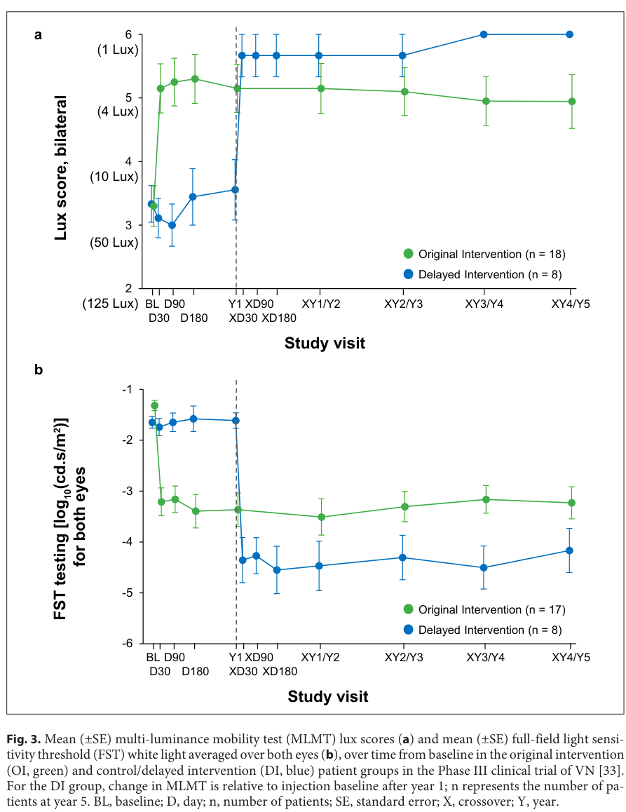
snippet: "13 (65%) of 20 intervention participants, but no control participants, passed MLMT at the lowest luminance level tested (1 lux), demonstrating maximum possible improvement"
explanation: >-
The primary efficacy endpoint showed that 65% of voretigene-treated participants
achieved maximum possible improvement on the multi-luminance mobility test
(passing at 1 lux), while no control participants did, demonstrating the
clinical magnitude of gene therapy benefit.
- reference: PMID:31725251
reference_title: "Autosomal Recessive RPE65-Related Retinal Degeneration."
supports: SUPPORT
evidence_source: OTHER
snippet: "Subretinal gene supplementation therapy for individuals between ages 12 months and 65 years with retinal degeneration known to be associated with biallelic RPE65 pathogenic variants."
explanation: >-
The GeneReview confirms the approved gene therapy indication and age range,
consistent with the subretinal delivery approach established in the
phase 3 trial.
- reference: DOI:10.1159/000526317
reference_title: "Gene Therapy for Inherited Retinal Disease: Long-Term Durability of Effect"
supports: SUPPORT
evidence_source: HUMAN_CLINICAL
snippet: "sustained results for up to 7.5 years for the full-field light sensitivity threshold test and 5 years for the multi-luminance mobility test in the Phase I and Phase III trials, respectively"
explanation: >-
This systematic review of voretigene neparvovec long-term outcomes documents
sustained functional improvements for up to 7.5 years (FST) and 5 years
(MLMT) across Phase I and Phase III clinical trials, confirming durable efficacy.
- reference: DOI:10.3390/biom14010122
reference_title: "Real-World Safety and Effectiveness of Voretigene Neparvovec: Results up to 2 Years from the Prospective, Registry-Based PERCEIVE Study"
supports: SUPPORT
evidence_source: HUMAN_CLINICAL
snippet: "Thirty-five patients (34%) experienced ocular treatment-emergent adverse events (TEAEs), most frequently related to chorioretinal atrophy"
explanation: >-
The prospective PERCEIVE real-world registry (n=103 patients) documents the
real-world safety profile of voretigene neparvovec; chorioretinal atrophy is
the most frequent ocular adverse event, occurring in 12.6% of patients, which
is important for post-treatment monitoring.
- reference: PMID:28712537
reference_title: "Efficacy and safety of voretigene neparvovec (AAV2-hRPE65v2) in patients with RPE65-mediated inherited retinal dystrophy: a randomised, controlled, open-label, phase 3 trial."
supports: SUPPORT
evidence_source: HUMAN_CLINICAL
snippet: "Improvements in both navigational abilities and light sensitivity were evident within the first 30 days after subretinal delivery and remained stable for 1 year, as they have for at least 3 years in the participants of the phase 1 follow-on trial."
explanation: >-
This states the limit of what the randomised comparison established. The
controlled endpoint was change at 1 year; stability beyond that point is
carried by the uncontrolled phase 1 follow-on cohort, not by the randomised
trial. Durability claims longer than a year in this entry therefore rest on
extension and registry data rather than on the pivotal trial.
- reference: PMID:28712537
reference_title: "Efficacy and safety of voretigene neparvovec (AAV2-hRPE65v2) in patients with RPE65-mediated inherited retinal dystrophy: a randomised, controlled, open-label, phase 3 trial."
supports: SUPPORT
evidence_source: HUMAN_CLINICAL
quote_role: BACKGROUND
snippet: "Other RPE65 gene therapy trials, which were administered to only one eye per individual using different gene constructs, vector formulations, or surgical approaches, have shown improvements in retinal function but variable durability of effect."
explanation: >-
The trial report's own introduction records that earlier RPE65 gene therapy
trials, using different constructs and surgical approaches, showed improved
retinal function but variable durability of effect. This is the caveat the
pivotal trial was not designed to resolve, and it is quoted from that paper's
background rather than from its results.
- reference: PMID:28689169
reference_title: "Leber congenital amaurosis/early-onset severe retinal dystrophy: clinical features, molecular genetics and therapeutic interventions."
supports: REFUTE
directness: INDIRECT
evidence_source: OTHER
quote_role: REVIEW_SYNTHESIS
snippet: "most studies at later intervals have shown a lack of durability of the improvements, with continuing retinal degeneration even in patients with improved retinal function."
explanation: >-
Recorded as a REFUTE against any reading of this treatment as arresting the
degeneration. Reviewing the four phase 1 RPE65 gene therapy trials, this paper
reports that most later follow-up showed a lack of durability of the functional
improvements, with retinal degeneration continuing even in eyes whose retinal
function had improved. Gene supplementation restores chromophore regeneration;
on this evidence it does not demonstrably stop the photoreceptor loss, which is
why treating while photoreceptors remain is the operative clinical point.
Graded INDIRECT because the cohorts quoted are the phase 1 trials rather than
the approved product's own programme.
- reference: DOI:10.1038/s41433-024-03065-6
reference_title: "Voretigene neparvovec for inherited retinal dystrophy due to RPE65 mutations: a scoping review of eligibility and treatment challenges from clinical trials to real practice"
supports: SUPPORT
evidence_source: HUMAN_CLINICAL
snippet: "paediatric patients retain more viable cells, present a less severe disease stage and show the highest potential for improvements, making them the most suitable candidates for treatment"
explanation: >-
This scoping review of real-world eligibility for voretigene neparvovec
identifies pediatric patients as optimal candidates due to greater photoreceptor
viability, supporting early evaluation and intervention in children with
confirmed biallelic RPE65 mutations.
- reference: PMID:42425207
reference_title: "Post-approval outcomes of voretigene neparvovec (Luxturna®) retinal gene therapy: A systematic review and meta-analysis"
supports: SUPPORT
evidence_source: HUMAN_CLINICAL
snippet: "Pooled evidence demonstrated statistically significant improvements in full-field stimulus threshold testing of 2.2 log10cd.s.m-2 (95% CI -2.87 to -1.53; P < 0.001) and in visual acuity of 0.08 logMAR (95% CI -0.16-0; P = 0.040) in patients post treatment. Chorioretinal atrophy, a complication not observed in clinical trials, was reported in a subset of studies and occurred in 0.30 (95% CI 0.10-0.51; P < 0.001) for chorioretinal atrophy located anywhere"
explanation: >-
A 2026 systematic review and meta-analysis of post-approval voretigene
neparvovec outcomes. The quoted sentences carry both halves of the claim:
pooled improvement in full-field stimulus threshold and in visual acuity,
and a pooled chorioretinal atrophy proportion of 0.30 for atrophy anywhere
in the retina - a complication the pivotal trials did not report. The
chorioretinal atrophy signal is independently carried by the PERCEIVE
registry item above.
- name: Supportive care and low vision rehabilitation
therapeutic_modality: OTHER
description: >-
Low vision aids, orientation and mobility training, and educational supports for
the visually impaired are core components of management for patients not eligible
for or not yet treated with gene therapy. Dietary interventions including
docosahexaenoic acid and lutein supplements have been considered supportively, and
children should be monitored with routine ophthalmological follow-up.
treatment_term:
preferred_term: supportive care
term:
id: NCIT:C15747
label: Supportive Care
evidence:
- reference: PMID:31725251
reference_title: "Autosomal Recessive RPE65-Related Retinal Degeneration."
supports: SUPPORT
evidence_source: OTHER
snippet: "Healthy balanced diet to reach the minimum reference daily intake (RDI) for nutrients. Docosahexaenoic acid / eicosapentaenoic acid supplements up to 500 mg/day as well as lutein supplements up to 10 mg/day can be considered if dietary intake is not sufficient."
explanation: >-
The GeneReview describes supportive management including dietary supplements
as standard of care alongside or in lieu of gene therapy, alongside early
referral to low vision services.
- name: Genetic counseling
therapeutic_modality: OTHER
description: >-
Genetic counseling is indicated for affected individuals and their families to
clarify the inheritance pattern (autosomal recessive), reproductive risk (25%
recurrence for parents of an affected child), and to identify at-risk siblings
who may benefit from early evaluation and timely gene therapy consideration.
treatment_term:
preferred_term: genetic counseling
term:
id: NCIT:C15240
label: Genetic Counseling
evidence:
- reference: PMID:31725251
reference_title: "Autosomal Recessive RPE65-Related Retinal Degeneration."
supports: SUPPORT
evidence_source: OTHER
snippet: "Evaluation of relatives at risk: It is appropriate to clarify the genetic status of apparently unaffected older and younger at-risk sibs in order to identify those who have biallelic RPE65 pathogenic variants and may benefit from early evaluation, counseling, and timely consideration of subretinal gene supplementation therapy."
explanation: >-
The GeneReview explicitly recommends genetic status clarification in
at-risk siblings to enable early evaluation and gene therapy consideration,
establishing genetic counseling as a core management component.
classifications:
harrisons_chapter:
- classification_value: GENETICS_ENVIRONMENT_DISEASE
- classification_value: NEUROLOGIC
diagnosis:
- name: Molecular genetic testing
diagnosis_term:
preferred_term: diagnostic procedure
term:
id: NCIT:C18020
label: Diagnostic Procedure
description: >-
Molecular confirmation of biallelic pathogenic RPE65 variants is required to
establish the diagnosis, guide prognosis, and determine eligibility for voretigene
neparvovec gene therapy (which requires confirmed biallelic RPE65 mutations).
Next-generation sequencing panels covering inherited retinal disease genes or
whole exome/genome sequencing are the primary diagnostic tools.
results: >-
Identification of biallelic pathogenic RPE65 variants confirms the diagnosis
and qualifies the patient for gene therapy evaluation; detection of a single
heterozygous variant in a clinically affected patient should prompt consideration
of a dominant form (MONDO:0100452) or a missed second allele.
evidence:
- reference: PMID:31725251
reference_title: "Autosomal Recessive RPE65-Related Retinal Degeneration."
supports: SUPPORT
evidence_source: OTHER
snippet: "The diagnosis of autosomal recessive RPE65-related retinal degeneration is established in a proband with suggestive findings and biallelic pathogenic variants in RPE65 identified by molecular genetic testing."
explanation: >-
The GeneReview specifies that molecular genetic testing confirming biallelic
RPE65 variants is the standard for establishing the diagnosis.
- name: Functional visual assessment and retinal imaging
diagnosis_term:
preferred_term: diagnostic procedure
term:
id: NCIT:C18020
label: Diagnostic Procedure
description: >-
Full-field light sensitivity threshold (FST) testing, Goldmann visual field
perimetry, electroretinography (ERG), and optical coherence tomography (OCT)
together characterize the degree of photoreceptor dysfunction and remaining viable
retinal tissue, both for diagnosis and for assessing gene therapy eligibility
(minimum retinal thickness on OCT is required).
results: >-
Dark-adapted ERG shows severely reduced or absent rod responses; light-adapted
ERG is also impaired. FST demonstrates markedly elevated light sensitivity
thresholds. OCT shows outer retinal layer preservation in gene-therapy-eligible
patients. Improvement in FST after voretigene neparvovec is an established
biomarker of restored RPE65 enzymatic activity.
evidence:
- reference: PMID:28712537
reference_title: "Efficacy and safety of voretigene neparvovec (AAV2-hRPE65v2) in patients with RPE65-mediated inherited retinal dystrophy: a randomised, controlled, open-label, phase 3 trial."
supports: SUPPORT
evidence_source: HUMAN_CLINICAL
snippet: "in the intervention group showed a rapid, greater than 2 log units improvement by day 30 in light sensitivity that remained stable over 1 year"
explanation: >-
The phase 3 trial establishes that FST improvement of more than 2 log units
after gene therapy directly reflects restoration of RPE65 enzymatic activity
in the RPE, validating FST as a key functional biomarker for this disease.
epidemiology:
- name: Prevalence among inherited retinal dystrophy subtypes
description: >-
Biallelic RPE65 mutations account for approximately 8% of Leber congenital
amaurosis cases and 2% of retinitis pigmentosa cases, making it one of the
more common single-gene causes within these rare inherited retinal dystrophy
categories. Absolute prevalence is rare; the gene therapy approval has made
genetic diagnosis of greater clinical relevance.
evidence:
- reference: DOI:10.1038/s41433-024-03065-6
reference_title: "Voretigene neparvovec for inherited retinal dystrophy due to RPE65 mutations: a scoping review of eligibility and treatment challenges from clinical trials to real practice"
supports: SUPPORT
evidence_source: HUMAN_CLINICAL
snippet: "Biallelic mutations in the RPE65 gene affect nearly 8% of Leber Congenital Amaurosis and 2% of Retinitis Pigmentosa cases."
explanation: >-
This scoping review of voretigene neparvovec eligibility provides the
prevalence figures for RPE65 mutations among the LCA and RP subtypes,
establishing the relative frequency of this treatable genetic cause.
progression:
- phase: Early-onset severe visual impairment with relative stability in first decade
age_range: Infancy to age 10
notes: >-
In the LCA2 form, visual manifestations arise in infancy or the first year.
Children aged 4–10 have severe mean visual acuity (20/126). Vision is
relatively stable through the first decade but already severely impaired.
evidence:
- reference: PMID:31725251
reference_title: "Autosomal Recessive RPE65-Related Retinal Degeneration."
supports: SUPPORT
evidence_source: OTHER
snippet: "In RPE65-related LCA, onset of visual manifestations frequently occurs in infancy during the first year of life. Central vision can be variable, but children ages four to ten years in the largest case series had severe visual impairment (mean visual acuity: 20/126). While vision is relatively stable in the first decade of life, it begins to decline again starting in the teens"
explanation: >-
The GeneReview documents the natural history of RPE65-LCA: early-onset
severe impairment with relative plateau in the first decade before
the accelerating teenage decline.
- phase: Accelerating decline leading to legal blindness in second and third decade
age_range: Age 10–40
notes: >-
Vision loss accelerates from the teenage years onward. By age 20, 50% of
LCA2 individuals meet legal blindness criteria. After age 20, progressive
loss leads to complete blindness by the fourth decade in virtually all cases.
Gene therapy can interrupt this trajectory when viable retina remains.
evidence:
- reference: PMID:31725251
reference_title: "Autosomal Recessive RPE65-Related Retinal Degeneration."
supports: SUPPORT
evidence_source: OTHER
snippet: "with 50% of individuals being legally blind (visual acuity of 20/200 and/or visual fields extending <20 degrees from fixation) by age 20 years. After age 20 years, vision loss is more rapidly progressive: all affected individuals are legally blind by the fourth decade and many have complete loss of vision (i.e., no light perception)."
explanation: >-
The GeneReview natural history data establish the biphasic decline pattern
with the critical therapeutic window — the decades before complete photoreceptor
loss — in which gene therapy can restore function.
clinical_trials:
- name: NCT00999609
phase: PHASE_III
status: COMPLETED
description: >-
Pivotal randomised, controlled, open-label Phase 3 trial of voretigene
neparvovec (AAV2-hRPE65v2) in participants aged 3 years or older with biallelic
RPE65 mutations and sufficient viable retina. Primary endpoint: 1-year change
in bilateral multi-luminance mobility test (MLMT) performance. Results
published as PMID:28712537 (Russell et al., Lancet 2017); the trial was the
basis for FDA approval of voretigene neparvovec (Luxturna) in December 2017.
target_phenotypes:
- preferred_term: Reduced visual acuity
term:
id: HP:0007663
label: Reduced visual acuity
- preferred_term: Constriction of peripheral visual field
term:
id: HP:0001133
label: Constriction of peripheral visual field
evidence:
- reference: clinicaltrials:NCT00999609
reference_title: "A Safety and Efficacy Study in Subjects With Leber Congenital Amaurosis (LCA) Using Adeno-Associated Viral Vector to Deliver the Gene for Human RPE65 to the Retinal Pigment Epithelium (RPE) [AAV2-hRPE65v2-301]"
supports: SUPPORT
evidence_source: HUMAN_CLINICAL
snippet: "The study is a Phase 3, open-label, randomized controlled trial of gene therapy intervention by subretinal administration of AAV2-hRPE65v2 (voretigene neparvovec-rzyl). At least twenty-four subjects, three years of age or older, will be recruited."
explanation: >-
ClinicalTrials.gov record for the pivotal Phase 3 voretigene neparvovec
trial that established efficacy and safety supporting FDA approval for
biallelic RPE65-mediated retinal dystrophy.
references:
- reference: PMID:31725251
title: "Autosomal Recessive RPE65-Related Retinal Degeneration."
tags:
- GeneReviews
References & Deep Research
References
1Deep Research
2Deep research results are used as seeds for research; they do not undergo the same validation as the main records and may contain errors. How we use deep research.
Evaluations and curation notes (1)
Curate Leber congenital amaurosis 2 (MONDO:0008765) as the LCA2 subtype of RPE65-Related Retinopathy · 2026-09-24T17:12:18Z · View source
Lump/split: MONDO:0008765 (Leber congenital amaurosis 2) was curated as a has_subtypes node plus a skos:narrowMatch mondo_mappings entry on the existing kb/disorders/RPE65-Related_Retinopathy.yaml (MONDO:0100368), not as a new Disease entry. MONDO files MONDO:0008765 beneath MONDO:0100368; the gene (RPE65, hgnc:10294) and the proximal lesion are identical, and the existing entry's own notes already recorded LCA2/EOSRD/juvenile RP as one genetic entity separated by residual enzyme activity. This differs from Leber_Congenital_Amaurosis_9/10/13, which are separate entries because each is a different gene with no gene-level entry to fold into. Gene confirmed against the primary source PMID:9326941 (Gu et al. 1997), not from the stub. Added: LCA2 subtype bound to MONDO:0008765 with AR inheritance and three evidence items; mappings.mondo_mappings narrowMatch; three LCA2-scoped pediatric phenotypes (oculodigital sign HP:0001483, high hypermetropia HP:0008499, abnormal pupillary light reflex HP:0007695); five new causal edges taking the pathograph from visual-cycle arrest through to nyctalopia, pupillary reflex, peripheral field loss and photophobia, plus two phenotype sequelae (nystagmus, hypermetropia), raising phenotype causal-inlink coverage from 1/6 to 7/9; a delivery_system block on voretigene neparvovec (VIRAL_VECTOR, AAV2, subretinal, RPE target cell) sourced to the phase 3 report; and three treatment evidence items recording the limits of the pivotal trial (1-year controlled endpoint) and a REFUTE item on durability from the phase 1 follow-up literature. Corrected an overstated nystagmus frequency claim. PMID:42425207 lost its quotable body under linkml-reference-validator 0.3.0rc1 (issue #12672) and its snippet was removed rather than paraphrased; the citation and an explicit gap statement were kept. Deep research: falcon requested, returned HTTP 402, claude_code fallback produced research/RPE65-Related_Retinopathy-deep-research-claude_code.md; preflight-dr PASS against MONDO:0100368. All gates run under linkml-reference-validator 0.3.0rc1.
RPE65-Related Retinopathy: Comprehensive Research Report
MONDO ID: MONDO:0100368 Category: Mendelian, autosomal recessive (rare autosomal dominant form also reported) Key OMIM entries: #204100 (Leber Congenital Amaurosis 2, LCA2), #613794 (Retinitis Pigmentosa 20, RP20), *180069 (Retinoid Isomerohydrolase RPE65, the gene locus)
1. Disease Information
Overview. RPE65-related retinopathy is the umbrella term for a spectrum of autosomal recessive (and, rarely, autosomal dominant) inherited retinal dystrophies caused by biallelic (or, for one specific variant, heterozygous) pathogenic variants in RPE65, which encodes the retinal pigment epithelium (RPE)-specific isomerohydrolase that regenerates the visual chromophore 11-cis-retinal. The clinical spectrum spans a continuum of severity: Leber congenital amaurosis (LCA2) at the severe end, early-onset severe retinal dystrophy (EOSRD) in the middle, and RPE65-associated retinitis pigmentosa (RP20), sometimes with juvenile onset, at the milder end — "the three phenotypes of autosomal recessive RPE65-related retinal degeneration, from most severe to mildest, are Leber congenital amaurosis (LCA), early-onset severe retinal dystrophy (EOSRD), and juvenile retinitis pigmentosa (RP)" (GeneReviews, NBK549574). This is the first inherited retinal disease with an FDA/EMA-approved gene-replacement therapy (voretigene neparvovec-rzyl, Luxturna).
Key identifiers:
| Resource | Identifier |
|---|---|
| OMIM (LCA2) | #204100 |
| OMIM (RP20) | #613794 |
| OMIM (gene) | *180069 |
| MONDO | MONDO:0100368 (per prompt); related MONDO:0018091 (LCA2) |
| Orphanet | ORPHA:65 (Leber congenital amaurosis, umbrella); RPE65-specific subtype pages exist under the LCA/EOSRD Orphanet classification |
| MeSH | Leber Congenital Amaurosis (D016954); Retinitis Pigmentosa (D012174) |
| Gene | HGNC:10294 (RPE65); NCBI Gene 6121; Ensembl ENSG00000116745 |
| ICD-10 | H35.5 (hereditary retinal dystrophy); ICD-11 9B76 |
Synonyms: RPE65-associated retinal dystrophy; RPE65-mediated inherited retinal disease (RPE65-IRD); Leber congenital amaurosis type 2 (LCA2); RPE65 deficiency; Retinitis pigmentosa 20 (RP20); autosomal recessive childhood-onset severe retinal dystrophy (RPE65 type); retinoid isomerohydrolase deficiency.
Evidence basis: This report draws on OMIM/GeneReviews disease-level syntheses, structured registries (Orphanet, ClinVar/ClinGen), large multi-center natural-history and clinical-trial cohorts (e.g., the Phase 1/3 voretigene neparvovec program), and individual case reports/small case series for variant-level detail — the evidence tiers differ and are noted per claim.
2. Etiology
Disease causal factor: RPE65-related retinopathy is monogenic. Biallelic loss-of-function or hypomorphic variants in RPE65 (chromosome 1p31.3) cause the autosomal recessive spectrum (LCA2/EOSRD/RP20). A single specific missense variant, p.Asp477Gly (D477G), causes a distinct autosomal dominant late-onset retinitis pigmentosa through a toxic gain-of-function/aggregation mechanism rather than simple haploinsufficiency (see §4 and §6).
Genetic risk factors: - Causal variants: >150 distinct pathogenic RPE65 variants reported across missense, nonsense, frameshift, splice-site, and small in-frame indel classes (ClinVar; PMC7016655 "Pathogenicity Reclassification of RPE65 Missense Variants"). - Modifier genes: No well-established modifier locus is confirmed for RPE65-retinopathy specifically, unlike CEP290-LCA (where AHI1 has been proposed as a modifier; PMC3048164). Phenotypic variability among biallelic-genotype-matched patients is well documented but its genetic basis is not fully resolved. - Consanguinity: Increases homozygosity for rare recessive RPE65 alleles in populations/families with high consanguinity rates; multiple case series (e.g., from the Russian Federation, PMC10671290; East Asian cohorts, PMC9644481) report founder/recurrent alleles. - Founder alleles: Population-specific recurrent variants are documented; East Asian carrier frequency was estimated at ~0.10% (11/10,919 alleles) in gnomAD/KRGDB-derived East Asian samples and ~0.06% in Koreans specifically (PMC9644481, Orphanet J Rare Dis 2022).
Environmental risk factors: None established as causal — this is a purely Mendelian disease. However, ambient light/luminance exposure modulates phenotype severity in the dominant D477G form: "The interplay of environmental luminance and genetics in the retinal dystrophy induced by the dominant RPE65 mutation" (PMC8931212, PNAS 2022) demonstrates that light exposure interacts with the D477G genotype to determine degeneration severity in animal models, a rare example of gene-environment interaction in an otherwise monogenic disease.
Protective factors: No genetic protective variants are established. Reduced UV/blue-light exposure is biologically plausible as ameliorating in the dominant form (lipofuscin/A2E-related photo-oxidative mechanisms, see §6) but is not established as a clinical protective intervention through controlled trials.
Gene-environment interactions: Best characterized for D477G — see PMC8931212 above; not established for the biallelic recessive forms, where disease is fully penetrant regardless of environment.
3. Phenotypes
Core phenotype set (by category)
Symptoms/signs — visual: | Phenotype | Frequency/notes | Suggested HP term | |---|---|---| | Severe congenital/infantile vision loss (LCA) or progressive vision loss (EOSRD/RP) | Nearly universal for LCA form; onset in infancy | HP:0000618 (Blindness) / HP:0000505 (Visual impairment) | | Nystagmus (typically pendular) | Very common, often presenting sign in infancy | HP:0000639 (Nystagmus) | | Nyctalopia (night blindness) | "Most patients with RPE65 mutations are profoundly night blind"; earliest symptom, often before central vision loss, even in RP-only phenotype | HP:0000662 (Nyctalopia) | | Photophobia | Common | HP:0000613 (Photophobia) | | Sluggish or near-absent pupillary light reflex | Frequent in LCA | HP:0007686 (Abnormal pupillary function) / HP:0008070 (Sluggish pupillary response) | | Oculodigital sign (eye-poking/pressing/rubbing) | Common in infantile LCA presentations | HP:0006990 (Eye poking) | | High hyperopia | Frequent | HP:0008515 (Hyperopia) | | Keratoconus | Reported, secondary to chronic eye rubbing | HP:0000563 (Keratoconus) | | Cataract | Reported as a secondary/late finding | HP:0000518 (Cataract) | | Strabismus | Common | HP:0000486 (Strabismus) | | Reduced/constricted visual field | Progressive | HP:0000603 (Constriction of peripheral visual field) | | Variable/abnormal fundus appearance | "Variable appearance to the fundus" — ranges from near-normal in infancy to bone-spicule pigmentary changes, attenuated vessels, optic disc pallor, and macular atrophy with progression | HP:0007663 (Reduced visual acuity) as functional correlate; HP:0000580 (Pigmentary retinopathy); HP:0000556 (Retinal dystrophy) | | Macular atrophy/coloboma-like macular changes | More characteristic in later stages and in some genotypes | HP:0007401 (Macular atrophy) |
Laboratory/electrophysiologic abnormality (functional): - Electroretinogram (ERG): severely reduced or non-recordable (rod and cone responses) — "the ERG is severely reduced or non-recordable" in RPE65-LCA/EOSRD. In the milder RP20 phenotype ERG may remain partially recordable, especially earlier in the disease course. Suggested term: HP:0000513 (Abnormal electroretinogram)/ more specific HP:0000662-associated functional test findings. - Full-field stimulus threshold (FFST): markedly elevated dark-adapted thresholds (used as a primary trial endpoint; log-unit improvements of ~2 log10 cd·s·m⁻² reported post-gene-therapy).
Phenotype characteristics: - Age of onset: LCA form — onset in infancy, typically within the first year of life (congenital or near-congenital); EOSRD — early childhood; RP20 (autosomal recessive juvenile/childhood RP) — onset from childhood into adolescence; the OMIM RP20 family description notes "onset of severe visual impairment was between 3 and 7 years of age, with night blindness being a typical and early symptom in all patients." The autosomal dominant D477G form is distinctly later-onset ("advanced late-onset retinitis pigmentosa," PMC7228561). - Severity: Generally severe (profound, near-blind) in LCA; variable but progressive in EOSRD/RP. - Progression: Progressive in all forms; central vision is often relatively preserved longer than peripheral/night vision early on, but declines with age — "night blindness is one of the earliest symptoms but patients tend to suffer central visual loss later." In a large natural-history case series, children aged 4–10 years already had severe visual impairment (mean visual acuity ~20/126). - Frequency among affected individuals: Night blindness and nystagmus are present in nearly all cases; keratoconus and cataract are present in a minority and tend to be later complications.
Quality of life impact: Profound vision loss from infancy has major developmental, educational, and psychosocial consequences (mobility, literacy via Braille/assistive technology, dependence). Gene-therapy trials formally captured functional vision (multi-luminance mobility testing, MLMT) and quality-of-life instruments, showing meaningful gains post-treatment, "particularly in younger patients" (per long-term follow-up data).
4. Genetic/Molecular Information
Gene: RPE65 — Retinoid Isomerohydrolase RPE65; HGNC:10294; chromosome 1p31.3 (varies slightly by source, commonly cited 1p31); NM_000329.3 is the reference transcript used in clinical variant nomenclature.
Causal genes/loci: RPE65 is the sole causal gene for this entity by definition. OMIM lists two disease associations: #204100 LCA2 (autosomal recessive) and #613794 RP20 (autosomal recessive); a separate autosomal dominant RP phenotype (sometimes designated RP87 in more recent OMIM nomenclature) is caused specifically by the heterozygous D477G variant.
Variant spectrum and classification: - Types: missense (the largest category), nonsense, frameshift (e.g., c.106del predicted to cause a frameshift and premature stop, leading to nonsense-mediated decay), splice-site, and small in-frame deletions/insertions. - ACMG/ClinVar classification: Variants are individually curated pathogenic/likely pathogenic/VUS per ACMG-AMP criteria; a ClinGen RPE65-specific Variant Curation Expert Panel specification exists (cspec.genome.network/cspec/ui/svi/doc/GN120), refining general ACMG rules for this gene. A dedicated reclassification effort addressed missense VUS: "Pathogenicity Reclassification of RPE65 Missense Variants Related to Leber Congenital Amaurosis and Early-Onset Retinal Dystrophy" (PMC7016655). - Population allele frequency: Individual pathogenic variants are each very rare in gnomAD (e.g., c.1039C>T p.Arg347Cys reported at ~0.00000124 allele frequency, 5/1,179,740 alleles, in European non-Finnish gnomAD), consistent with a rare recessive disease; aggregate carrier frequency in East Asians was estimated at ~0.10%. - Somatic vs. germline: Exclusively germline; no somatic mosaicism mechanism described for this disease. - Functional consequences: - Most recessive loss-of-function alleles (nonsense, frameshift, canonical splice-site) abolish or near-abolish isomerohydrolase enzymatic activity — complete loss of function. - Many missense alleles are hypomorphic (partial loss of function), correlating with the milder EOSRD/RP20 end of the phenotypic spectrum rather than LCA — see the "novel RPE65 hypomorph" functional study (PMC5015590). - The dominant D477G variant does not act by simple haploinsufficiency: functional studies show "the adverse cellular consequence of the D477G mutant coexpressed with WT-RPE65 far exceeded that of losing a single allele of WT-RPE65," and structural/biochemical work indicates D477G forms an aggregation-prone surface that induces abnormal protein–protein interactions with wild-type RPE65, driving RPE65 protein degradation and reduced isomerase activity beyond what simple heterozygous loss would predict — i.e., a toxic/dominant-negative gain-of-function mechanism (PMID:29659842, Hum Mol Genet 2018; PMC7760593/PMID:33261050).
Modifier genes: None definitively established for the RPE65-recessive spectrum.
Epigenetic information: No disease-specific DNA methylation/histone modification mechanism is established as causal; this is a classic monogenic enzymatic deficiency disease.
Chromosomal abnormalities: Not a recognized mechanism — disease results from point mutations/small indels within RPE65, not large structural rearrangements, though whole-gene or exonic deletions have occasionally been reported as pathogenic alleles.
Ontology suggestions: Gene — hgnc:10294 (RPE65); Molecular function — GO:0003920 (retinoid isomerohydrolase activity / GO:0033760 "retinal isomerase activity" umbrella term), GO:0033748 (bisretinoid catabolism, indirectly relevant); relevant CHEBI terms: CHEBI:17561 (retinol), CHEBI:17278 (all-trans-retinyl ester), CHEBI:17885 (11-cis-retinal).
5. Environmental Information
RPE65-related retinopathy is not an infectious, toxin-induced, or classically "environmental" disease — it is monogenic. The only substantiated environmental interaction is:
- Ambient luminance/light exposure as a phenotype modifier specifically for the dominant D477G allele (PMC8931212, PNAS 2022) — higher-light environments were associated with greater retinal dystrophy severity in genetically modeled systems, implicating light-driven photo-oxidative stress (via the retinoid/lipofuscin cycle, see §6) as an aggravating factor superimposed on the genetic lesion.
- Vitamin A/retinoid status: Because RPE65 sits in the core vitamin A (retinoid) visual cycle, systemic vitamin A metabolism is mechanistically intertwined with the RPE65 defect, though dietary vitamin A supplementation has not been shown to correct the enzymatic block (unlike some other IRDs, e.g., Refsum disease with dietary phytanic acid restriction). This is the pharmacological rationale instead for exogenous synthetic retinoid replacement therapy (QLT091001/9-cis-retinyl acetate; see §12).
- No infectious agents are implicated.
- Lifestyle factors: No behavioral risk-modifying factor (smoking, diet, exercise) has controlled-trial evidence in this specific disease, though general retinal-health advice (UV protection) is plausible extrapolation from the light-luminance mechanistic data above, primarily relevant to D477G carriers.
Suggested ECTO term if curating an environmental modifier edge: an ambient/ocular light-exposure term (e.g., ECTO class for "light exposure") linked specifically to the D477G pathophysiology node, with environmental_effect: EXACERBATES.
6. Mechanism / Pathophysiology
Ordered causal chain (recessive/loss-of-function form — LCA2/EOSRD/RP20)
- Biallelic loss-of-function or hypomorphic pathogenic variants in RPE65 lead to absent or markedly reduced RPE65 retinoid isomerohydrolase enzymatic activity in the retinal pigment epithelium (demonstrated biochemically; PMID:16116091, PNAS 2005 — "RPE65 is the isomerohydrolase in the retinoid visual cycle").
- Loss of isomerohydrolase activity results in failure to convert all-trans-retinyl ester to 11-cis-retinol within the RPE cell, the rate-limiting step of the retinoid ("visual") cycle.
- Absence of 11-cis-retinol/11-cis-retinal leads to depletion of functional visual chromophore available to combine with opsin apoprotein in rod and cone outer segments, so rhodopsin (rods) and cone opsins cannot be regenerated after bleaching.
- Chromophore-free ("apo-") opsin causes two convergent downstream consequences, demonstrated to be mechanistically distinct in rods versus cones:
- 4a. Rods: unliganded rod opsin is intrinsically unstable/constitutively signals abnormally and triggers a Bax-dependent apoptotic pathway in rod photoreceptors (PMC2720534, "Bax-Induced Apoptosis in Leber's Congenital Amaurosis: A Dual Role in Rod and Cone Degeneration"), together with upregulation of lysosomal-mediated autophagic gene expression contributing to early rod apoptosis in Rpe65⁻/⁻ mice (PMID:22227450).
- 4b. Cones: cone photoreceptor death proceeds via a Bax-independent mechanism — "early loss of cones was not prevented in Rpe65⁻/⁻/Bax⁻/⁻ mice, indicating that pro-apoptotic Bax was not involved in the pathogenesis of cone cell death in Rpe65-deficient mice" — implicating a separate degenerative cascade (proposed to relate to chronic unliganded-cone-opsin signaling/ER stress rather than classical Bax-mediated apoptosis) and explaining the disproportionately early, severe cone dysfunction seen clinically even though rods normally vastly outnumber cones.
- Progressive rod and (via the separate pathway) cone photoreceptor apoptosis results in outer-segment shortening/disorganization and eventual photoreceptor cell loss, detectable clinically as thinning of the outer nuclear layer/ellipsoid zone on OCT and as non-recordable or severely reduced ERG amplitudes.
- Concurrently, accumulation of un-isomerized all-trans-retinyl esters and derived bis-retinoid byproducts (notably A2E, a diretinal-phosphatidylethanolamine conjugate formed from all-trans-retinal condensing with phosphatidylethanolamine in photoreceptor outer segments and subsequently phagocytosed into the RPE) accumulates as RPE lipofuscin. A2E cannot be enzymatically degraded, perturbs RPE lysosomal membrane integrity and phagolysosomal function (inhibiting phagolysosomal degradation of photoreceptor phospholipid), and confers susceptibility to blue-light-induced RPE apoptosis — providing a secondary, light-exacerbated toxic mechanism to RPE and photoreceptor injury superimposed on the primary chromophore-deprivation mechanism. (Note: in the RPE65-null state itself, lipofuscin/A2E accumulation is reduced relative to wild type because retinyl-ester substrate is not being processed through the normal cycle at all — the Leu450Met Rpe65 hypomorphic mouse variant, by contrast, shows reduced A2E via slowed cycle flux, PMID:15277666 — so the lipofuscin/A2E axis is most clinically relevant to milder hypomorphic and to the light-modulated dominant D477G phenotype rather than to null LCA2.)
- Clinically, this cascade manifests as profound infantile/childhood visual impairment, nystagmus, night blindness, and a non-recordable or severely subnormal ERG (steps 1→3 dominate the earliest, most severe LCA presentation; steps 4→6 drive the progressive component seen even in successfully gene-corrected retina, explaining why continued photoreceptor loss can occur after gene therapy despite restored chromophore supply — see §12).
Branch: dominant D477G mechanism
1′. The heterozygous D477G missense variant produces a structurally abnormal RPE65 protein with an aggregation-prone surface. 2′. This mutant protein engages in atypical protein–protein interactions with co-expressed wild-type RPE65, causing wild-type RPE65 protein degradation and a net reduction in cellular isomerase activity that exceeds the reduction expected from simple loss of one functional allele (i.e., not classical haploinsufficiency) — a dominant-negative/toxic gain-of-function mechanism (PMID:29659842; PMID:33261050). 3′. The resulting partial visual-cycle impairment, superimposed on and exacerbated by ambient light/luminance exposure (PMC8931212), leads to a milder, later-onset retinitis pigmentosa phenotype than the biallelic null state.
Categories covered
- Molecular pathways: Retinoid (visual) cycle — RPE65 catalyzes the committed isomerization/hydrolysis step converting all-trans-retinyl ester → 11-cis-retinol (KEGG: Retinol metabolism, hsa00830; Reactome: "Visual phototransduction").
- Cellular processes: Apoptosis (rod: Bax-dependent, GO:0006915/GO:0043523 negative regulation of neuron apoptotic process is disrupted; cone: Bax-independent, distinct pathway); autophagy/lysosomal gene upregulation (GO:0006914 autophagy); phagolysosomal dysfunction in RPE.
- Protein dysfunction: Loss of enzymatic function (null/hypomorphic alleles) vs. misfolding/aggregation-driven dominant-negative toxicity (D477G).
- Metabolic changes: Disrupted vitamin A/retinoid metabolism; secondary accumulation of retinyl esters and altered bis-retinoid (A2E) flux.
- Tissue damage mechanisms: Chronic photo-oxidative stress from lipofuscin/A2E accumulation, particularly light-exposure-dependent.
- Biochemical abnormality: Isomerohydrolase enzyme deficiency — the defining lesion.
- Molecular profiling: Biochemical/functional assays (HPLC-MS/MS quantification of retinoids) are used for variant functional characterization (e.g., PMC7138296-style approach applied across LCA genes); RPE65 protein expression studies in HEK293/insect-cell heterologous systems and iPSC-derived RPE/retinal organoids from patients (PMC6749091) recapitulate reduced isomerase activity and photoreceptor phenotypes in vitro.
Suggested GO terms: GO:0003920 (retinal isomerase activity — closest functional GO term for the isomerohydrolase activity), GO:0009416 (response to light stimulus), GO:0007601 (visual perception), GO:0006915 (apoptotic process), GO:0043524 (negative regulation of neuron apoptotic process). Suggested CL terms: CL:0000210 (photoreceptor cell), CL:0000604 (retinal rod cell), CL:0000573 (retinal cone cell), CL:0002586 (retinal pigment epithelial cell). Suggested CHEBI terms: CHEBI:17561 (retinol), CHEBI:17885 (11-cis-retinal), CHEBI:17278 (retinyl ester, generic), lipofuscin/A2E lacks a precise CHEBI ID commonly used in curation — record as free text if no exact match.
7. Anatomical Structures Affected
Organ level: - Primary organ: eye — specifically the neurosensory retina and retinal pigment epithelium (UBERON:0000966 retina; UBERON:0002050 retinal pigment epithelium is not a standard distinct UBERON ID — RPE is typically captured via CL:0002586 cell type plus UBERON:0000966 retina, or UBERON:0001782 (choroid)-adjacent structures depending on ontology version). - Secondary involvement: cornea (keratoconus secondary to chronic eye-rubbing, UBERON:0000362 cornea), lens (cataract, UBERON:0001845), extraocular muscles (strabismus, secondary to poor fixation). - Body systems: primarily the visual/sensory system (nervous system extension); no systemic organ involvement — RPE65-retinopathy is a non-syndromic, eye-limited disease (distinguishing it from syndromic ciliopathy-associated LCA genes such as CEP290).
Tissue and cell level: - Neurosensory retina — outer nuclear layer (photoreceptor cell bodies), photoreceptor inner/outer segments. - RPE monolayer — site of the primary enzymatic defect (CL:0002586, retinal pigment epithelial cell). - Affected cell populations: rod photoreceptors (CL:0000604) and cone photoreceptors (CL:0000573), both degenerate, via distinct mechanisms (§6); RPE cells (CL:0002586) accumulate lipofuscin/A2E.
Subcellular level: - RPE smooth endoplasmic reticulum — site of RPE65 enzymatic activity (GO Cellular Component: GO:0005783 endoplasmic reticulum; RPE65 is a microsomal membrane-associated protein). - RPE lysosomes — site of A2E/lipofuscin accumulation and impaired phagolysosomal degradation (GO:0005764 lysosome). - Photoreceptor outer segment discs — site of opsin/chromophore complex formation and of A2E precursor formation (GO:0001750 photoreceptor outer segment).
Localization: Bilateral, generally symmetric involvement (as expected for a systemic monogenic enzymatic deficiency rather than a focal lesion); no lateralization pattern reported.
8. Temporal Development
Onset: - LCA2: congenital/infantile — visual manifestations typically apparent within the first year of life; frequently the presenting complaint is roving eye movements/nystagmus and lack of visual fixation noticed by caregivers in early infancy. - EOSRD: early childhood onset, intermediate severity. - RP20 (recessive, juvenile RP phenotype): onset in one documented family series between ages 3–7 years, with night blindness as the earliest and most consistent symptom. - Dominant D477G RP: distinctly later-onset, described in the literature as "advanced late-onset retinitis pigmentosa" (PMC7228561), i.e., adult-onset relative to the recessive forms. - Onset pattern: insidious/progressive rather than acute, though the profound congenital deficit in LCA2 can appear as an abrupt discovery at birth/infancy even though the underlying enzymatic lesion is present from conception.
Progression: - Disease course is chronic and progressive across the spectrum; there is no spontaneous remission. - Natural history: night blindness (rod-pathway dysfunction) typically precedes and is more severe/earlier than central (cone-mediated) visual loss, which develops later but can still occur in the first decade — "children ages four to ten years in the largest case series had severe visual impairment (mean visual acuity: 20/126)." - Progression rate: variable across the recessive spectrum (LCA fastest/most severe from birth; RP20 slower); within any one genotype, phenotype severity can still vary between patients, suggesting genetic background or environmental modifiers. - Disease duration: lifelong, chronic — no natural resolution; some patients with milder hypomorphic alleles retain functional (if severely reduced) vision into adulthood, providing the therapeutic window exploited by gene therapy. - Even after gene augmentation therapy restores chromophore-generating capacity, continued photoreceptor degeneration has been documented in surviving but already-compromised retina (see §12) — i.e., the enzymatic correction does not fully halt an already-initiated degenerative process in retina beyond a certain disease stage, underscoring the importance of the "critical period"/treatment-timing concept below.
Patterns: - No remission pattern (progressive monogenic disease). - Critical period for intervention: A central concept in this disease's management is that gene therapy is most effective when administered to retina with sufficient viable photoreceptors remaining — "treated locations having more than 63% of normal photoreceptors showed robust treatment-related retention of photoreceptors in the long term, while treated regions with less retained photoreceptors at the time of the intervention showed progressive degeneration similar to untreated regions" — establishing a structural/temporal window beyond which restoring the enzyme cannot rescue already-committed-to-death photoreceptors. This underlies current gene-therapy eligibility criteria requiring sufficient viable retinal area (see §10, §12).
9. Inheritance and Population
Epidemiology: - LCA overall (all genes combined) affects an estimated 1 in 30,000 to 1 in 81,000 live births; RPE65 accounts for roughly 5–10% of LCA cases. - Retinitis pigmentosa overall affects 1 in 3,000–7,000 people; autosomal recessive RP accounts for ~5–20% of RP, and RPE65 accounts for roughly 2–5% of autosomal recessive RP. - In a multi-center inherited-retinal-dystrophy cohort of 2,240 patients, 18 (0.8%) had RPE65-related disease: 38.8% presenting as LCA, 22.2% as RP, 33.3% as severe early-onset retinal dystrophy, and 5.5% as congenital stationary night blindness-like presentation — illustrating the phenotypic breadth captured under one genotype. - In a Russian Federation cohort, RPE65-associated retinopathy accounted for 5.3% (25/474) of molecularly confirmed inherited retinal disease cases (PMC10671290). - Global disease burden: given the rarity, RPE65-retinopathy is classified as an ultra-rare disease; precise worldwide point-prevalence figures analogous to Orphanet's numeric prevalence bands are not consistently published as a single figure, but the above LCA/RP fraction data support an estimated worldwide prevalence in the range of roughly 1–2 per 100,000 to 1 per 1,000,000 (consistent with an Orphanet "1-9 / 1,000,000" or similarly rare band; a precise Orphanet numeric class was not independently confirmed in this search).
Inheritance pattern: Predominantly autosomal recessive (biallelic pathogenic variants — homozygous or compound heterozygous); a distinct, well-characterized autosomal dominant form exists caused specifically by the heterozygous D477G variant.
Penetrance: The recessive forms are essentially fully penetrant given biallelic pathogenic genotypes (consistent with a classic enzyme-deficiency Mendelian disease). Penetrance/expressivity of D477G dominant disease is modulated by environmental light exposure (§2, §6), suggesting incomplete or variable penetrance/expressivity for that specific allele depending on environmental luminance history — an unusual and specifically documented gene-environment penetrance modifier for a monogenic retinal disease.
Expressivity: Variable — even among patients with identical or similar biallelic genotypes, phenotype severity (LCA vs. EOSRD vs. RP20) and rate of progression differ, and macular morphology differs by causal gene even within the LCA differential (RPE65 vs. CEP290 vs. GUCY2D vs. AIPL1 patients show distinguishable macular phenotypes), suggesting genotype is only a partial predictor.
Genetic anticipation: Not described — RPE65-retinopathy is not a repeat-expansion disorder.
Germline mosaicism: Not specifically documented as a recurring feature in RPE65-retinopathy literature reviewed; general recessive-disease recurrence-risk counseling principles apply (25% recurrence risk per pregnancy for carrier parents of an affected homozygous/compound heterozygous child).
Founder effects: Documented at the population level — recurrent/founder pathogenic alleles reported in specific populations (East Asian, Russian, and other cohorts cited above), though no single globally dominant founder allele analogous to some other IRD genes has been established across all populations.
Consanguinity role: As with other rare autosomal recessive IRDs, consanguineous unions increase homozygosity for rare pathogenic RPE65 alleles and are commonly noted in case series from populations with higher consanguinity rates.
Carrier frequency: Estimated ~0.10% in East Asians (gnomAD/KRGDB combined) and ~0.06% in Koreans specifically (PMC9644481); carrier frequencies elsewhere are lower given the gene's overall rarity and the population-specific nature of most pathogenic alleles.
Population demographics: No strong sex predilection is reported (autosomal, not X-linked); no major geographic endemicity beyond population-specific founder-allele effects; age distribution of affected individuals spans from infancy (LCA presentation) through adulthood (dominant D477G presentation), reflecting the phenotypic spectrum rather than a single onset age.
10. Diagnostics
Clinical tests: - Electroretinography (ERG): central diagnostic test — severely reduced or non-recordable scotopic and photopic responses in LCA/EOSRD; may show residual but markedly subnormal responses in milder RP20/dominant D477G presentations, especially earlier in the disease course. - Optical coherence tomography (OCT): assesses outer nuclear layer/ellipsoid zone thickness and residual photoreceptor structure — critical for gene-therapy eligibility assessment (identifying sufficient viable retina) and for longitudinal monitoring of structural decline post-treatment. - Fundus autofluorescence: can show altered patterns related to lipofuscin/RPE status, useful in phenotyping and monitoring. - Full-field stimulus threshold (FFST) testing: dark-adapted psychophysical threshold test — the primary functional endpoint in the pivotal voretigene neparvovec trials, given the non-recordable ERG in many LCA patients makes ERG unsuitable as a treatment-response endpoint. - Multi-luminance mobility testing (MLMT): a functional-vision obstacle-course assay developed specifically for the voretigene neparvovec trials, assessing ability to navigate a course at graded light levels — became a novel, disease-specific, FDA-recognized functional endpoint. - Visual field testing: Goldmann or other perimetry to assess peripheral field constriction, where central fixation/cooperation allow. - Biopsy/histopathology: not routinely performed given the availability of genetic diagnosis; historical/postmortem histopathology contributed to early understanding of photoreceptor/RPE degeneration.
Genetic testing: - Recommended approach: targeted RPE65 single-gene sequencing when clinical phenotype (early nyctalopia, ERG pattern) is suggestive, or more commonly as part of a multi-gene inherited retinal disease (IRD)/LCA panel given overlapping phenotypes among the ~20+ LCA-associated genes (see differential diagnosis below) — panel-based next-generation sequencing (NGS) is now first-line given genetic and phenotypic heterogeneity of LCA/EOSRD, with whole exome sequencing (WES) as a common escalation when panels are non-diagnostic, given the diagnostic yield reported (e.g., PMID:28966547, "Diagnostic application of clinical exome sequencing in Leber congenital amaurosis"). - Whole genome sequencing (WGS): used in research/tertiary settings when exome-negative, to capture deep intronic or structural variants. - Chromosomal microarray/karyotype/FISH: not primary tools for this specific monogenic point-mutation disease; not typically indicated unless a contiguous gene deletion is suspected. - Confirmatory requirement for gene therapy: biallelic RPE65 pathogenic/likely pathogenic variant confirmation is an absolute prerequisite for voretigene neparvovec eligibility per FDA label and the international consensus eligibility recommendations (PMID:34088339, Orphanet J Rare Dis 2021), which formalized clinical, electrophysiological, imaging, and genetic criteria (including required retinal-area/viable-photoreceptor thresholds) for patient selection.
Clinical criteria/differential diagnosis: RPE65-retinopathy must be distinguished from other LCA/EOSRD genes on the basis of genotype (phenotype overlap is substantial). Per GeneReviews-style LCA gene lists, at least ~20 genes cause LCA, accounting for ~70% of cases, including:
| Gene | LCA subtype | Distinguishing notes |
|---|---|---|
| GUCY2D | LCA1 | Most common LCA gene overall in many populations; near-normal fundus early, preserved photoreceptor structure with severe functional loss ("cone-rod dystrophy dissociation") |
| RPE65 | LCA2 | This entry; eligible for approved gene therapy |
| CRX | LCA7 | Can have dominant or recessive inheritance |
| CRB1 | LCA8 | Often with preserved paravascular retinal pigment, nanophthalmos/high hyperopia, Coats-like exudative vasculopathy |
| NMNAT1 | LCA9 | Characteristic macular coloboma-like atrophy |
| CEP290 | LCA10 | Most common single LCA gene in many Western cohorts (up to ~21% of LCA); syndromic ciliopathy associations (Joubert, Senior-Løken, Bardet-Biedl, Meckel) must be excluded |
| RDH12 | LCA13 | |
| LRAT | LCA14 | Same visual-cycle pathway as RPE65 (upstream enzyme); phenotypically similar and also responds to oral synthetic retinoid in trials |
| AIPL1 | LCA4 | Severe cone-rod phenotype |
| others | SPATA7, LCA5, RPGRIP1, IMPDH1, RD3, TULP1, IQCB1, CLUAP1, PRPH2, KCNJ13, IFT140 | Rarer causes |
Clinical phenotyping can help direct genetic testing strategy, since "different genes are associated with differential macular morphology in patients with RPE65-, CEP290-, GUCY2D-, and AIPL1-related Leber congenital amaurosis," but molecular confirmation remains the diagnostic gold standard given overlap.
Screening: No universal newborn screening program specifically targets RPE65-retinopathy (unlike metabolic newborn screening panels); diagnosis is prompted by parental/clinical recognition of poor visual behavior, nystagmus, or abnormal red reflex in infancy, followed by ophthalmologic and electrophysiologic evaluation and confirmatory genetic testing. Given the availability of approved gene therapy, early genetic diagnosis is now clinically urgent (to enable treatment while sufficient viable photoreceptors remain), which has driven increasing use of rapid genetic panels/WES in infants with suspected LCA.
11. Outcome/Prognosis
Survival and mortality: RPE65-retinopathy is an ocular-only, non-syndromic disease with no reduction in life expectancy or systemic mortality attributable to the condition itself. This distinguishes it from syndromic LCA genes (e.g., CEP290-associated ciliopathies) that carry systemic morbidity/mortality risk.
Morbidity and function: - Without treatment, the natural history is progressive visual decline toward severe visual impairment/legal blindness, typically well established by the first decade of life in the LCA/EOSRD forms. - Functional/disability burden is substantial: profound low vision from infancy affects developmental milestones, education, and independence; quality-of-life measures were formally incorporated into gene-therapy trial endpoints, which showed "meaningful benefits in selected functional outcomes and quality of life, particularly in younger patients" following treatment.
Disease course/complications: - Secondary ocular complications include keratoconus (from chronic eye-rubbing/oculodigital sign) and cataract. - Post-gene-therapy, a notable complication not observed in the original clinical trials but reported in post-approval real-world data is chorioretinal atrophy at or around the subretinal injection site — the PERCEIVE registry study (103 patients, mean age 19.5 years) found "35 patients (34%) experienced ocular treatment-emergent adverse events, most frequently related to chorioretinal atrophy" (PMC10813228), and a post-approval systematic review/meta-analysis likewise noted chorioretinal atrophy as a complication not seen pre-approval. This is an important, evolving pharmacovigilance signal.
Recovery potential: - Without treatment: no spontaneous recovery; progressive decline is the rule. - With gene therapy: substantial, rapid (within ~30 days), and durable (documented to at least 3–4 years, with some cohorts followed 7+ years) improvement in dark-adapted/light-sensitivity function and functional mobility vision is achievable, though structural (OCT-measured) photoreceptor decline can continue even in treated, functionally-improved eyes — "photoreceptors continue to degenerate years after gene therapy... despite continued clinical benefit in some patients," and long-term structural outcome studies (PMID:31604676, Molecular Therapy) documented an overall decrease in SD-OCT structural outcomes over 24 months of follow-up despite functional benefit. This dissociation between structural and functional trajectories is an active area of ongoing research and patient counseling importance.
Prognostic factors: - Genotype severity (null vs. hypomorphic allele) correlates with phenotype severity (LCA vs. EOSRD vs. RP) and likely with residual photoreceptor reserve at any given age. - Age/disease stage at treatment is the single most important modifiable prognostic factor for gene-therapy response: retinal regions retaining >~63% of normal photoreceptor density at treatment show robust long-term rescue, while more advanced regions continue to degenerate similarly to untreated retina — underscoring "earlier is better" for intervention. - Baseline visual function (visual acuity, FFST threshold) at treatment predicts magnitude of measurable improvement in trials.
12. Treatment
Approved gene therapy — the practice-defining treatment for this disease
Voretigene neparvovec-rzyl (Luxturna®): a recombinant AAV2 vector carrying a functional human RPE65 cDNA, delivered by subretinal injection, one eye at a time (bilateral sequential administration).
- Mechanism: restores RPE65 isomerohydrolase expression in transduced RPE cells, re-enabling the visual (retinoid) cycle and chromophore regeneration.
- Pivotal evidence: Phase 3 randomized, controlled, open-label trial (Russell et al., Lancet 2017) in patients ≥3 years with confirmed biallelic RPE65 mutations, best-corrected visual acuity 20/60 or worse or visual field <20°, receiving bilateral subretinal injection of 1.5×10¹¹ vector genomes in 0.3 mL — demonstrated significant improvement in the primary MLMT endpoint. Efficacy and durability further reported through pooled Phase 1/3 follow-up (PMID:31443789, "Efficacy, Safety, and Durability of Voretigene Neparvovec-rzyl... Results of Phase 1 and 3 Trials") — "the therapeutic effect is nearly maximal by 30 days after administration and is durable for 4 years, with observation ongoing," and pooled analyses show statistically significant gains in full-field stimulus threshold (2.2 log₁₀ cd·s·m⁻² improvement) and visual acuity (0.08 logMAR).
- Regulatory status: FDA approval December 19, 2017 (first FDA-approved gene therapy for a genetic disease and the first for an inherited retinal disease); European Commission approval November 23, 2018.
- Long-term data: Long-term structural outcome studies (PMID:31604676) and durability reviews (PMID:36103843, Ophthalmic Research 2022) show sustained functional benefit alongside variable/progressive structural (OCT) decline, particularly in more advanced treated regions.
- Real-world post-approval data: PERCEIVE registry study (up to 2 years, 103 patients; PMC10813228) confirms real-world safety/effectiveness while flagging chorioretinal atrophy as an emergent adverse event category; additional single-center case series (PMID:39900645, 2025, 14 eyes/8 patients, 26-month follow-up) report sustained visual acuity gains (mean +2 lines) and functional test improvements; a broader post-approval systematic review/meta-analysis has further quantified pooled outcomes across the post-marketing literature.
- NCIT term suggestion: NCIT:C15238 (Gene Therapy) for treatment_term; specific agent could be recorded via therapeutic_agent — voretigene neparvovec has an NCIT concept (search the NCIT adapter for the specific C-code at curation time; not independently confirmed here).
- Eligibility criteria: formalized by international consensus (PMID:34088339, Orphanet J Rare Dis 2021) — genetically confirmed biallelic RPE65 pathogenic variants, sufficient viable retinal area/photoreceptors (informed by the structural-durability data above), and specific visual acuity/visual field thresholds; surgical and post-operative management guidance is also addressed by this consensus document.
Investigational/historical pharmacotherapy
QLT091001 (9-cis-retinyl acetate, an oral synthetic retinoid): bypasses the enzymatic block by supplying a pre-formed chromophore precursor directly. - Phase 1b trials in LCA/RP due to RPE65 or LRAT mutations (same visual-cycle pathway) showed: "10 (77%) of 13 LCA patients and 12 (86%) of 14 RP patients were 'responders'... in at least 1 eye in either functional retinal area or ETDRS visual acuity" (PMID:25030840, and PMC4687523/PLOS ONE). - Also shown effective specifically in the dominant D477G late-onset RP phenotype: "Advanced late-onset retinitis pigmentosa with dominant-acting D477G RPE65 mutation is responsive to oral synthetic retinoid therapy" (PMC7228561) — notable because gene-replacement therapy (which adds more RPE65 protein) is mechanistically inappropriate for a toxic gain-of-function dominant allele, making a chromophore-replacement strategy a rational alternative for this specific genotype. - Adverse effects: headache, fatigue, photophobia, photopsia, erythema, flushing, nausea/vomiting; reversible elevations in triglycerides, LDL, AST/ALT and reductions in HDL and thyroxine — consistent with the retinoid drug class; effects were transient/reversible. - Not currently FDA-approved (development status per available search results is Phase 1b-level historical trial data; current regulatory status should be re-verified at curation time).
Supportive/adjunctive care
- Low-vision rehabilitation: low-vision aids, orientation and mobility training, educational support (NCIT:C15315 Rehabilitation).
- Genetic counseling: essential given autosomal recessive (25% recurrence risk) or, for D477G, autosomal dominant (50% recurrence risk, variable penetrance by light exposure) inheritance (NCIT:C15240 Genetic Counseling).
- Management of secondary complications: keratoconus and cataract management via standard ophthalmic surgical approaches as needed (NCIT:C16186 Orthopedic/ophthalmic surgical procedure category as applicable; NCIT:C15329 Surgical Procedure generic).
- UV/blue-light protection: plausible adjunct particularly for D477G carriers given the light-luminance interaction data, though not established via controlled trial as a formal preventive intervention.
Treatment strategy / personalized medicine
- Genotype-guided treatment is central to this disease: biallelic null/hypomorphic genotypes with adequate retinal reserve → voretigene neparvovec; the specific dominant D477G genotype → oral synthetic retinoid is mechanistically preferred over gene augmentation, illustrating a clear precision-medicine bifurcation within a single gene's disease spectrum.
- No combination-therapy regimens are established.
Experimental pipeline (general orientation; verify current ClinicalTrials.gov status at curation time)
Beyond the above, general inherited-retinal-disease pipeline approaches with potential relevance include next-generation AAV capsids/alternate delivery routes (e.g., suprachoroidal injection, being explored broadly across IRD gene therapies to reduce surgical morbidity relative to subretinal injection) and optogenetic or photoreceptor-replacement strategies applicable at very advanced disease stages where no endogenous photoreceptors remain to rescue. Specific RPE65-dedicated trials of these newer modalities were not independently confirmed in this search session and should be verified via ClinicalTrials.gov/WHO ICTRP before citation in a KB entry.
13. Prevention
Primary prevention: Not applicable in the vaccination/risk-factor-modification sense, since this is a fully genetically determined (recessive) or genetically triggered (dominant, light-modulated) disease. The principal "primary prevention" avenue is reproductive/genetic: - Genetic/carrier screening: carrier screening in populations/families with known RPE65 pathogenic alleles, particularly relevant given documented founder alleles in specific populations. - Prenatal diagnosis and preimplantation genetic testing (PGT-M): available for families with a previously identified proband/known biallelic pathogenic genotype, per standard rare-monogenic-disease reproductive genetics practice (ACMG/ACOG-aligned). - Genetic counseling: central to family planning guidance given the 25% recurrence risk for recessive disease (or variable-penetrance 50% risk for D477G-associated dominant disease).
Secondary prevention (early detection): - Early genetic diagnosis in infancy is now itself a preventive strategy in a specific sense — it enables gene therapy while sufficient photoreceptors remain viable, effectively "preventing" the otherwise inevitable progression to severe, irreversible vision loss. This reframes early diagnosis as time-critical secondary prevention rather than passive diagnostic labeling. - No population-based newborn screening program specifically targets RPE65 currently (verify against current national newborn-screening panels at curation time, as genomic newborn screening pilots are expanding rapidly and may include IRD genes in some programs by the time of curation).
Tertiary prevention (preventing complications): - Managing chronic eye-rubbing/oculodigital behavior to reduce secondary keratoconus risk. - Routine surveillance for cataract and keratoconus. - Post-gene-therapy structural surveillance (OCT) to detect and manage emergent chorioretinal atrophy.
Immunization: Not applicable — non-infectious disease.
Behavioral interventions: Plausible light/UV exposure moderation for D477G carriers (extrapolated from the luminance-interaction mechanistic data), not established via formal behavioral-intervention trial.
Public health/environmental interventions: Not applicable in the traditional sanitation/pollution-control sense; the relevant "public health" dimension is orphan-disease-specific: rare-disease registries (e.g., PERCEIVE), expanded newborn/early genetic testing infrastructure, and gene-therapy-center accreditation/access programs given the specialized surgical delivery required.
Prophylaxis: No pharmacologic prophylactic regimen is established; gene therapy and synthetic retinoid therapy are disease-modifying/restorative rather than prophylactic in the classical sense.
14. Other Species / Natural Disease
Taxonomy of affected species: Dogs (Canis lupus familiaris, NCBITaxon:9615) and mice (Mus musculus, NCBITaxon:10090) are the principal model/naturally-occurring-disease species; no other companion-animal or wildlife species with naturally occurring RPE65 deficiency was identified in this search.
Breed: Briard dog — naturally occurring RPE65-mutant canine model, historically first described as causing congenital stationary night blindness (CSNB)-like/severe early retinal dystrophy in this breed; a null mutation in canine RPE65 causes a severe retinal dystrophy phenotype. (VBO identifier for Briard breed should be looked up at curation time — not independently confirmed here.)
Gene: Canine RPE65 ortholog (NCBI Gene — canine RPE65 gene ID to be confirmed at curation time); murine Rpe65 (MGI ortholog).
Natural disease: - Briard dog RPE65-CSNB/retinal dystrophy is the paradigm naturally-occurring large-animal model and was the substrate for the field-defining proof-of-concept gene therapy experiments: Acland and colleagues (2001, Nature Genetics) showed that subretinal AAV-RPE65 gene augmentation restored visual function in RPE65-mutant dogs — "gene therapy restores vision in a canine model of childhood blindness," widely credited as the foundational large-animal proof-of-concept that catalyzed the entire RPE65 human gene-therapy development program culminating in voretigene neparvovec. Multiple subsequent canine trials refined vector dose, delivery route, and long-term durability assessment ("Gene Augmentation Trials Using the Rpe65-Deficient Dog: Contributions Towards Development and Refinement of Human Clinical Trials"). - Veterinary relevance: Briard-breed RPE65-CSNB is recognized in veterinary ophthalmology/OMIA as a hereditary canine eye disease of breed-health importance (consult OMIA for the specific entry at curation time).
Comparative biology: - rd12 mouse: a spontaneously arising naturally occurring recessive nonsense Rpe65 mutant mouse strain — "displays a profoundly diminished rod electroretinogram, an absence of 11-cis-retinaldehyde and rhodopsin, and photoreceptor degeneration," serving as the standard small-animal model for human RPE65-LCA and used extensively in gene-therapy dose-response and mechanistic (e.g., Bax-dependence) studies. - Rpe65⁻/⁻ knockout mouse (engineered, distinct from rd12): the primary engineered null model used in the Bax-dependent rod-apoptosis and Bax-independent cone-death mechanistic studies cited in §6. - Evolutionary conservation: RPE65's isomerohydrolase role in the vertebrate visual cycle is deeply conserved, with comparative work tracing "origin and evolution of retinoid isomerization machinery in vertebrate visual cycle" even to jawless vertebrates (PMC3507948), underscoring the gene's fundamental, ancient role in vertebrate vision.
Transmission: Not applicable — non-infectious, non-zoonotic monogenic disease; no cross-species transmission relevance.
15. Model Organisms
| Model | Type | Genetic basis | Key phenotype/utility | Reference |
|---|---|---|---|---|
| rd12 mouse | Naturally occurring, spontaneous mutant (Mus musculus) | Recessive nonsense Rpe65 mutation | Profoundly diminished rod ERG, absent 11-cis-retinal and rhodopsin, progressive photoreceptor degeneration; standard model for "midcourse RPE65 LCA" — used to test gene therapy at intermediate disease stages (IOVS, "Gene Therapy Rescues Cone Structure and Function in the 3-Month-Old rd12 Mouse") | multiple, incl. IOVS study above |
| Rpe65⁻/⁻ knockout mouse | Engineered knockout | Complete Rpe65 ablation | Used to dissect Bax-dependent rod apoptosis vs. Bax-independent cone death (PMC2720534); lysosomal-autophagic gene upregulation in early rod apoptosis (PMID:22227450); reduced A2E/lipofuscin due to blocked substrate flux | PMC2720534; PMID:22227450 |
| Rpe65 Leu450Met hypomorphic mouse variant | Engineered/natural hypomorphic allele | Partial-activity missense variant | Slowed visual cycle flux with reduced A2E/iso-A2E lipofuscin fluorophore accumulation — models the milder, hypomorphic end of the human allelic spectrum and the lipofuscin/A2E toxicity mechanism | PMID:15277666 |
| Briard dog | Naturally occurring (large animal) | Canine RPE65 null mutation | Severe early retinal dystrophy/CSNB-like phenotype; historically the first large-animal model in which AAV-RPE65 subretinal gene therapy restored visual function (Acland et al. 2001), directly enabling translation to human trials; extensively used for dose/vector/delivery optimization | multiple canine gene-therapy trial reports |
| Dominant D477G mouse/cell models | Engineered knock-in / heterologous expression | Heterozygous D477G | Recapitulates toxic dominant-negative mechanism and light-luminance × genotype interaction on degeneration severity | PMID:29659842; PMC8931212 |
| Patient-derived iPSC/retinal organoids | Human cellular model | Patient-specific compound heterozygous RPE65 genotypes | Recapitulates reduced RPE65 expression/isomerase activity and photoreceptor abnormalities in a human genetic background; used for functional variant characterization and (potentially) autologous/personalized therapeutic testing | PMC6749091 |
| Heterologous expression systems (HEK293, insect cells) | In vitro biochemical model | Wild-type and mutant recombinant RPE65 | Established RPE65's isomerohydrolase enzymatic activity directly (PMID:16116091) and is the standard assay platform for functional classification of novel missense variants (e.g., PMC5015590 hypomorph characterization; PMC7016655 reclassification effort) | PMID:16116091; PMC5015590; PMC7016655 |
Phenotype recapitulation: Both the canine and murine models faithfully recapitulate the core biochemical lesion (absent/reduced 11-cis-retinal, non-recordable or severely reduced ERG) and the therapeutic response to AAV-RPE65 gene augmentation, which is why they were sufficient to support translation to human trials without an intermediate large-animal efficacy gap — a relatively unusual and favorable translational history among inherited retinal diseases.
Model limitations: Mouse retina is rod-dominated (very few cones relative to human macula-containing retina), limiting direct extrapolation of cone-specific degeneration kinetics and any future macula-specific therapeutic endpoints to the human macula-bearing eye; the Briard dog model, while a valuable large-animal proof-of-concept, does not have a macula either (dogs lack a true fovea/macula), so neither major model organism fully recapitulates the human macular cone biology most relevant to central visual acuity outcomes measured in human trials — a caveat regulatory reviewers and treating clinicians have noted when interpreting preclinical durability data relative to human long-term structural (OCT) findings.
Research applications: Mechanistic dissection of rod vs. cone death pathways (Bax-dependence), lipofuscin/A2E toxicity biology, gene-therapy vector/dose/route optimization, disease-stage-dependent treatment-window characterization (photoreceptor-reserve threshold for rescue), and — via the D477G models — dominant-negative toxic-aggregation mechanisms and light-modulated penetrance, informing both mechanistic understanding and differential treatment-selection strategy (gene augmentation vs. chromophore replacement) described in §12.
Resources: MGI (Mouse Genome Informatics) for Rpe65 mouse allele records including rd12; IMSR (International Mouse Strain Resource) for rd12 and knockout strain availability; OMIA (Online Mendelian Inheritance in Animals) for the canine Briard RPE65 entry (specific OMIA accession not independently confirmed in this search — verify at curation time).
Summary Table: Suggested Ontology Bindings for KB Curation
| Concept | Suggested term | Notes |
|---|---|---|
| Gene | hgnc:10294 (RPE65) | lowercase hgnc: per repo convention |
| Disease (LCA form) | MONDO:0018091 or the provided MONDO:0100368 | verify exact MONDO scope/preferred ID via OAK lookup before binding |
| Night blindness | HP:0000662 (Nyctalopia) | core, earliest phenotype |
| Nystagmus | HP:0000639 | |
| Photophobia | HP:0000613 | |
| Reduced/non-recordable ERG | HP:0000513-family (abnormal ERG) | verify exact HPO term at curation time |
| Keratoconus | HP:0000563 | secondary complication |
| Cataract | HP:0000518 | secondary complication |
| Photoreceptor cell | CL:0000210 (general) / CL:0000604 (rod) / CL:0000573 (cone) | |
| RPE cell | CL:0002586 | |
| Retina | UBERON:0000966 | |
| Retinoid isomerohydrolase activity | GO term for the enzymatic function | confirm exact GO ID via OAK before binding |
| Apoptotic process | GO:0006915 | |
| Gene therapy (treatment) | NCIT:C15238 | for voretigene neparvovec |
| Pharmacotherapy (oral retinoid) | NCIT:C15986 | for QLT091001, with therapeutic_agent detail |
| Genetic counseling | NCIT:C15240 |
Curatorial caveat: All ontology term IDs above should be independently verified via runoak/the repo's cache-first lookup workflow before being written into a KB entry — per the Ontology Term Contract, no CURIE should be bound from this report's memory-recall without a fresh lookup, and several IDs above (exact ERG-abnormality HPO term, precise MONDO scope, canine breed/gene identifiers, RPE65 NCIT drug-concept code) were explicitly flagged as unconfirmed and need lookup, not direct reuse.
Sources
- Entry - #204100 - LEBER CONGENITAL AMAUROSIS 2; LCA2 - OMIM
- Entry - *180069 - RETINOID ISOMEROHYDROLASE RPE65 - OMIM
- Entry - #613794 - RETINITIS PIGMENTOSA 20; RP20 - OMIM
- Autosomal Recessive RPE65-Related Retinal Degeneration - GeneReviews
- RPE65-associated LCA - CHRD, UPenn
- RPE65 gene - Gene Vision
- RPE65 is the isomerohydrolase in the retinoid visual cycle - PubMed (PMID:16116091)
- RPE65 is the isomerohydrolase in the retinoid visual cycle - PNAS
- RPE65: role in the visual cycle, human retinal disease, and gene therapy - PubMed
- Voretigene neparvovec-rzyl (Luxturna) for inherited retinal dystrophy - PubMed (PMID:29635265)
- Efficacy and safety of voretigene neparvovec... phase 3 trial - The Lancet
- Efficacy, Safety, and Durability of Voretigene Neparvovec-rzyl... Phase 1 and 3 Trials - PubMed (PMID:31443789)
- Efficacy and Safety of Voretigene Neparvovec in RPE65-Retinopathy: Phase III Trial in Japan - PMC
- Post-approval outcomes of voretigene neparvovec: systematic review and meta-analysis - PubMed
- Real-World Safety and Effectiveness of Voretigene Neparvovec: PERCEIVE Study - PMC
- Real-world outcomes of Voretigene Neparvovec: single-centre case series - PubMed (PMID:39900645)
- Long-Term Structural Outcomes of Late-Stage RPE65 Gene Therapy - PubMed (PMID:31604676)
- Gene Therapy for Inherited Retinal Disease: Long-Term Durability of Effect - PubMed (PMID:36103843)
- RPE65-associated inherited retinal diseases: consensus recommendations for eligibility to gene therapy - PubMed (PMID:34088339)
- Carrier frequency and incidence estimation of RPE65-associated IRD in East Asian population - PMC
- A Molecular Genetic Analysis of RPE65-Associated Forms of IRD in the Russian Federation - PMC
- Prevalence and genetic characteristics of RPE65-associated retinal disease - IOVS
- Genetics and Phenotypes of RPE65 Mutations in Inherited Retinal Degeneration - IOVS
- Pathogenicity Reclassification of RPE65 Missense Variants - PMC
- A novel RPE65 hypomorph expands the clinical phenotype - PMC
- Insights into the pathogenesis of dominant retinitis pigmentosa D477G - PubMed (PMID:29659842)
- Properties and Therapeutic Implications of D477G RPE65 Variant - PubMed (PMID:33261050)
- The interplay of environmental luminance and genetics in D477G RPE65 dystrophy - PMC
- Advanced late-onset RP with dominant D477G RPE65 responsive to oral synthetic retinoid - PMC
- Oral 9-cis retinoid for childhood blindness due to LCA (RPE65/LRAT) - PubMed (PMID:25030840)
- Safety and Proof-of-Concept Study of Oral QLT091001 - PMC
- Early apoptosis of rod photoreceptors in Rpe65-/- mice - PubMed (PMID:22227450)
- Bax-Induced Apoptosis in Leber's Congenital Amaurosis - PMC
- Rpe65 Leu450Met variant and reduced lipofuscin A2E/iso-A2E - PubMed (PMID:15277666)
- The lipofuscin component A2E selectively inhibits phagolysosomal degradation - PNAS
- Origin and Evolution of Retinoid Isomerization Machinery in Vertebrate Visual Cycle - PMC
- Gene Augmentation Trials Using the Rpe65-Deficient Dog - Springer
- Human gene therapy for RPE65 isomerase deficiency activates the retinoid cycle - PNAS
- Gene Therapy Rescues Cone Structure and Function in 3-Month-Old rd12 Mouse - IOVS
- Diagnostic application of clinical exome sequencing in Leber congenital amaurosis - PubMed
- Leber Congenital Amaurosis - American Academy of Ophthalmology
- Generation and Characterization of iPSC and Retinal Organoids from RPE65 LCA Patient - PMC
- RPE65-associated Leber Congenital Amaurosis - EyeRounds, University of Iowa
Reference Validation
Checked with linkml-reference-validator 0.3.0rc1.
| Outcome | Count |
|---|---|
| References checked | 33 |
| Resolved | 33 |
| Unresolved (possible confabulation) | 0 |
| Unverifiable | 0 |
| Quoted claims checked | 7 |
| Quoted claims found in source | 3 |
| Quoted claims not found in source | 4 |
| References weighed for topical relevance | 33 |
| On topic | 28 |
| Off topic | 0 |
Quotes not found in the cited source
Searched the abstract, any retrieved full text, and the title. A quote drawn from a part of the paper that was not retrieved will appear here too, so check before treating one as invented:
Every one of these was searched against an abstract alone, with no full text retrieved - marked abstract only below. Where full text can be fetched, re-running with it will settle them; where the source publishes only a summary to PubMed, as GeneReviews chapters do, it will not, and the quote has to be checked by hand against the chapter itself.
PMC:PMC7016655(abstract only): "Pathogenicity Reclassification of RPE65 Missense Variants Related to Leber Congenital Amaurosis and Early-Onset Retinal Dystrophy"- closest text in source: "Two other pieces of evidence were accepted after further analysis of these Brazilian families: (i) p.Phe83Leu and p.Gly187Glu segregate with childhood retinal dystrophy within families, and (ii) their prevalence in Leber congenital amaurosis (LCA)/early-onset retinal dystrophy (EORD) patients can be considered higher than in other inherited retinal dystrophy patients"
PMC:PMC10813228(abstract only): "35 patients (34%) experienced ocular treatment-emergent adverse events, most frequently related to chorioretinal atrophy"- closest text in source: "Thirty-five patients (34%) experienced ocular treatment-emergent adverse events (TEAEs), most frequently related to chorioretinal atrophy (n = 13 [12.6%])"
PMID:25030840(abstract only): "10 (77%) of 13 LCA patients and 12 (86%) of 14 RP patients were 'responders'... in at least 1 eye in either functional retinal area or ETDRS visual acuity"- closest text in source: "We regarded patients as having an improvement in vision if we noted at least a 20% improvement in retinal area on GVF compared with baseline or a visual acuity improvement of five or more letters compared with baseline in two consecutive study visits (or any improvement from no vision at baseline)"
PMC:PMC4687523(abstract only): "10 (77%) of 13 LCA patients and 12 (86%) of 14 RP patients were 'responders'... in at least 1 eye in either functional retinal area or ETDRS visual acuity"- closest text in source: "Eight of 18 patients (44%) showed a ≥20% increase and 4 of 18 (22%) showed a ≥40% increase in functional retinal area determined from Goldmann visual fields; 12 (67%) and 5 (28%) of 18 patients showed a ≥5 and ≥10 ETDRS letter score increase of visual acuity, respectively, in one or both eyes at two or more visits within 2 months of treatment"
Term Validation
Checked with linkml-term-validator 0.4.5, through the ols: adapter.
| Outcome | Count |
|---|---|
| Terms checked | 54 |
| Resolved | 51 |
| Unresolved (possible confabulation) | 1 |
| Obsolete | 0 |
| Unverifiable | 2 |
| Terms whose name was checked | 28 |
| Terms named correctly | 15 |
| Terms named as a different term | 9 |
| Terms whose name is worth a second look | 4 |
Terms the report names something else
These identifiers resolve, so nothing about them looks wrong, and the ontology calls them something unrelated to what the report calls them. That usually means the identifier is not the one the sentence needs:
HP:0006990(1 mention) - the report calls it "Eye poking"; HP calls it Myelin-dependent gliosisHP:0008515(1 mention) - the report calls it "Hyperopia"; HP calls it Aplasia/Hypoplasia of the vertebraeHP:0000603(1 mention) - the report calls it "Constriction of peripheral visual field"; HP calls it Central scotomaGO:0003920(2 mentions) - the report calls it "retinal isomerase activity — closest functional GO term for the isomerohydrolase activity"; GO calls it GMP reductase activityGO:0033748(1 mention) - the report calls it "bisretinoid catabolism, indirectly relevant"; GO calls it hydrogenase (acceptor) activityCHEBI:17278(2 mentions) - the report calls it "all-trans-retinyl ester", "retinyl ester, generic"; CHEBI calls it 5beta-cholestane-3alpha,7alpha,12alpha,26-tetrolCHEBI:17885(2 mentions) - the report calls it "11-cis-retinal"; CHEBI calls it CDP-glycerolCL:0000604(3 mentions) - the report calls it "retinal rod cell", "Affected cell populations: rod photoreceptors"; CL calls it retinal rod cellUBERON:0001782(1 mention) - the report calls it "choroid"; UBERON calls it pigmented layer of retina
Unresolved terms
These identifiers do not exist in an ontology that resolved other terms from the same prefix, so they were most likely invented:
HP:0000513(2 mentions), reported as "Abnormal electroretinogram" - HP does not contain this term
Terms whose name is worth a second look
The report's name for these is recognisably related to the term's own name without being one of them. A loose paraphrase reads the same way as a citation of the wrong sibling term - and so does a related synonym, which the ontology records precisely because it names something adjacent rather than the same thing - so these are listed rather than judged:
CHEBI:17561(2 mentions) - the report calls it "retinol"; CHEBI calls it L-cysteine, and lists "FREE CYSTEINE" among its other namesCL:0002586(5 mentions) - the report calls it "retinal pigment epithelial cell", "RPE cell"; CL calls it retinal pigment epithelial cell, and lists "RPE" among its other namesNCIT:C15238(2 mentions) - the report calls it "Gene therapy (treatment)"; NCIT calls it Gene TherapyNCIT:C15986(1 mention) - the report calls it "Pharmacotherapy (oral retinoid)"; NCIT calls it Pharmacotherapy
Terms named inconsistently
The report gives these identifiers more than one name of its own:
CHEBI:17278- called "all-trans-retinyl ester", "retinyl ester, generic"GO:0006915- called "apoptotic process", "Apoptotic process"CL:0000604- called "retinal rod cell", "Affected cell populations: rod photoreceptors"CL:0002586- called "retinal pigment epithelial cell", "RPE cell"
Prefixes with no resolver
Terms carrying these prefixes were not checked either way, because no configured ontology covers them. An unrecognised prefix may name an ontology this run could not reach as easily as one that does not exist, so nothing here is evidence of fabrication: ORPHA.
Executive Summary
RPE65-related retinopathy is an inherited retinal degeneration caused predominantly by biallelic pathogenic variants in RPE65, which encodes the retinoid isomerohydrolase required for 11-cis-retinoid regeneration in the visual cycle in retinal pigment epithelium (RPE). Biochemically, loss of RPE65 reduces 11-cis-retinoids and leads to retinyl-ester accumulation, resulting in rod dysfunction (early nyctalopia) and progressive photoreceptor degeneration with severe early-onset visual impairment. Clinically it spans Leber congenital amaurosis (LCA), early-onset severe retinal dystrophy (EOSRD), and early/severe retinitis pigmentosa (RP) phenotypes. The main real-world therapy is voretigene neparvovec (Luxturna®), a subretinal AAV gene-augmentation treatment, which improves light sensitivity and functional vision in selected patients with viable retinal cells, with durability measured to 5 years (Phase III MLMT) and 7.5 years (Phase I FST) in follow-up publications and reviews. (stepanova2023amoleculargenetic pages 1-2, han2023voretigeneneparvovecfor pages 1-2, leroy2023genetherapyfor pages 1-2)
1. Disease Information
1.1 Definition and overview
RPE65-associated retinal dystrophy/retinopathy refers to retinal degenerations caused by RPE65 mutations and presenting clinically as LCA, EOSRD, and early/severe RP. (han2023voretigeneneparvovecfor pages 1-2)
Direct abstract quote (2023 consensus): “Mutations in the RPE65 gene… share common clinical characteristics, such as early-onset severe nyctalopia, nystagmus, low vision, and progressive visual field constriction…” (han2023voretigeneneparvovecfor pages 1-2)
1.2 Key identifiers (available from retrieved evidence)
- MONDO (OpenTargets):
- RPE65-related recessive retinopathy: MONDO:0100368 (OpenTargets Search: RPE65-related retinopathy,Leber congenital amaurosis,retinitis pigmentosa-RPE65)
- RPE65-related dominant retinopathy: MONDO:0100452 (OpenTargets Search: RPE65-related retinopathy,Leber congenital amaurosis,retinitis pigmentosa-RPE65)
- Leber congenital amaurosis: MONDO:0018998 (OpenTargets Search: RPE65-related retinopathy,Leber congenital amaurosis,retinitis pigmentosa-RPE65)
- OMIM (explicitly cited in retrieved text):
- LCA (OMIM 204100) (stepanova2023amoleculargenetic pages 1-2)
- Early-onset RP20 (OMIM 613794) (stepanova2023amoleculargenetic pages 1-2)
Not retrieved in the available excerpts: Orphanet IDs, ICD-10/ICD-11 codes, and MeSH identifiers specific to “RPE65-related retinopathy” (trial excerpts did include MeSH terms for LCA and RP but without stable identifiers in the extracted snippet). (NCT00999609 chunk 2)
1.3 Common synonyms / alternative names (as used in the literature)
- “RPE65-associated retinal dystrophy” (han2023voretigeneneparvovecfor pages 1-2)
- “RPE65-mediated inherited retinal dystrophy” (testa2024voretigeneneparvovecfor pages 1-2, fischer2024realworldsafetyand pages 1-2)
- “RPE65-associated retinopathy/retinopathies” (stepanova2023amoleculargenetic pages 1-2, testa2022rpe65associatedretinopathiesin pages 1-2)
- “LCA2” / “LCA type 2” (RPE65-related LCA subtype) (chiu2021anupdateon pages 5-6)
1.4 Evidence sources: aggregated vs individual
Most information in this report is derived from aggregated disease-level sources: consensus statements and scoping reviews (2023–2024), registry-based post-authorization studies (2024), and multicenter natural history cohorts (2022), rather than EHR-only single-patient sources. (han2023voretigeneneparvovecfor pages 1-2, testa2024voretigeneneparvovecfor pages 1-2, fischer2024realworldsafetyand pages 1-2, testa2022rpe65associatedretinopathiesin pages 1-2)
| Concept | Identifier system | Identifier | Evidence/notes | Source (with year and URL) |
|---|---|---|---|---|
| RPE65-related recessive retinopathy | MONDO | MONDO:0100368 | OpenTargets lists disease-target association for RPE65; useful umbrella disease mapping term for biallelic RPE65 disease (OpenTargets Search: RPE65-related retinopathy,Leber congenital amaurosis,retinitis pigmentosa-RPE65) | OpenTargets, accessed in this session: https://platform.opentargets.org |
| RPE65-related dominant retinopathy | MONDO | MONDO:0100452 | OpenTargets also lists a distinct dominant entity; relevant for differential classification because most therapeutic literature here concerns recessive/biallelic disease (OpenTargets Search: RPE65-related retinopathy,Leber congenital amaurosis,retinitis pigmentosa-RPE65) | OpenTargets, accessed in this session: https://platform.opentargets.org |
| Leber congenital amaurosis | MONDO | MONDO:0018998 | OpenTargets lists LCA as associated with RPE65; many RPE65 cases present clinically as LCA/LCA2 (OpenTargets Search: RPE65-related retinopathy,Leber congenital amaurosis,retinitis pigmentosa-RPE65, chiu2021anupdateon pages 5-6) | OpenTargets; Chiu et al. 2021, https://doi.org/10.3390/ijms22094534 |
| Leber congenital amaurosis | OMIM | OMIM:204100 | Russian cohort review explicitly states that biallelic RPE65 variants cause LCA (OMIM 204100) (stepanova2023amoleculargenetic pages 1-2) | Stepanova et al. 2023, https://doi.org/10.3390/genes14112056 |
| Severe early-onset retinitis pigmentosa / RP20 | OMIM | OMIM:613794 | Same review explicitly maps severe early-onset RP due to RPE65 to RP20 (OMIM 613794) (stepanova2023amoleculargenetic pages 1-2) | Stepanova et al. 2023, https://doi.org/10.3390/genes14112056 |
| LCA type 2 / LCA2 | Disease subtype term | Not explicitly identified in evidence by OMIM/MONDO | Review states “LCA type 2 (LCA2) is caused by the mutation in the RPE65 gene on chromosome 1p31” (chiu2021anupdateon pages 5-6) | Chiu et al. 2021, https://doi.org/10.3390/ijms22094534 |
| RPE65-associated retinal dystrophy | Synonym / disease label | — | Used in Korean consensus; encompasses LCA, EOSRD, and early/severe RP phenotypes due to RPE65 mutations (han2023voretigeneneparvovecfor pages 1-2) | Han et al. 2023, https://doi.org/10.3341/kjo.2023.0008 |
| RPE65-mediated inherited retinal dystrophy | Synonym / disease label | — | Used in treatment and registry literature, especially for voretigene neparvovec eligibility and outcomes (testa2024voretigeneneparvovecfor pages 1-2, fischer2024realworldsafetyand pages 1-2) | Testa et al. 2024, https://doi.org/10.1038/s41433-024-03065-6; Fischer et al. 2024, https://doi.org/10.3390/biom14010122 |
| RPE65-associated retinopathy / retinopathies | Synonym / disease label | — | Used in natural history and molecular epidemiology studies for biallelic RPE65 disease spectrum (stepanova2023amoleculargenetic pages 1-2, testa2022rpe65associatedretinopathiesin pages 1-2) | Stepanova et al. 2023, https://doi.org/10.3390/genes14112056; Testa et al. 2022, https://doi.org/10.1167/iovs.63.2.13 |
| Early-onset severe retinal dystrophy | Phenotypic classification term | EOSRD | Frequently used clinical classification overlapping with LCA in RPE65 disease (han2023voretigeneneparvovecfor pages 1-2, testa2022rpe65associatedretinopathiesin pages 1-2) | Han et al. 2023, https://doi.org/10.3341/kjo.2023.0008; Testa et al. 2022, https://doi.org/10.1167/iovs.63.2.13 |
| Safety and Efficacy Study in Subjects With Leber Congenital Amaurosis | ClinicalTrials.gov | NCT00999609 | Pivotal phase 3 voretigene neparvovec study; trial excerpt specifies molecular confirmation of RPE65 mutations and viable retinal cells (NCT00999609 chunk 2) | ClinicalTrials.gov, https://clinicaltrials.gov/study/NCT00999609 |
| Safety Study in Subjects With Leber Congenital Amaurosis | ClinicalTrials.gov | NCT00516477 | Phase 1 RPE65 gene therapy study referenced in trial search results (OpenTargets Search: RPE65-related retinopathy,Leber congenital amaurosis,retinitis pigmentosa-RPE65) | ClinicalTrials.gov, https://clinicaltrials.gov/study/NCT00516477 |
| Phase 1 Follow-on Study of AAV2-hRPE65v2 Vector in Subjects With LCA2 | ClinicalTrials.gov | NCT01208389 | Follow-on bilateral/second-eye study after initial phase 1 treatment (OpenTargets Search: RPE65-related retinopathy,Leber congenital amaurosis,retinitis pigmentosa-RPE65) | ClinicalTrials.gov, https://clinicaltrials.gov/study/NCT01208389 |
| Long-term Follow-up Study in Subjects Who Received Voretigene Neparvovec-rzyl | ClinicalTrials.gov | NCT03602820 | Long-term observational follow-up after VN treatment (OpenTargets Search: RPE65-related retinopathy,Leber congenital amaurosis,retinitis pigmentosa-RPE65) | ClinicalTrials.gov, https://clinicaltrials.gov/study/NCT03602820 |
| Patient Registry Study for Patients Treated With Voretigene Neparvovec in US | ClinicalTrials.gov | NCT03597399 | US registry-based observational study of treated patients (OpenTargets Search: RPE65-related retinopathy,Leber congenital amaurosis,retinitis pigmentosa-RPE65) | ClinicalTrials.gov, https://clinicaltrials.gov/study/NCT03597399 |
| Study of Efficacy and Safety of Voretigene Neparvovec in Japanese Patients With Biallelic RPE65 Mutation-associated Retinal Dystrophy | ClinicalTrials.gov | NCT04516369 | Japanese phase 3 study of VN in genetically confirmed biallelic RPE65 disease (OpenTargets Search: RPE65-related retinopathy,Leber congenital amaurosis,retinitis pigmentosa-RPE65) | ClinicalTrials.gov, https://clinicaltrials.gov/study/NCT04516369 |
Table: This table compiles the main disease names and formal identifiers explicitly present in the retrieved evidence for RPE65-related retinopathy. It is useful for harmonizing terminology across natural history studies, treatment trials, and ontology-based knowledge bases.
2. Etiology
2.1 Disease causal factors
Primary cause: germline RPE65 variants, typically biallelic (autosomal recessive) causing RPE65-associated retinopathies, including LCA and early-onset RP. (stepanova2023amoleculargenetic pages 1-2, testa2024voretigeneneparvovecfor pages 1-2)
2.2 Risk factors
- Genetic risk factors (causal variants): biallelic pathogenic/likely pathogenic variants in RPE65. (stepanova2023amoleculargenetic pages 1-2)
- Genotype–phenotype trend (severity timing): individuals with two missense alleles tend to present later (≥1 year) than those with one/two truncating variants (<1 year). (han2023voretigeneneparvovecfor pages 2-4)
Environmental risk factors: no specific environmental toxins/lifestyle factors were identified as causal in the retrieved evidence; disease is primarily monogenic.
2.3 Protective factors
No definitive genetic or environmental “protective factors” for preventing disease onset were identified in the retrieved clinical evidence. (Limit: not exhaustively searched beyond retrieved sources.)
2.4 Gene–environment interactions
No gene–environment interaction evidence specific to RPE65 retinopathy was retrieved.
3. Phenotypes
RPE65-related retinopathy typically presents with night blindness, nystagmus, severe early visual impairment, and progressive visual field constriction; ERG is often markedly reduced/absent, and fundus findings may be minimal early but evolve to retinal degeneration. (han2023voretigeneneparvovecfor pages 1-2, testa2024voretigeneneparvovecfor pages 1-2, kumaran2017lebercongenitalamaurosisearlyonset pages 1-2)
| Phenotype (plain language) | Phenotype type | Typical onset | Progression | Frequency/notes with quantitative values when available | Suggested HPO term(s) |
|---|---|---|---|---|---|
| Severe visual impairment / low visual acuity | Symptom/sign | Birth, infancy, or early childhood; mean self-reported symptom onset 2.2 ± 2.1 years in one natural-history cohort | Usually progressive, though acuity decline may be slow early; median age to low vision 33.8 years and blindness 41.4 years by BCVA in Italian cohort (testa2022rpe65associatedretinopathiesin pages 3-4, testa2022rpe65associatedretinopathiesin pages 1-2, testa2022rpe65associatedretinopathiesin pages 4-6) | Reported in 32/43 (74.4%) in Italian cohort; phase/phenotype labels include LCA and EOSRD; BCVA often severely reduced, but some residual vision may persist into adulthood (testa2022rpe65associatedretinopathiesin pages 3-4, testa2022rpe65associatedretinopathiesin pages 1-2, kumaran2017lebercongenitalamaurosisearlyonset pages 1-2) | HP:0000505 Visual impairment; HP:0000518 Cataract not primary; HP:0000572 Reduced visual acuity |
| Night blindness / severe nyctalopia | Symptom | Early childhood to infancy; often among earliest symptoms | Progressive, reflecting early rod dysfunction | Reported in 28/43 (65.1%) in Italian cohort; described as a characteristic early feature across RPE65 disease and often severe (testa2022rpe65associatedretinopathiesin pages 3-4, han2023voretigeneneparvovecfor pages 1-2, testa2024voretigeneneparvovecfor pages 1-2) | HP:0000662 Nyctalopia |
| Nystagmus / roving eye movements | Sign | Congenital or infancy | Often persistent; may accompany severe early vision loss | Reported in 24/43 (55.8%) in Italian cohort; classic early LCA/EOSRD sign noted in foundational reviews (testa2022rpe65associatedretinopathiesin pages 3-4, kumaran2017lebercongenitalamaurosisearlyonset pages 1-2, testa2022rpe65associatedretinopathiesin pages 1-2) | HP:0000639 Nystagmus |
| Constricted peripheral visual fields / visual field loss | Symptom/test | Childhood to adolescence, sometimes recognized later than nyctalopia | Progressive constriction | Reported in 18/43 (41.9%) in Italian cohort; Korean/Testa reviews describe progressive visual field constriction as a core feature; pivotal VN studies also used residual field as part of viability/eligibility assessment (testa2022rpe65associatedretinopathiesin pages 3-4, han2023voretigeneneparvovecfor pages 1-2, testa2024voretigeneneparvovecfor pages 1-2, NCT00999609 chunk 2) | HP:0001133 Constricted visual field |
| Poor pupillary light responses / abnormal pupils | Sign | Infancy | Usually persistent | Classic LCA/EOSRD feature in broader review literature; often accompanies severe congenital/early visual dysfunction (kumaran2017lebercongenitalamaurosisearlyonset pages 1-2, testa2022rpe65associatedretinopathiesin pages 1-2) | HP:0000613 Photophobia overlaps; HP:0007690 Abnormal pupillary light reflex |
| Photophobia / photoaversion | Symptom | Childhood | Variable; may persist | Reported in 20/43 (46.5%) in Italian cohort; also included among variable LCA manifestations in review literature (testa2022rpe65associatedretinopathiesin pages 3-4, chiu2021anupdateon pages 5-6) | HP:0000613 Photophobia |
| Markedly reduced or absent ERG | Test abnormality | Detectable at diagnostic testing in infancy/childhood | Typically severe and persistent; reflects generalized rod-cone dysfunction | ERG undetectable in 26/34 (76.5%) in Italian cohort; Kumaran review describes ERG as typically undetectable or severely abnormal in LCA/EOSRD; Testa 2024 notes reduced/non-detectable ERG as typical in RPE65 disease (testa2022rpe65associatedretinopathiesin pages 1-2, testa2022rpe65associatedretinopathiesin pages 4-6, kumaran2017lebercongenitalamaurosisearlyonset pages 1-2, testa2024voretigeneneparvovecfor pages 1-2) | HP:0030533 Abnormal electroretinogram; HP:0000550 Reduced retinal function |
| Minimal or normal early fundus, later retinal degeneration | Sign/imaging | Early childhood may have minimal abnormalities; later childhood/adulthood show degeneration | Progressive | Early fundus may be normal/minimally abnormal; later findings can include vessel attenuation, disc pallor, peripheral pigmentary change, salt-and-pepper change, or RP-like fundus (han2023voretigeneneparvovecfor pages 1-2, kumaran2017lebercongenitalamaurosisearlyonset pages 1-2, testa2022rpe65associatedretinopathiesin pages 7-8, testa2022rpe65associatedretinopathiesin pages 1-2) | HP:0000520 Prolonged dark adaptation not fundus; HP:0001103 Abnormality of the retina; HP:0000548 Retinal degeneration |
| Reduced/absent fundus autofluorescence | Imaging finding | Childhood to adulthood when imaged | Usually reflects progressive retinal/RPE dysfunction | Testa 2024 review describes markedly reduced/absent FAF as typical; useful in structural assessment and treatment selection (testa2024voretigeneneparvovecfor pages 1-2) | HP:0030610 Abnormal fundus autofluorescence |
| Retinal thinning / reduced central foveal thickness | Imaging finding | Usually documented from childhood onward | Progressive overall; cross-sectional decline with age | Central foveal thickness declined at about −0.6%/year cross-sectionally in Italian natural history study; ONL thinning common (~79% of eyes in excerpted analysis) (testa2022rpe65associatedretinopathiesin pages 1-2, testa2022rpe65associatedretinopathiesin pages 4-6) | HP:0030829 Retinal thinning; HP:0000546 Retinal atrophy |
| Epiretinal membrane | Imaging/sign | Later childhood to adulthood | Variable | Seen in 5/31 (16.1%) on OCT in Italian cohort; secondary rather than defining phenotype (testa2022rpe65associatedretinopathiesin pages 1-2) | HP:0011505 Epiretinal membrane |
| Oculodigital sign / eye-poking behavior | Behavioral sign | Infancy/early childhood | Can persist | Classic LCA/EOSRD feature emphasized in foundational review, though not quantified in RPE65-specific natural-history excerpt (kumaran2017lebercongenitalamaurosisearlyonset pages 1-2) | HP:0000657 Oculodigital sign |
Table: This table summarizes the main clinical phenotypes reported for RPE65-related retinopathy, including onset, progression, and quantitative natural-history details where available. It is useful for structuring phenotype annotations and mapping them to HPO terms.
Quality-of-life impact (inferred from functional endpoints): Functional vision deficits are severe enough that pivotal and real-world studies use mobility and light-sensitivity endpoints (e.g., MLMT and FST) to quantify daily function changes after treatment. (leroy2023genetherapyfor pages 8-9, fischer2024realworldsafetyand pages 1-2)
4. Genetic / Molecular Information
4.1 Causal gene
- Gene: RPE65 (retinoid isomerohydrolase RPE65), chromosome region 1p31 (reported as 1p31.3 in consensus). (han2023voretigeneneparvovecfor pages 1-2, stepanova2023amoleculargenetic pages 1-2)
- Encodes a 533-aa (~65 kDa) RPE-specific protein. (han2023voretigeneneparvovecfor pages 1-2, stepanova2023amoleculargenetic pages 1-2)
4.2 Pathogenic variant landscape (recent database snapshots)
As of March 7, 2023, one consensus report summarizes: - ClinVar: 776 RPE65 variants (162 pathogenic, 65 likely pathogenic, 231 VUS); most are SNVs (n=671). (han2023voretigeneneparvovecfor pages 2-4) - LOVD: 364 variations (280 pathogenic/likely pathogenic, 60 VUS). (han2023voretigeneneparvovecfor pages 2-4) - HGMD: 292 disease-causing entries. (han2023voretigeneneparvovecfor pages 2-4)
4.3 Population-specific variant spectra (recent cohorts)
- Russian IRD cohort (2023): among 1053 unrelated IRD patients, 25/474 molecularly diagnosed IRD cases (5.3%) had RPE65-associated retinopathy; 26 variants detected, 9 novel; three common alleles (c.304G>T p.Glu102, c.370C>T p.Arg124, c.272G>A p.Arg91Gln) accounted for 41.8% of affected chromosomes. (stepanova2023amoleculargenetic pages 1-2)
- Danish LCA cohort: RPE65 was the most frequently mutated LCA gene (16%). Literature aggregation highlighted recurrent variants p.(R91W), p.(Y368H), and c.11+5G>A as major contributors; an estimate of RPE65 carrier frequency 1/158 was reported. (astuti2016comprehensivegenotypingreveals pages 6-7, astuti2016comprehensivegenotypingreveals pages 8-9)
4.4 Modifier genes / epigenetics / chromosomal abnormalities
No specific modifier genes, epigenetic mechanisms, or large chromosomal abnormalities were identified in the retrieved excerpts.
5. Environmental Information
No specific non-genetic environmental contributors were identified in the retrieved sources; the disorder is primarily monogenic.
6. Mechanism / Pathophysiology
6.1 Causal chain (molecular defect → clinical manifestations)
- Normal visual cycle: after photon absorption, 11-cis-retinal is converted to all-trans-retinal; all-trans-retinol is esterified in RPE; RPE65 converts all-trans-retinyl esters → 11-cis-retinol, later oxidized to 11-cis-retinal to regenerate photopigment. (stepanova2023amoleculargenetic pages 1-2)
- RPE65 loss of function: pathogenic variants reduce/abolish isomerohydrolase activity; biochemical consequences include accumulation of all-trans-retinyl esters and decrease/absence of visual pigment. (stepanova2023amoleculargenetic pages 1-2)
- Physiologic consequence: impaired photopigment regeneration produces rod-mediated night blindness and broader retinal dysfunction; over time, the consensus describes this disruption as leading to “progressive loss of photoreceptors.” (han2023voretigeneneparvovecfor pages 1-2)
Direct text quote (mechanism; 2023 cohort paper): “RPE65… plays a vital role in the regeneration of 11-cis-retinol in the visual cycle… [and] converts all-trans-retinyl esters into 11-cis-retinol…” (stepanova2023amoleculargenetic pages 1-2)
6.2 Cell types and anatomical substrates
- Primary cell type: retinal pigment epithelial cell (RPE) (RPE65 expression “exclusively in RPE”). (han2023voretigeneneparvovecfor pages 2-4)
- Downstream affected cells: rod and cone photoreceptors (functional loss and progressive degeneration are described; ERG often absent and nyctalopia prominent). (han2023voretigeneneparvovecfor pages 1-2, testa2024voretigeneneparvovecfor pages 1-2)
6.3 Ontology term suggestions
- GO (Biological Process) suggestions: visual perception; visual cycle; retinoid metabolic process; phototransduction-related processes (supported by RPE65’s role in 11-cis-retinoid regeneration). (stepanova2023amoleculargenetic pages 1-2, han2023voretigeneneparvovecfor pages 2-4)
- CL (Cell Ontology) suggestions: retinal pigment epithelial cell; rod photoreceptor cell; cone photoreceptor cell. (han2023voretigeneneparvovecfor pages 2-4, testa2024voretigeneneparvovecfor pages 1-2)
- UBERON suggestions: retina; retinal pigment epithelium; photoreceptor layer. (han2023voretigeneneparvovecfor pages 2-4, testa2024voretigeneneparvovecfor pages 1-2)
7. Anatomical Structures Affected
- Primary organ/system: eye/visual system; retina. (testa2024voretigeneneparvovecfor pages 1-2)
- Primary tissues: retina and retinal pigment epithelium. (han2023voretigeneneparvovecfor pages 2-4)
- Localization: typically bilateral retinal disease (implicitly in IRD cohorts and bilateral treatment paradigms). (kiraly2023outcomesandadverse pages 5-8, NCT00999609 chunk 2)
8. Temporal Development
8.1 Onset
- Frequently congenital/infantile (LCA) with severe visual loss from birth/early infancy and nystagmus. (kumaran2017lebercongenitalamaurosisearlyonset pages 1-2, testa2022rpe65associatedretinopathiesin pages 1-2)
- EOSRD onset overlaps but can present “between early childhood and age five,” often with milder residual function compared with classic LCA. (testa2022rpe65associatedretinopathiesin pages 1-2)
8.2 Progression
- Progressive constriction of visual fields and photoreceptor degeneration are core features. (han2023voretigeneneparvovecfor pages 1-2)
- Quantitative natural history (Italian cohort): median age to low vision 33.8 years and blindness 41.4 years (BCVA-based). (testa2022rpe65associatedretinopathiesin pages 1-2)
- Retinal structural decline: central foveal thickness decreased ~0.6% per year cross-sectionally with age. (testa2022rpe65associatedretinopathiesin pages 1-2)
9. Inheritance and Population
9.1 Inheritance
- Predominantly autosomal recessive (biallelic) in most clinical series and in Luxturna eligibility framing. (testa2024voretigeneneparvovecfor pages 1-2, stepanova2023amoleculargenetic pages 1-2)
- Rare dominant RPE65 retinopathy exists as a distinct MONDO entity. (OpenTargets Search: RPE65-related retinopathy,Leber congenital amaurosis,retinitis pigmentosa-RPE65)
9.2 Epidemiology (recent quantitative statements)
- LCA prevalence reported as 1.20–2.37 per 100,000 in one consensus summary. (han2023voretigeneneparvovecfor pages 1-2)
- One scoping review states LCA prevalence ~1:300,000. (testa2024voretigeneneparvovecfor pages 1-2)
- Contribution of RPE65 to disease categories:
- “Nearly 8% of LCA and 2% of RP cases” in the 2024 scoping review. (testa2024voretigeneneparvovecfor pages 1-2)
- Estimated global prevalence among LCA ≈5–10%, versus <5% in RP, in 2023 consensus text. (han2023voretigeneneparvovecfor pages 1-2)
- Example regional frequency: in a Korean survey, biallelic RPE65 variants were found in 6/2,140 IRD patients (0.28%). (han2023voretigeneneparvovecfor pages 1-2)
9.3 Population genetics / founder effects
- Denmark: RPE65 was 16% of LCA in one national cohort; major recurrent variants p.(R91W), p.(Y368H), and c.11+5G>A were highlighted in literature aggregation; estimated RPE65 carrier frequency 1/158. (astuti2016comprehensivegenotypingreveals pages 6-7, astuti2016comprehensivegenotypingreveals pages 8-9)
- Russia: common alleles c.304G>T, c.370C>T, c.272G>A comprised 41.8% of affected chromosomes. (stepanova2023amoleculargenetic pages 1-2)
10. Diagnostics
10.1 Clinical/functional testing used in practice and trials
Common modalities include: - Visual acuity (BCVA), visual fields (e.g., Goldmann), OCT, ERG, fundus autofluorescence, and psychophysical tests such as full-field stimulus threshold (FST). (testa2022rpe65associatedretinopathiesin pages 1-2, testa2024voretigeneneparvovecfor pages 1-2, fischer2024realworldsafetyand pages 1-2)
10.2 Genetic testing
- Diagnostic emphasis: phenotypic overlap with other IRDs makes molecular diagnosis essential; “appropriate genetic testing is essential to make a correct diagnosis.” (han2023voretigeneneparvovecfor pages 1-2)
- In one consensus summary, genetic testing can identify underlying causes in “up to 76% of IRD cases.” (han2023voretigeneneparvovecfor pages 2-4)
- Example testing approach used in a 2023 national cohort: targeted massive-parallel sequencing (211-gene panel), confirmatory Sanger sequencing for biallelic status, and MLPA for exon-level copy number. (stepanova2023amoleculargenetic pages 1-2)
10.3 Eligibility/viability criteria for gene therapy (real-world implementations)
Trial inclusion criteria and real-world decisions use evidence of viable retinal cells, including OCT/ophthalmoscopy; one trial excerpt explicitly references thresholds such as >100 µm retinal thickness at the posterior pole or alternative criteria. (NCT00999609 chunk 2)
11. Outcome / Prognosis
- Natural history suggests severe functional impairment early, with many patients meeting blindness criteria in adulthood; in the Italian cohort, 67.4% met blindness criteria at baseline. (testa2022rpe65associatedretinopathiesin pages 4-6)
- ERG is often nonrecordable: 76.5% (26/34) undetectable in one cohort. (testa2022rpe65associatedretinopathiesin pages 1-2)
- Genotype severity association: patients stratified by loss-of-function allele burden showed worse BCVA with more LoF alleles. (testa2022rpe65associatedretinopathiesin pages 1-2)
12. Treatment
12.1 Approved advanced therapeutic: voretigene neparvovec (Luxturna®)
Mechanism and delivery: AAV2-mediated delivery of human RPE65 cDNA by subretinal injection after vitrectomy, for patients with biallelic RPE65 mutations and sufficient viable retinal cells. (testa2024voretigeneneparvovecfor pages 1-2, fischer2024realworldsafetyand pages 1-2)
Recent real-world effectiveness (2024 PERCEIVE registry): - n=103 patients; mean age 19.5 years; mean follow-up 0.8 years (max 2.3). (fischer2024realworldsafetyand pages 1-2) - FST (white) mean change from baseline: −16.59 dB (month 1), −18.24 dB (month 6), −15.84 dB (year 1), −13.67 dB (year 2) in available eyes, indicating substantial light-sensitivity improvements through 2 years. (fischer2024realworldsafetyand pages 1-2) - Visual acuity change: “not clinically significant.” (fischer2024realworldsafetyand pages 1-2)
Safety (2024 PERCEIVE): - 34% experienced ocular TEAEs; most frequent was chorioretinal atrophy 12.6% (13/103). (fischer2024realworldsafetyand pages 1-2) - TEAEs of special interest in 17.5% (including procedure-related inflammation/infection). (fischer2024realworldsafetyand pages 1-2)
Single-center real-world safety signals (2023 Oxford cohort, 6 patients/12 eyes, mean follow-up 8.2 months): - Cataracts in 4 eyes, mild intraocular inflammation in 2 eyes, retinal atrophy in 10 eyes (some severe), and increased IOP in 6 eyes with glaucoma surgery in 4 eyes. (kiraly2023outcomesandadverse pages 1-2, kiraly2023outcomesandadverse pages 5-8)
12.2 Durability of effect (clinical trial follow-up)
A 2023 durability review reports sustained outcomes in human trials: - “sustained results for up to 7.5 years for the full-field light sensitivity threshold test and 5 years for the multi-luminance mobility test” in Phase I and Phase III trials. (leroy2023genetherapyfor pages 1-2) - Trial program summary: Phase I included 12 subjects with dose escalation and second-eye treatment; Phase III enrolled 31 randomized participants. (leroy2023genetherapyfor pages 7-8)
Figure evidence: Phase III durability of MLMT and FST trajectories through year 5 is shown in a reproduced figure. (leroy2023genetherapyfor media 51c99434)
12.3 Treatment strategy / patient selection (expert analysis)
A 2024 scoping review emphasizes that no single structural cutoff defines eligibility, but functional rescue is linked to photoreceptor preservation and that pediatric patients often have more viable cells and better potential for improvements. (testa2024voretigeneneparvovecfor pages 1-2)
12.4 Experimental / ongoing studies (examples)
ClinicalTrials.gov studies retrieved in this session include the pivotal trial and long-term follow-up/registry studies (e.g., NCT00999609, NCT03602820, NCT03597399, NCT04516369). (NCT00999609 chunk 2, OpenTargets Search: RPE65-related retinopathy,Leber congenital amaurosis,retinitis pigmentosa-RPE65)
12.5 MAXO suggestions
- Gene replacement therapy / gene augmentation therapy (for AAV-mediated subretinal delivery of RPE65). (testa2024voretigeneneparvovecfor pages 1-2, fischer2024realworldsafetyand pages 1-2)
- Vitrectomy and subretinal injection as procedural components of delivery. (testa2024voretigeneneparvovecfor pages 1-2)
13. Prevention
No primary prevention (in the sense of preventing disease onset) is established for this monogenic condition in the retrieved evidence. Secondary/tertiary prevention centers on: - Genetic counseling and cascade testing in families (implied by autosomal recessive inheritance and diagnostic emphasis). (stepanova2023amoleculargenetic pages 1-2, han2023voretigeneneparvovecfor pages 1-2) - Early diagnosis and referral to assess eligibility for gene therapy while retinal cells remain viable. (testa2024voretigeneneparvovecfor pages 1-2, NCT00999609 chunk 2)
14. Other Species / Natural Disease
The retrieved evidence references long-duration treatment effects in canine disease models for RPE65 gene replacement (effects lasting nearly a decade), supporting comparative biology, but does not provide explicit taxonomy identifiers in the excerpt. (leroy2023genetherapyfor pages 1-2)
15. Model Organisms
The retrieved evidence indicates extensive animal-model work and reports long-term efficacy in canine models; a 2023 review included 71 animal-model gene-therapy publications and notes rAAV genome episomal persistence. (leroy2023genetherapyfor pages 1-2)
Recent Developments (2023–2024 highlights)
- Real-world registry scale-up: PERCEIVE provides the largest prospective post-authorization real-world dataset to date (103 patients) with quantified safety (chorioretinal atrophy 12.6%) and sustained FST improvements to 2 years. (fischer2024realworldsafetyand pages 1-2)
- Eligibility and implementation challenges: a 2024 scoping review synthesizes real-world variability and underscores that photoreceptor viability and pediatric timing are key determinants, with no universal structural cutoff for eligibility. (testa2024voretigeneneparvovecfor pages 1-2)
- Population-specific variant catalogs expanding: 2023 national cohort work adds novel variants and defines high-frequency alleles in Russia (three variants = 41.8% of affected chromosomes), supporting country-level screening strategies. (stepanova2023amoleculargenetic pages 1-2)
Limitations of this Report
- Some requested identifiers (Orphanet, ICD-10/ICD-11, MeSH) and several categories (environmental risk factors, epigenetics, modifier genes) were not explicitly present in the retrieved full-text excerpts, and are therefore not asserted here.
- This report emphasizes sources successfully retrieved in this tool session; it is not an exhaustive systematic review of all PubMed literature.
References
-
(stepanova2023amoleculargenetic pages 1-2): Anna Stepanova, Natalya Ogorodova, Vitaly Kadyshev, Olga Shchagina, Sergei Kutsev, and Aleksandr Polyakov. A molecular genetic analysis of rpe65-associated forms of inherited retinal degenerations in the russian federation. Genes, 14:2056, Nov 2023. URL: https://doi.org/10.3390/genes14112056, doi:10.3390/genes14112056. This article has 5 citations.
-
(han2023voretigeneneparvovecfor pages 1-2): Jinu Han, Kwangsic Joo, Ungsoo Samuel Kim, Se Joon Woo, Eun Kyoung Lee, Joo Yong Lee, Tae Kwann Park, Sang Jin Kim, and Suk Ho Byeon. Voretigene neparvovec for the treatment of rpe65-associated retinal dystrophy: consensus and recommendations from the korea rpe65-ird consensus paper committee. Korean Journal of Ophthalmology, 37:166-186, Apr 2023. URL: https://doi.org/10.3341/kjo.2023.0008, doi:10.3341/kjo.2023.0008. This article has 6 citations.
-
(leroy2023genetherapyfor pages 1-2): Bart P. Leroy, M. Dominik Fischer, John G. Flannery, Robert E. MacLaren, Deniz Dalkara, Hendrik P.N. Scholl, Daniel C. Chung, Claudio Spera, Daniel Viriato, and Judit Banhazi. Gene therapy for inherited retinal disease: long-term durability of effect. Ophthalmic Research, 66:179-196, Sep 2023. URL: https://doi.org/10.1159/000526317, doi:10.1159/000526317. This article has 81 citations and is from a peer-reviewed journal.
-
(OpenTargets Search: RPE65-related retinopathy,Leber congenital amaurosis,retinitis pigmentosa-RPE65): Open Targets Query (RPE65-related retinopathy,Leber congenital amaurosis,retinitis pigmentosa-RPE65, 3 results). Buniello, A. et al. (2025). Open Targets Platform: facilitating therapeutic hypotheses building in drug discovery. Nucleic Acids Research.
-
(NCT00999609 chunk 2): Safety and Efficacy Study in Subjects With Leber Congenital Amaurosis. Spark Therapeutics, Inc.. 2012. ClinicalTrials.gov Identifier: NCT00999609
-
(testa2024voretigeneneparvovecfor pages 1-2): Francesco Testa, Giacomo Bacci, Benedetto Falsini, Giancarlo Iarossi, Paolo Melillo, Dario Pasquale Mucciolo, Vittoria Murro, Anna Paola Salvetti, Andrea Sodi, Giovanni Staurenghi, and Francesca Simonelli. Voretigene neparvovec for inherited retinal dystrophy due to rpe65 mutations: a scoping review of eligibility and treatment challenges from clinical trials to real practice. Eye, 38:2504-2515, Apr 2024. URL: https://doi.org/10.1038/s41433-024-03065-6, doi:10.1038/s41433-024-03065-6. This article has 38 citations and is from a peer-reviewed journal.
-
(fischer2024realworldsafetyand pages 1-2): M. Dominik Fischer, Francesca Simonelli, Jayashree Sahni, Frank G. Holz, Rainer Maier, Christina Fasser, Andrea Suhner, Daniel P. Stiehl, Bee Chen, Isabelle Audo, and Bart P. Leroy. Real-world safety and effectiveness of voretigene neparvovec: results up to 2 years from the prospective, registry-based perceive study. Biomolecules, 14:122, Jan 2024. URL: https://doi.org/10.3390/biom14010122, doi:10.3390/biom14010122. This article has 64 citations.
-
(testa2022rpe65associatedretinopathiesin pages 1-2): Francesco Testa, Vittoria Murro, Sabrina Signorini, Leonardo Colombo, Giancarlo Iarossi, Francesco Parmeggiani, Benedetto Falsini, Anna Paola Salvetti, Raffaella Brunetti-Pierri, Giorgia Aprile, Chiara Bertone, Agnese Suppiej, Francesco Romano, Marianthi Karali, Simone Donati, Paolo Melillo, Andrea Sodi, Luciano Quaranta, Luca Rossetti, Luca Buzzonetti, Marzio Chizzolini, Stanislao Rizzo, Giovanni Staurenghi, Sandro Banfi, Claudio Azzolini, and Francesca Simonelli. rpe65-associated retinopathies in the italian population: a longitudinal natural history study. Investigative Opthalmology & Visual Science, 63:13, Feb 2022. URL: https://doi.org/10.1167/iovs.63.2.13, doi:10.1167/iovs.63.2.13. This article has 23 citations.
-
(chiu2021anupdateon pages 5-6): Wei Chiu, Ting-Yi Lin, Yun-Chia Chang, Henkie Isahwan-Ahmad Mulyadi Lai, Shen-Che Lin, Chun Ma, Aliaksandr A. Yarmishyn, Shiuan-Chen Lin, Kao-Jung Chang, Yu-Bai Chou, Chih-Chien Hsu, Tai-Chi Lin, Shih-Jen Chen, Yueh Chien, Yi-Ping Yang, and De-Kuang Hwang. An update on gene therapy for inherited retinal dystrophy: experience in leber congenital amaurosis clinical trials. International Journal of Molecular Sciences, 22:4534, Apr 2021. URL: https://doi.org/10.3390/ijms22094534, doi:10.3390/ijms22094534. This article has 128 citations.
-
(han2023voretigeneneparvovecfor pages 2-4): Jinu Han, Kwangsic Joo, Ungsoo Samuel Kim, Se Joon Woo, Eun Kyoung Lee, Joo Yong Lee, Tae Kwann Park, Sang Jin Kim, and Suk Ho Byeon. Voretigene neparvovec for the treatment of rpe65-associated retinal dystrophy: consensus and recommendations from the korea rpe65-ird consensus paper committee. Korean Journal of Ophthalmology, 37:166-186, Apr 2023. URL: https://doi.org/10.3341/kjo.2023.0008, doi:10.3341/kjo.2023.0008. This article has 6 citations.
-
(kumaran2017lebercongenitalamaurosisearlyonset pages 1-2): Neruban Kumaran, Anthony T Moore, Richard G Weleber, and Michel Michaelides. Leber congenital amaurosis/early-onset severe retinal dystrophy: clinical features, molecular genetics and therapeutic interventions. The British Journal of Ophthalmology, 101:1147-1154, Jul 2017. URL: https://doi.org/10.1136/bjophthalmol-2016-309975, doi:10.1136/bjophthalmol-2016-309975. This article has 412 citations.
-
(testa2022rpe65associatedretinopathiesin pages 3-4): Francesco Testa, Vittoria Murro, Sabrina Signorini, Leonardo Colombo, Giancarlo Iarossi, Francesco Parmeggiani, Benedetto Falsini, Anna Paola Salvetti, Raffaella Brunetti-Pierri, Giorgia Aprile, Chiara Bertone, Agnese Suppiej, Francesco Romano, Marianthi Karali, Simone Donati, Paolo Melillo, Andrea Sodi, Luciano Quaranta, Luca Rossetti, Luca Buzzonetti, Marzio Chizzolini, Stanislao Rizzo, Giovanni Staurenghi, Sandro Banfi, Claudio Azzolini, and Francesca Simonelli. rpe65-associated retinopathies in the italian population: a longitudinal natural history study. Investigative Opthalmology & Visual Science, 63:13, Feb 2022. URL: https://doi.org/10.1167/iovs.63.2.13, doi:10.1167/iovs.63.2.13. This article has 23 citations.
-
(testa2022rpe65associatedretinopathiesin pages 4-6): Francesco Testa, Vittoria Murro, Sabrina Signorini, Leonardo Colombo, Giancarlo Iarossi, Francesco Parmeggiani, Benedetto Falsini, Anna Paola Salvetti, Raffaella Brunetti-Pierri, Giorgia Aprile, Chiara Bertone, Agnese Suppiej, Francesco Romano, Marianthi Karali, Simone Donati, Paolo Melillo, Andrea Sodi, Luciano Quaranta, Luca Rossetti, Luca Buzzonetti, Marzio Chizzolini, Stanislao Rizzo, Giovanni Staurenghi, Sandro Banfi, Claudio Azzolini, and Francesca Simonelli. rpe65-associated retinopathies in the italian population: a longitudinal natural history study. Investigative Opthalmology & Visual Science, 63:13, Feb 2022. URL: https://doi.org/10.1167/iovs.63.2.13, doi:10.1167/iovs.63.2.13. This article has 23 citations.
-
(testa2022rpe65associatedretinopathiesin pages 7-8): Francesco Testa, Vittoria Murro, Sabrina Signorini, Leonardo Colombo, Giancarlo Iarossi, Francesco Parmeggiani, Benedetto Falsini, Anna Paola Salvetti, Raffaella Brunetti-Pierri, Giorgia Aprile, Chiara Bertone, Agnese Suppiej, Francesco Romano, Marianthi Karali, Simone Donati, Paolo Melillo, Andrea Sodi, Luciano Quaranta, Luca Rossetti, Luca Buzzonetti, Marzio Chizzolini, Stanislao Rizzo, Giovanni Staurenghi, Sandro Banfi, Claudio Azzolini, and Francesca Simonelli. rpe65-associated retinopathies in the italian population: a longitudinal natural history study. Investigative Opthalmology & Visual Science, 63:13, Feb 2022. URL: https://doi.org/10.1167/iovs.63.2.13, doi:10.1167/iovs.63.2.13. This article has 23 citations.
-
(leroy2023genetherapyfor pages 8-9): Bart P. Leroy, M. Dominik Fischer, John G. Flannery, Robert E. MacLaren, Deniz Dalkara, Hendrik P.N. Scholl, Daniel C. Chung, Claudio Spera, Daniel Viriato, and Judit Banhazi. Gene therapy for inherited retinal disease: long-term durability of effect. Ophthalmic Research, 66:179-196, Sep 2023. URL: https://doi.org/10.1159/000526317, doi:10.1159/000526317. This article has 81 citations and is from a peer-reviewed journal.
-
(astuti2016comprehensivegenotypingreveals pages 6-7): Galuh D N Astuti, Mette Bertelsen, Markus N Preising, Muhammad Ajmal, Birgit Lorenz, Sultana M H Faradz, Raheel Qamar, Rob W J Collin, Thomas Rosenberg, and Frans P M Cremers. Comprehensive genotyping reveals rpe65 as the most frequently mutated gene in leber congenital amaurosis in denmark. European Journal of Human Genetics, 24:1071-1079, Dec 2016. URL: https://doi.org/10.1038/ejhg.2015.241, doi:10.1038/ejhg.2015.241. This article has 98 citations and is from a domain leading peer-reviewed journal.
-
(astuti2016comprehensivegenotypingreveals pages 8-9): Galuh D N Astuti, Mette Bertelsen, Markus N Preising, Muhammad Ajmal, Birgit Lorenz, Sultana M H Faradz, Raheel Qamar, Rob W J Collin, Thomas Rosenberg, and Frans P M Cremers. Comprehensive genotyping reveals rpe65 as the most frequently mutated gene in leber congenital amaurosis in denmark. European Journal of Human Genetics, 24:1071-1079, Dec 2016. URL: https://doi.org/10.1038/ejhg.2015.241, doi:10.1038/ejhg.2015.241. This article has 98 citations and is from a domain leading peer-reviewed journal.
-
(kiraly2023outcomesandadverse pages 5-8): Peter Kiraly, Charles L. Cottriall, Laura J. Taylor, Jasleen K. Jolly, Jasmina Cehajic-Kapetanovic, Imran H. Yusuf, Cristina Martinez-Fernandez de la Camara, Morag Shanks, Susan M. Downes, Robert E. MacLaren, and M. Dominik Fischer. Outcomes and adverse effects of voretigene neparvovec treatment for biallelic rpe65-mediated inherited retinal dystrophies in a cohort of patients from a single center. Biomolecules, 13:1484, Oct 2023. URL: https://doi.org/10.3390/biom13101484, doi:10.3390/biom13101484. This article has 28 citations.
-
(kiraly2023outcomesandadverse pages 1-2): Peter Kiraly, Charles L. Cottriall, Laura J. Taylor, Jasleen K. Jolly, Jasmina Cehajic-Kapetanovic, Imran H. Yusuf, Cristina Martinez-Fernandez de la Camara, Morag Shanks, Susan M. Downes, Robert E. MacLaren, and M. Dominik Fischer. Outcomes and adverse effects of voretigene neparvovec treatment for biallelic rpe65-mediated inherited retinal dystrophies in a cohort of patients from a single center. Biomolecules, 13:1484, Oct 2023. URL: https://doi.org/10.3390/biom13101484, doi:10.3390/biom13101484. This article has 28 citations.
-
(leroy2023genetherapyfor pages 7-8): Bart P. Leroy, M. Dominik Fischer, John G. Flannery, Robert E. MacLaren, Deniz Dalkara, Hendrik P.N. Scholl, Daniel C. Chung, Claudio Spera, Daniel Viriato, and Judit Banhazi. Gene therapy for inherited retinal disease: long-term durability of effect. Ophthalmic Research, 66:179-196, Sep 2023. URL: https://doi.org/10.1159/000526317, doi:10.1159/000526317. This article has 81 citations and is from a peer-reviewed journal.
-
(leroy2023genetherapyfor media 51c99434): Bart P. Leroy, M. Dominik Fischer, John G. Flannery, Robert E. MacLaren, Deniz Dalkara, Hendrik P.N. Scholl, Daniel C. Chung, Claudio Spera, Daniel Viriato, and Judit Banhazi. Gene therapy for inherited retinal disease: long-term durability of effect. Ophthalmic Research, 66:179-196, Sep 2023. URL: https://doi.org/10.1159/000526317, doi:10.1159/000526317. This article has 81 citations and is from a peer-reviewed journal.