Posterior Polymorphous Corneal Dystrophy
Posterior polymorphous corneal dystrophy (PPCD) is a rare, usually autosomal-dominant, bilateral posterior corneal endothelial dystrophy. Its defining pathology is an abnormal transdifferentiation of the corneal endothelium toward an epithelial-like phenotype: endothelial cells acquire epithelial morphology, gene expression (E-cadherin, cytokeratins), and proliferative behavior, and deposit an aberrant, multilaminar Descemet membrane. Clinically this produces the characteristic vesicular, band, and diffuse opacities at the level of Descemet membrane. Most patients are asymptomatic, but a subset develop corneal edema, iridocorneal adhesions, corectopia, and secondary glaucoma. PPCD is genetically heterogeneous and the recognized subtypes are caused by transcription-factor dysregulation that converges on the ZEB1 epithelial-mesenchymal transition (EMT) axis: OVOL2 promoter mutations (PPCD1), ZEB1 loss-of-function (PPCD3), and GRHL2 non-coding regulatory mutations (PPCD4). It is distinct from Fuchs endothelial corneal dystrophy, which is an attritional (cell-loss) endothelial dystrophy rather than an endothelial-to-epithelial transdifferentiation disorder.
Ask OpenScientist
Ask a research question about Posterior Polymorphous Corneal Dystrophy. OpenScientist will conduct autonomous deep research using the Disorder Mechanisms Knowledge Base and PubMed literature (typically 10-30 minutes).
Do not include personal health information in your question. Questions and results are cached in your browser's local storage.
Inheritance
1Show evidence (1 reference)
Subtypes
3Show evidence (1 reference)
Show evidence (1 reference)
Show evidence (1 reference)
Pathophysiology
5Show evidence (4 references)
Show evidence (3 references)
Show evidence (2 references)
Show evidence (1 reference)
Show evidence (1 reference)
Pathograph
Phenotypes
9Digestive 1
Show evidence (1 reference)
Eye 2
Show evidence (1 reference)
Show evidence (1 reference)
Other 6
Show evidence (1 reference)
Show evidence (1 reference)
Show evidence (1 reference)
Show evidence (1 reference)
Show evidence (1 reference)
Show evidence (1 reference)
Genetic Associations
3Show evidence (1 reference)
Show evidence (2 references)
Show evidence (1 reference)
Medical Actions
4Show evidence (1 reference)
Show evidence (1 reference)
Show evidence (1 reference)
Show evidence (1 reference)
Source YAML
click to showname: Posterior Polymorphous Corneal Dystrophy
creation_date: "2026-06-15T00:00:00Z"
category: Mendelian
description: >-
Posterior polymorphous corneal dystrophy (PPCD) is a rare, usually
autosomal-dominant, bilateral posterior corneal endothelial dystrophy. Its
defining pathology is an abnormal transdifferentiation of the corneal
endothelium toward an epithelial-like phenotype: endothelial cells acquire
epithelial morphology, gene expression (E-cadherin, cytokeratins), and
proliferative behavior, and deposit an aberrant, multilaminar Descemet
membrane. Clinically this produces the characteristic vesicular, band, and
diffuse opacities at the level of Descemet membrane. Most patients are
asymptomatic, but a subset develop corneal edema, iridocorneal adhesions,
corectopia, and secondary glaucoma. PPCD is genetically heterogeneous and the
recognized subtypes are caused by transcription-factor dysregulation that
converges on the ZEB1 epithelial-mesenchymal transition (EMT) axis:
OVOL2 promoter mutations (PPCD1), ZEB1 loss-of-function (PPCD3), and GRHL2
non-coding regulatory mutations (PPCD4). It is distinct from Fuchs endothelial
corneal dystrophy, which is an attritional (cell-loss) endothelial dystrophy
rather than an endothelial-to-epithelial transdifferentiation disorder.
disease_term:
preferred_term: posterior polymorphous corneal dystrophy
term:
id: MONDO:0020364
label: posterior polymorphous corneal dystrophy
synonyms:
- PPCD
- PPMD
- Schlichting dystrophy
- Posterior polymorphous dystrophy
parents:
- Corneal endothelial dystrophy
- Posterior corneal dystrophy
- Hereditary disease
has_subtypes:
- name: PPCD1
display_name: PPCD1 (OVOL2-related, 20p11.2)
description: >-
PPCD subtype mapped to chromosome 20p11.2 and caused by mutations in the
proximal promoter of OVOL2, a gene not normally expressed in the corneal
endothelium. The mutations drive aberrant ectopic OVOL2 expression; OVOL2
promotes mesenchymal-to-epithelial transition and directly represses ZEB1.
PPCD1 is allelic with the autosomal-dominant congenital hereditary
endothelial dystrophy CHED1.
evidence:
- reference: PMID:26749309
reference_title: "Autosomal-Dominant Corneal Endothelial Dystrophies CHED1 and PPCD1 Are Allelic Disorders Caused by Non-coding Mutations in the Promoter of OVOL2."
supports: SUPPORT
evidence_source: HUMAN_CLINICAL
snippet: "the identification of pathogenic variants in a conserved region of the OVOL2 proximal promoter sequence in the index families (c.-339_361dup for CHED1 and c.-370T>C for PPCD1)"
explanation: Identifies OVOL2 promoter mutations as the cause of PPCD1.
- name: PPCD3
display_name: PPCD3 (ZEB1/TCF8-related, 10p11.2)
description: >-
PPCD subtype caused by heterozygous loss-of-function (nonsense and
frameshift) mutations in ZEB1 (formerly TCF8), accounting for roughly a
quarter to a third of screened PPCD families. ZEB1 haploinsufficiency
derepresses epithelial genes in the endothelium. PPCD3 is also associated
with extraocular abnormalities, notably inguinal and abdominal hernias.
evidence:
- reference: PMID:16252232
reference_title: "Mutations in TCF8 cause posterior polymorphous corneal dystrophy and ectopic expression of COL4A3 by corneal endothelial cells."
supports: SUPPORT
evidence_source: HUMAN_CLINICAL
snippet: "we report a heterozygous frameshift mutation in TCF8 that segregates with PPCD in the family used to map PPCD3 and four different heterozygous nonsense and frameshift mutations in TCF8 in four other PPCD probands"
explanation: Establishes ZEB1 (TCF8) loss-of-function mutations as the cause of PPCD3.
- name: PPCD4
display_name: PPCD4 (GRHL2-related, 8q22.3-q24.12)
description: >-
PPCD subtype caused by non-coding (intronic/regulatory) mutations in GRHL2
that induce ectopic GRHL2 expression in the corneal endothelium. GRHL2 is a
transcription factor that suppresses EMT and directly represses ZEB1; its
ectopic expression drives the endothelium toward an epithelial-like state.
evidence:
- reference: PMID:29499165
reference_title: "Ectopic GRHL2 Expression Due to Non-coding Mutations Promotes Cell State Transition and Causes Posterior Polymorphous Corneal Dystrophy 4."
supports: SUPPORT
evidence_source: HUMAN_CLINICAL
snippet: "Whole-genome sequencing identified a unique variant (c.20+544G>T) in this locus, within an intronic regulatory region of GRHL2."
explanation: Identifies non-coding GRHL2 regulatory mutations as the cause of PPCD4.
notes: >-
Subtype-to-gene assignment follows the 2020 Clinical Utility Gene Card (CUGC)
for PPCD (OVOL2/PPCD1, ZEB1/PPCD3, GRHL2/PPCD4). Historical assignments of
VSX1 to PPCD1 and COL8A2 to PPCD2 (OMIM 609140) are now regarded as
unconfirmed or of uncertain significance; evidence-based reviews support a
segregating causal role only for ZEB1 in PPCD3 among the early candidates.
Many PPCD pedigrees remain genetically unresolved. PPCD is mechanistically and
classificationally distinct from Fuchs endothelial corneal dystrophy.
inheritance:
- name: Autosomal dominant inheritance
inheritance_term:
preferred_term: autosomal dominant inheritance
term:
id: HP:0000006
label: Autosomal dominant inheritance
evidence:
- reference: PMID:29499165
reference_title: "Ectopic GRHL2 Expression Due to Non-coding Mutations Promotes Cell State Transition and Causes Posterior Polymorphous Corneal Dystrophy 4."
supports: SUPPORT
evidence_source: HUMAN_CLINICAL
snippet: "we mapped a locus for an autosomal-dominant corneal endothelial dystrophy, posterior polymorphous corneal dystrophy 4 (PPCD4), to 8q22.3-q24.12"
explanation: PPCD is described as an autosomal-dominant corneal endothelial dystrophy.
pathophysiology:
- name: Corneal Endothelial-to-Epithelial Transdifferentiation
description: >-
The central, convergent mechanism of PPCD. Normal corneal endothelial cells
are post-mitotic, neural-crest-derived cells with a hexagonal monolayer
morphology. In PPCD, the endothelium undergoes metaplasia toward an
epithelial-like phenotype, acquiring epithelial morphology, epithelial gene
expression (E-cadherin, cytokeratin 7), microvilli, and an aberrant
proliferative/migratory behavior. These cells can overgrow the trabecular
meshwork and iris. The cell-state switch reflects dysregulation of the
ZEB1 EMT axis: ZEB1 normally maintains the mesenchymal/endothelial state by
repressing epithelial genes, while OVOL2 and GRHL2 are EMT-suppressing
transcription factors that repress ZEB1 and drive
mesenchymal-to-epithelial transition (MET). Loss of ZEB1 (PPCD3) or ectopic
activation of OVOL2 (PPCD1) or GRHL2 (PPCD4) all push the endothelium toward
the epithelial state, explaining the indistinguishable phenotypes.
cell_types:
- preferred_term: Corneal endothelial cell undergoing epithelial-like transdifferentiation
term:
id: CL:0000132
label: corneal endothelial cell
modifier: ABNORMAL
biological_processes:
- preferred_term: Aberrant mesenchymal-to-epithelial transition of the endothelium
term:
id: GO:0060231
label: mesenchymal to epithelial transition
modifier: ABNORMAL
- preferred_term: Dysregulated epithelial-to-mesenchymal transition control
term:
id: GO:0001837
label: epithelial to mesenchymal transition
modifier: ABNORMAL
evidence:
- reference: PMID:16252232
reference_title: "Mutations in TCF8 cause posterior polymorphous corneal dystrophy and ectopic expression of COL4A3 by corneal endothelial cells."
supports: SUPPORT
evidence_source: HUMAN_CLINICAL
snippet: "a rare disease involving metaplasia and overgrowth of corneal endothelial cells. In patients with PPCD, these cells manifest in an epithelial morphology and gene expression pattern"
explanation: Directly describes the endothelial-to-epithelial metaplasia central to PPCD.
- reference: PMID:29499165
reference_title: "Ectopic GRHL2 Expression Due to Non-coding Mutations Promotes Cell State Transition and Causes Posterior Polymorphous Corneal Dystrophy 4."
supports: SUPPORT
evidence_source: HUMAN_CLINICAL
snippet: "These cells were also positive for epithelial markers E-Cadherin and Cytokeratin 7, indicating they have transitioned to an epithelial-like cell type. We suggest that mutations inducing MET within the corneal endothelium are a convergent pathogenic mechanism"
explanation: Demonstrates epithelial-marker expression in PPCD endothelium and the convergent MET mechanism.
- reference: PMID:31194824
reference_title: "ZEB1 insufficiency causes corneal endothelial cell state transition and altered cellular processing."
supports: SUPPORT
evidence_source: IN_VITRO
snippet: "Transcriptomic and functional studies support the hypothesis that CEnC undergo a MET-like transition in PPCD, termed endothelial to epithelial transition (EnET)"
explanation: A CRISPR ZEB1-knockout corneal endothelial cell model defines the endothelial-to-epithelial transition (EnET) as the cell-state mechanism of PPCD.
- reference: PMID:31194824
reference_title: "ZEB1 insufficiency causes corneal endothelial cell state transition and altered cellular processing."
supports: SUPPORT
evidence_source: IN_VITRO
snippet: "PPCD is characterized by a cadherin-switch and transition to an epithelial-like transcriptomic and cellular phenotype"
explanation: Documents the cadherin switch and epithelial-like transcriptomic phenotype underlying PPCD transdifferentiation.
downstream:
- target: Aberrant Descemet Membrane Deposition
causal_link_type: DIRECT
- target: Iridocorneal Adhesion and Anterior Chamber Distortion
causal_link_type: DIRECT
- name: ZEB1 EMT-Axis Transcription Factor Dysregulation
description: >-
PPCD-associated genes encode transcription factors that regulate
cell-state transitions and converge on ZEB1. ZEB1 (PPCD3) loss-of-function
causes haploinsufficiency that derepresses epithelial genes. OVOL2 (PPCD1)
and GRHL2 (PPCD4) are EMT-suppressing factors that directly repress ZEB1
and are normally absent from the corneal endothelium; PPCD mutations cause
their ectopic expression. Transcriptomic and immunohistochemical studies of
PPCD endothelium show decreased ZEB1, increased OVOL2/GRHL2, and aberrant
activation of the Wnt signaling pathway (nuclear accumulation of
phospho-beta-catenin), consistent with a shared GRHL2-OVOL2-ZEB1 axis
disturbance.
biological_processes:
- preferred_term: Dysregulated RNA polymerase II transcriptional regulation
term:
id: GO:0006357
label: regulation of transcription by RNA polymerase II
modifier: ABNORMAL
- preferred_term: Aberrant Wnt signaling activation
term:
id: GO:0016055
label: Wnt signaling pathway
modifier: INCREASED
evidence:
- reference: PMID:31233731
reference_title: "Alterations in GRHL2-OVOL2-ZEB1 axis and aberrant activation of Wnt signaling lead to altered gene transcription in posterior polymorphous corneal dystrophy."
supports: SUPPORT
evidence_source: HUMAN_CLINICAL
snippet: "Each of these genes encodes a transcription factor that regulates cell-state transitions."
explanation: Confirms the three PPCD genes are cell-state-transition transcription factors converging on a shared axis.
- reference: PMID:31233731
reference_title: "Alterations in GRHL2-OVOL2-ZEB1 axis and aberrant activation of Wnt signaling lead to altered gene transcription in posterior polymorphous corneal dystrophy."
supports: SUPPORT
evidence_source: HUMAN_CLINICAL
snippet: "indicating aberrant activation of Wnt signaling, which was not observed in control corneal endothelium"
explanation: Demonstrates aberrant Wnt signaling activation in PPCD endothelium.
- reference: PMID:26749309
reference_title: "Autosomal-Dominant Corneal Endothelial Dystrophies CHED1 and PPCD1 Are Allelic Disorders Caused by Non-coding Mutations in the Promoter of OVOL2."
supports: SUPPORT
evidence_source: HUMAN_CLINICAL
snippet: "OVOL2 encodes ovo-like zinc finger 2, a C2H2 zinc-finger transcription factor that regulates mesenchymal-to-epithelial transition and acts as a direct transcriptional repressor of the established PPCD-associated gene ZEB1"
explanation: Establishes OVOL2 as a direct repressor of ZEB1, anchoring the convergent axis.
downstream:
- target: Corneal Endothelial-to-Epithelial Transdifferentiation
causal_link_type: DIRECT
- name: Aberrant Descemet Membrane Deposition
description: >-
The transdifferentiated, epithelial-like endothelium produces an abnormal,
thickened, multilaminar basement membrane (Descemet membrane) with
posterior collagenous layers. In ZEB1-related PPCD, loss of ZEB1
transcriptional repression leads to ectopic expression of basement-membrane
collagen COL4A3 (collagen type IV alpha 3) by corneal endothelial cells, a
target shared with the Alport syndrome COL4A3 gene. This aberrant matrix
underlies the characteristic vesicular, band-like, and geographic opacities
at the level of Descemet membrane seen clinically.
cell_types:
- preferred_term: Corneal endothelial cell
term:
id: CL:0000132
label: corneal endothelial cell
biological_processes:
- preferred_term: Aberrant basement membrane assembly
term:
id: GO:0070831
label: basement membrane assembly
modifier: ABNORMAL
evidence:
- reference: PMID:16252232
reference_title: "Mutations in TCF8 cause posterior polymorphous corneal dystrophy and ectopic expression of COL4A3 by corneal endothelial cells."
supports: SUPPORT
evidence_source: HUMAN_CLINICAL
snippet: "produce an aberrant basement membrane"
explanation: PPCD endothelial cells deposit an aberrant basement membrane (Descemet membrane).
- reference: PMID:16252232
reference_title: "Mutations in TCF8 cause posterior polymorphous corneal dystrophy and ectopic expression of COL4A3 by corneal endothelial cells."
supports: SUPPORT
evidence_source: HUMAN_CLINICAL
snippet: "we present immunohistochemical evidence of ectopic expression of COL4A3 in corneal endothelium of the proband of the original PPCD3 family"
explanation: Documents ectopic COL4A3 basement-membrane collagen expression in PPCD3 endothelium.
- name: Iridocorneal Adhesion and Anterior Chamber Distortion
description: >-
The proliferative, migratory epithelial-like endothelium can extend across
the iridocorneal angle and over the iris surface, producing
iridocorneal/peripheral anterior synechiae, corectopia (pupillary
distortion), and pseudopolycoria. Overgrowth and membrane formation across
the trabecular meshwork impede aqueous outflow, increasing the risk of
secondary glaucoma.
cell_types:
- preferred_term: Epithelial-like (transdifferentiated) corneal endothelial cell
term:
id: CL:0000132
label: corneal endothelial cell
modifier: ABNORMAL
evidence:
- reference: PMID:16252232
reference_title: "Mutations in TCF8 cause posterior polymorphous corneal dystrophy and ectopic expression of COL4A3 by corneal endothelial cells."
supports: SUPPORT
evidence_source: HUMAN_CLINICAL
snippet: "sometimes, spread over the iris and nearby structures in a way that increases the risk for glaucoma"
explanation: Endothelial overgrowth onto the iris and angle increases glaucoma risk.
downstream:
- target: Secondary Glaucoma
causal_link_type: DIRECT
- name: Secondary Glaucoma
description: >-
Obstruction of aqueous humor outflow by the abnormal endothelium and
associated iridocorneal adhesions/membranes raises intraocular pressure,
leading to secondary (often angle-closure or membrane-associated) glaucoma,
a major cause of visual morbidity in PPCD.
evidence:
- reference: PMID:16252232
reference_title: "Mutations in TCF8 cause posterior polymorphous corneal dystrophy and ectopic expression of COL4A3 by corneal endothelial cells."
supports: SUPPORT
evidence_source: HUMAN_CLINICAL
snippet: "spread over the iris and nearby structures in a way that increases the risk for glaucoma"
explanation: Links the endothelial overgrowth mechanism to secondary glaucoma.
phenotypes:
- name: Polymorphous posterior corneal dystrophy
category: Ophthalmologic
description: >-
The defining clinical sign: polymorphous (vesicular, band-like, and
geographic/diffuse) opacities at the level of Descemet membrane and the
posterior cornea, reflecting the abnormal transdifferentiated endothelium
and aberrant Descemet membrane.
phenotype_term:
preferred_term: Polymorphous posterior corneal dystrophy
term:
id: HP:0007915
label: Polymorphous posterior corneal dystrophy
evidence:
- reference: PMID:16252232
reference_title: "Mutations in TCF8 cause posterior polymorphous corneal dystrophy and ectopic expression of COL4A3 by corneal endothelial cells."
supports: SUPPORT
evidence_source: HUMAN_CLINICAL
snippet: "Posterior polymorphous corneal dystrophy (PPCD, also known as PPMD) is a rare disease involving metaplasia and overgrowth of corneal endothelial cells."
explanation: Establishes the disease entity defined by the posterior polymorphous corneal lesions.
- name: Abnormal corneal endothelium morphology
category: Ophthalmologic
description: >-
The corneal endothelium shows metaplastic, epithelial-like morphology with
loss of the normal hexagonal monolayer architecture.
phenotype_term:
preferred_term: Abnormal corneal endothelium morphology
term:
id: HP:0011488
label: Abnormal corneal endothelium morphology
evidence:
- reference: PMID:16252232
reference_title: "Mutations in TCF8 cause posterior polymorphous corneal dystrophy and ectopic expression of COL4A3 by corneal endothelial cells."
supports: SUPPORT
evidence_source: HUMAN_CLINICAL
snippet: "these cells manifest in an epithelial morphology and gene expression pattern"
explanation: PPCD endothelial cells show abnormal epithelial-like morphology.
- name: Reduced corneal endothelial cell density
category: Ophthalmologic
description: >-
Quantitative reduction in corneal endothelial cell density (ECD). A
longitudinal pediatric case-control study found markedly lower ECD in PPCD
than in age-matched controls, with the lowest densities in diffuse-type
disease and eyes with many posterior corneal lesions.
phenotype_term:
preferred_term: Reduced corneal endothelial cell density
term:
id: HP:0011491
label: Reduced number of corneal endothelial cells
evidence:
- reference: PMID:33608638
reference_title: "Endothelial cell density in children with posterior polymorphous corneal dystrophy: a longitudinal case-control study."
supports: SUPPORT
evidence_source: HUMAN_CLINICAL
snippet: "Patients with PPCD had significantly lower endothelial cell densities (ECD) at recruitment (1918.9 ± 666.3 vs. 3340.1 ± 286.5 cells/mm2, p < 0.007)"
explanation: Quantitatively documents reduced corneal endothelial cell density in PPCD versus controls.
- name: Abnormal Descemet membrane morphology
category: Ophthalmologic
description: >-
Aberrant, thickened, multilaminar Descemet membrane produced by the
transdifferentiated endothelium.
phenotype_term:
preferred_term: Abnormal Descemet membrane morphology
term:
id: HP:0011490
label: Abnormal Descemet membrane morphology
evidence:
- reference: PMID:16252232
reference_title: "Mutations in TCF8 cause posterior polymorphous corneal dystrophy and ectopic expression of COL4A3 by corneal endothelial cells."
supports: SUPPORT
evidence_source: HUMAN_CLINICAL
snippet: "produce an aberrant basement membrane"
explanation: The aberrant basement membrane corresponds to abnormal Descemet membrane.
- name: Corneal opacity
category: Ophthalmologic
description: >-
Posterior and, in severe cases, diffuse corneal opacity from the abnormal
endothelium, Descemet changes, and corneal edema.
phenotype_term:
preferred_term: Corneal opacity
term:
id: HP:0007957
label: Corneal opacity
evidence:
- reference: PMID:26749309
reference_title: "Autosomal-Dominant Corneal Endothelial Dystrophies CHED1 and PPCD1 Are Allelic Disorders Caused by Non-coding Mutations in the Promoter of OVOL2."
supports: SUPPORT
evidence_source: HUMAN_CLINICAL
snippet: "CHED1 represents the extreme of what can be considered a disease spectrum"
explanation: PPCD1 lies on a spectrum with CHED1, whose severe extreme presents with corneal clouding/opacity.
- name: Glaucoma
category: Ophthalmologic
description: >-
Secondary glaucoma arising from endothelial overgrowth across the
iridocorneal angle and iris with impaired aqueous outflow.
phenotype_term:
preferred_term: Glaucoma
term:
id: HP:0000501
label: Glaucoma
evidence:
- reference: PMID:16252232
reference_title: "Mutations in TCF8 cause posterior polymorphous corneal dystrophy and ectopic expression of COL4A3 by corneal endothelial cells."
supports: SUPPORT
evidence_source: HUMAN_CLINICAL
snippet: "spread over the iris and nearby structures in a way that increases the risk for glaucoma"
explanation: Directly links PPCD pathology to increased glaucoma risk.
- name: Corectopia
category: Ophthalmologic
description: >-
Corectopia (displaced/distorted pupil) caused by iridocorneal adhesions and
iris traction from endothelial overgrowth.
phenotype_term:
preferred_term: Corectopia
term:
id: HP:0009918
label: Ectopia pupillae
evidence:
- reference: PMID:16252232
reference_title: "Mutations in TCF8 cause posterior polymorphous corneal dystrophy and ectopic expression of COL4A3 by corneal endothelial cells."
supports: SUPPORT
evidence_source: HUMAN_CLINICAL
snippet: "spread over the iris and nearby structures"
explanation: Endothelial spread over the iris underlies iris distortion/corectopia; the abstract documents the iris involvement that produces this sign.
- name: Iridocorneal adhesions
category: Ophthalmologic
description: >-
Iridocorneal/peripheral anterior synechiae from migration of the abnormal
endothelium across the angle and onto the iris.
phenotype_term:
preferred_term: Iridocorneal adhesions
term:
id: HP:0011483
label: Anterior synechiae of the anterior chamber
evidence:
- reference: PMID:16252232
reference_title: "Mutations in TCF8 cause posterior polymorphous corneal dystrophy and ectopic expression of COL4A3 by corneal endothelial cells."
supports: SUPPORT
evidence_source: HUMAN_CLINICAL
snippet: "spread over the iris and nearby structures"
explanation: Endothelial spread over the iris and angle produces iridocorneal adhesions (anterior synechiae).
- name: Inguinal hernia
category: Extraocular
subtype: PPCD3
description: >-
Inguinal/abdominal hernias occur at significantly increased frequency in
individuals with ZEB1 (TCF8)-associated PPCD3, reflecting a role for ZEB1 in
tissues lining body cavities beyond the eye.
phenotype_term:
preferred_term: Inguinal hernia
term:
id: HP:0000023
label: Inguinal hernia
evidence:
- reference: PMID:17935237
reference_title: "Posterior polymorphous corneal dystrophy is associated with TCF8 gene mutations and abdominal hernia."
supports: SUPPORT
evidence_source: HUMAN_CLINICAL
snippet: "the presence of apparently causative TCF8 mutations is associated with abdominal and inguinal hernias"
explanation: Establishes increased inguinal/abdominal hernia frequency in ZEB1-related PPCD3.
genetic:
- name: OVOL2
subtype: PPCD1
notes: >-
Mutations in the proximal promoter of OVOL2 cause PPCD1 by driving ectopic
OVOL2 expression in the corneal endothelium. OVOL2 promotes
mesenchymal-to-epithelial transition and directly represses ZEB1.
gene_term:
preferred_term: OVOL2
term:
id: hgnc:15804
label: OVOL2
evidence:
- reference: PMID:26749309
reference_title: "Autosomal-Dominant Corneal Endothelial Dystrophies CHED1 and PPCD1 Are Allelic Disorders Caused by Non-coding Mutations in the Promoter of OVOL2."
supports: SUPPORT
evidence_source: HUMAN_CLINICAL
snippet: "we postulate that the mutations identified create cryptic cis-acting regulatory sequence binding sites that drive aberrant OVOL2 expression during endothelial cell development"
explanation: OVOL2 promoter mutations drive aberrant endothelial OVOL2 expression, causing PPCD1.
- name: ZEB1
subtype: PPCD3
notes: >-
Heterozygous nonsense/frameshift loss-of-function mutations in ZEB1
(formerly TCF8) cause PPCD3 via haploinsufficiency, accounting for about a
quarter to a third of PPCD families and associated with extraocular hernias.
gene_term:
preferred_term: ZEB1
term:
id: hgnc:11642
label: ZEB1
evidence:
- reference: PMID:16252232
reference_title: "Mutations in TCF8 cause posterior polymorphous corneal dystrophy and ectopic expression of COL4A3 by corneal endothelial cells."
supports: SUPPORT
evidence_source: HUMAN_CLINICAL
snippet: "this study has identified TCF8 as the gene responsible for approximately half of the cases of PPCD"
explanation: Establishes ZEB1 (TCF8) as a major PPCD (PPCD3) gene.
- reference: PMID:33946386
reference_title: "Non-Penetrance for Ocular Phenotype in Two Individuals Carrying Heterozygous Loss-of-Function ZEB1 Alleles."
supports: SUPPORT
evidence_source: HUMAN_CLINICAL
snippet: "ZEB1 loss-of-function (LoF) alleles are known to cause a rare autosomal dominant disorder-posterior polymorphous corneal dystrophy type 3 (PPCD3)."
explanation: Confirms ZEB1 loss-of-function alleles as the cause of PPCD3; the study also documents reduced/incomplete penetrance.
- name: GRHL2
subtype: PPCD4
notes: >-
Non-coding (intronic regulatory) mutations in GRHL2 cause PPCD4 by inducing
ectopic GRHL2 expression in the corneal endothelium. GRHL2 suppresses EMT
and is a direct transcriptional repressor of ZEB1.
gene_term:
preferred_term: GRHL2
term:
id: hgnc:2799
label: GRHL2
evidence:
- reference: PMID:29499165
reference_title: "Ectopic GRHL2 Expression Due to Non-coding Mutations Promotes Cell State Transition and Causes Posterior Polymorphous Corneal Dystrophy 4."
supports: SUPPORT
evidence_source: HUMAN_CLINICAL
snippet: "GRHL2 is a transcription factor that suppresses epithelial-to-mesenchymal transition (EMT) and is a direct transcriptional repressor of ZEB1."
explanation: Establishes GRHL2 as a PPCD4 gene acting through the EMT/ZEB1 axis.
treatments:
- name: Observation
description: >-
Most individuals with PPCD are asymptomatic and require only periodic
monitoring of corneal status and intraocular pressure rather than active
intervention.
treatment_term:
preferred_term: watchful waiting
term:
id: NCIT:C15747
label: Supportive Care
evidence:
- reference: PMID:33608638
reference_title: "Endothelial cell density in children with posterior polymorphous corneal dystrophy: a longitudinal case-control study."
supports: SUPPORT
evidence_source: HUMAN_CLINICAL
snippet: "children with PPCD were followed with slit-lamp photography and non-contact specular microscopy"
explanation: Reflects real-world longitudinal monitoring (slit-lamp and specular microscopy follow-up) of PPCD patients, consistent with observation as the baseline management of mild/asymptomatic disease.
- name: Corneal Transplantation
description: >-
For symptomatic PPCD with corneal decompensation/edema, corneal
transplantation (endothelial keratoplasty such as DSEK/DMEK, or penetrating
keratoplasty) replaces the dysfunctional endothelium and Descemet membrane.
treatment_term:
preferred_term: corneal transplantation
term:
id: NCIT:C210959
label: Corneal Transplantation
evidence:
- reference: PMID:29499165
reference_title: "Ectopic GRHL2 Expression Due to Non-coding Mutations Promotes Cell State Transition and Causes Posterior Polymorphous Corneal Dystrophy 4."
supports: SUPPORT
evidence_source: HUMAN_CLINICAL
snippet: "leading to dysfunction of the endothelial barrier and disease"
explanation: PPCD produces endothelial-barrier dysfunction; when this decompensates the cornea, corneal transplantation (endothelial keratoplasty or penetrating keratoplasty) replaces the dysfunctional endothelium and Descemet membrane (clinical-management context).
- name: Antiglaucoma Therapy
description: >-
Medical and/or surgical management of secondary glaucoma to control
intraocular pressure in PPCD patients with angle involvement.
treatment_term:
preferred_term: antiglaucoma agent therapy
term:
id: NCIT:C15986
label: Pharmacotherapy
evidence:
- reference: PMID:16252232
reference_title: "Mutations in TCF8 cause posterior polymorphous corneal dystrophy and ectopic expression of COL4A3 by corneal endothelial cells."
supports: SUPPORT
evidence_source: HUMAN_CLINICAL
snippet: "increases the risk for glaucoma"
explanation: Increased glaucoma risk in PPCD motivates antiglaucoma management of affected patients.
- name: Genetic Counseling
description: >-
Genetic counseling for affected families given autosomal-dominant
inheritance and the availability of diagnostic gene testing for OVOL2,
ZEB1, and GRHL2.
treatment_term:
preferred_term: Genetic Counseling
term:
id: NCIT:C15240
label: Genetic Counseling
evidence:
- reference: PMID:31201376
reference_title: "CUGC for posterior polymorphous corneal dystrophy (PPCD)."
supports: SUPPORT
evidence_source: OTHER
snippet: "DNA-based testing for variants in theOVOL2, ZEB1andGRHL2gene(s) in a diagnostic setting, predictive and parental settings and for risk assesment in relatives"
explanation: The Clinical Utility Gene Card supports diagnostic/predictive gene testing and counseling for PPCD families.
References & Deep Research
Deep Research
11. Disease Information
1.1 Concise overview
Posterior polymorphous corneal dystrophy (PPCD) is a rare autosomal dominant corneal endothelial dystrophy characterized by abnormal corneal endothelial cell morphology and posterior corneal/Descemet membrane changes that can be clinically visible as vesicular lesions, band/“snail‑track/rail‑track” changes, and/or diffuse posterior opacities, with variable severity from asymptomatic findings to corneal edema, secondary glaucoma, and need for corneal transplantation. (Human clinical) (fung2021endothelialcelldensity pages 1-2, davidson2016autosomaldominantcornealendothelial pages 1-3, liskova2018ectopicgrhl2expression pages 6-7, davidson2020cugcforposterior pages 1-2)
1.2 Key identifiers
- OMIM disease IDs (PPCD subtypes): 122000; 609141; 618031 (davidson2020cugcforposterior pages 1-2)
- Genes and OMIM gene IDs: OVOL2 (616441), ZEB1 (189909), GRHL2 (608576) (davidson2020cugcforposterior pages 1-2)
- IC3D classification context: IC3D “Edition 3” is a 2024 update that evaluated peer‑reviewed publications from 2014–2023 and provides standardized corneal dystrophy templates and a management table. URL in abstract: https://corneasociety.org/publications/ic3d (Published Feb 2024) (fung2021endothelialcelldensity pages 1-2)
MONDO / MeSH / ICD / Orphanet IDs: Not reliably extractable from the retrieved documents in this session; therefore they are not asserted here.
1.3 Common synonyms / alternative names
- Posterior polymorphous corneal dystrophy
- Often discussed by genetic subtypes: PPCD1, PPCD3, PPCD4 (davidson2020cugcforposterior pages 1-2)
1.4 Source type (aggregated vs patient-level)
- Aggregated resources: IC3D classification (2024) (fung2021endothelialcelldensity pages 1-2)
- Aggregated clinical genetics guidance: CUGC for PPCD (2020) (davidson2020cugcforposterior pages 1-2)
- Patient-level primary data: OVOL2 families (AJHG 2016), GRHL2 families (AJHG 2018), ZEB1 non‑penetrance family (Genes 2021), pediatric longitudinal cohort (Eye 2021), family case report with variants (Frontiers Genet 2025) (fung2021endothelialcelldensity pages 2-4, davidson2016autosomaldominantcornealendothelial pages 6-8, liskova2018ectopicgrhl2expression pages 6-7, dudakova2021nonpenetranceforocular pages 1-2, lin2025polymorphouscornealdystrophy pages 2-3)
2. Etiology
2.1 Primary causes (genetic)
PPCD is predominantly caused by autosomal dominant variants affecting transcriptional regulators of epithelial/mesenchymal cell state: - PPCD1: non‑coding promoter mutations in OVOL2 (gain of promoter activity) (davidson2016autosomaldominantcornealendothelial pages 1-3, chung2017confirmationofthe pages 5-7) - PPCD3: ZEB1 haploinsufficiency / loss‑of‑function variants (dudakova2021nonpenetranceforocular pages 1-2, siddiqui2016geneticanalysisof pages 42-46) - PPCD4: non‑coding regulatory variants in GRHL2 causing increased transcription and ectopic endothelial expression (liskova2018ectopicgrhl2expression pages 1-2, liskova2018ectopicgrhl2expression pages 8-9)
Historically proposed loci such as COL8A2 have shown inconsistent replication across cohorts, suggesting weaker/uncertain evidence for a general PPCD2 mechanism in many populations. (siddiqui2016geneticanalysisof pages 42-46)
2.2 Risk factors
- Genetic risk factors: carrying a pathogenic/likely pathogenic variant in OVOL2 promoter, ZEB1 LoF, or GRHL2 regulatory region (davidson2016autosomaldominantcornealendothelial pages 1-3, liskova2018ectopicgrhl2expression pages 1-2, dudakova2021nonpenetranceforocular pages 1-2)
- Iatrogenic/clinical risk context: Reduced endothelial reserve may increase vulnerability to intraocular surgery; pediatric cohort emphasizes lower baseline endothelial cell density (ECD). (Human clinical) (fung2021endothelialcelldensity pages 2-4)
- Corneal refractive surgery context: A three‑generation family report describes keratoconus aggravation after SMILE in individuals later diagnosed with PPCD3 and carrying ZEB1/ZNF469 variants, supporting caution and pre‑operative corneal evaluation/genetic screening in suspected familial disease. (Human clinical) (lin2025polymorphouscornealdystrophy pages 2-3)
2.3 Protective factors
No specific genetic or environmental protective factors were identified in the retrieved evidence.
2.4 Gene–environment interactions
No PPCD-specific gene–environment interaction studies were identified in the retrieved evidence.
3. Phenotypes (with suggested HPO terms)
3.1 Core corneal/endothelial phenotypes
Posterior corneal lesions / Descemet abnormalities - Clinical lesion patterns: vesicular, band/snail-track, diffuse posterior corneal opacities (fung2021endothelialcelldensity pages 1-2, fung2021endothelialcelldensity pages 2-4) - Suggested HPO (examples): - Abnormality of the cornea (HP:0000481) - Corneal opacity (HP:0007957) - Corneal dystrophy (HP:0001117)
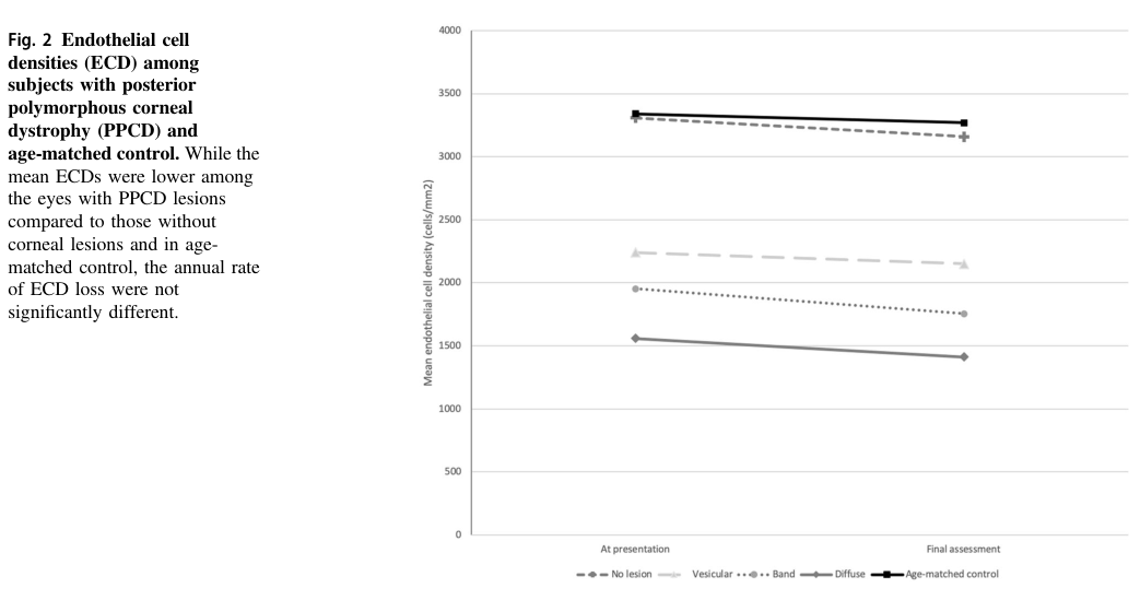
Reduced corneal endothelial cell density (ECD) (key quantitative phenotype) - Pediatric longitudinal case-control study (mean age 10.5 years; follow-up ~3 years): - Baseline ECD: 1918.9 ± 666.3 cells/mm² (PPCD) vs 3340.1 ± 286.5 cells/mm² (controls) - Final ECD: 1793.1 ± 684.6 (PPCD) vs 3265.2 ± 304.3 (controls) - Annual ECD loss: 1.9 ± 3.7% per year, not significantly different from controls (p=0.95) (Human clinical) (fung2021endothelialcelldensity pages 2-4) - Suggested HPO: - Abnormality of the corneal endothelium (no specific code provided here; map at curation time)
Visual impairment / amblyopia risk - PPCD can be asymmetric in children and may contribute to amblyopia via unilateral/asymmetric involvement. (Human clinical) (fung2021endothelialcelldensity pages 5-6) - Suggested HPO: - Reduced visual acuity (HP:0007663) - Amblyopia (HP:0000649)
3.2 Glaucoma and anterior segment abnormalities
- OVOL2-linked families: secondary glaucoma reported at approximately ~30% in one dataset excerpt. (Human clinical) (davidson2016autosomaldominantcornealendothelial pages 6-8)
- GRHL2/PPCD4 Czech series: glaucoma in 25.9% (mean diagnosis ~46 years). (Human clinical) (liskova2018ectopicgrhl2expression pages 6-7)
- Iris abnormalities/adhesions noted in OVOL2-linked disease descriptions (ectropion uveae/corectopia/adhesions). (Human clinical) (davidson2016autosomaldominantcornealendothelial pages 1-3)
- Suggested HPO:
- Glaucoma (HP:0000501)
- Corectopia (HP:0000579)
- Anterior segment dysgenesis (HP:0000591)
3.3 Course, onset, progression
- Disease expression ranges from asymptomatic to severe corneal edema requiring transplantation; infants may rarely present with early corneal edema/haze. (Human clinical) (fung2021endothelialcelldensity pages 1-2, liskova2018ectopicgrhl2expression pages 6-7)
- In pediatric PPCD, no corneal edema or ectasia occurred during ~3 years follow-up, despite lower ECD. (Human clinical) (fung2021endothelialcelldensity pages 1-2)
3.4 Quality of life impact
A prospective case-control study of non-Fuchs corneal dystrophies (2021–2024 recruitment; included 3 PPCD patients) found significantly worse quality of life scores vs controls using VF-14 and NEI-VFQ, correlated with visual acuity and higher-order aberrations. (Human clinical) (elhardt2025 study retrieval; PPCD-specific subgroup results not extractable in retrieved snippets) (lin2025polymorphouscornealdystrophy pages 5-7)
4. Genetic / Molecular Information
4.1 Causal genes (established)
- OVOL2 (PPCD1): promoter mutations (non-coding) (davidson2016autosomaldominantcornealendothelial pages 1-3, chung2017confirmationofthe pages 5-7)
- ZEB1 (PPCD3): LoF/haploinsufficiency (dudakova2021nonpenetranceforocular pages 1-2, siddiqui2016geneticanalysisof pages 42-46)
- GRHL2 (PPCD4): intronic/5′ regulatory mutations (liskova2018ectopicgrhl2expression pages 1-2, liskova2018ectopicgrhl2expression pages 6-7)
4.2 Pathogenic variant classes and examples
- OVOL2 promoter: c.-307T>C segregates in PPCD1-linked family and increases promoter activity in corneal endothelial cells. (Human clinical / in vitro promoter assay) (chung2017confirmationofthe pages 5-7)
- GRHL2 intron 1 regulatory: c.20+544G>T; c.20+257delT; c.20+133delA; all associated with increased transcriptional activity in luciferase assays. (Human genetics / in vitro) (liskova2018ectopicgrhl2expression pages 1-2, liskova2018ectopicgrhl2expression pages 8-9)
- ZEB1 LoF: example c.1279C>T p.(Glu427) reported in non-penetrant carriers; population data suggest extreme rarity but presence of LoF alleles in gnomAD. (Human genetics*) (dudakova2021nonpenetranceforocular pages 1-2)
4.3 Inheritance, penetrance, expressivity
- Inheritance: autosomal dominant (davidson2020cugcforposterior pages 1-2)
- Penetrance: Familial studies for ZEB1 LoF suggest ~95% penetrance, but documented non-penetrance indicates true penetrance may be lower. (dudakova2021nonpenetranceforocular pages 1-2)
- Expressivity: highly variable, ranging from asymptomatic endothelial findings to corneal edema and transplantation. (davidson2016autosomaldominantcornealendothelial pages 1-3, liskova2018ectopicgrhl2expression pages 6-7)
4.4 Modifier genes
A 2025 family report suggests potential interaction of ZEB1 variants with ZNF469 (ECM regulation; brittle cornea syndrome gene) in a family with PPCD3 and keratoconus aggravation; ZEB1 variant allele frequency noted as ~1e-5 in gnomAD EAS. (Human clinical) (lin2025polymorphouscornealdystrophy pages 3-5, lin2025polymorphouscornealdystrophy pages 2-3)
4.5 Epigenetic / chromosomal abnormalities
No PPCD-specific epigenetic signatures were identified in retrieved evidence; however, disease causality is frequently driven by cis-regulatory non-coding variants (OVOL2, GRHL2) (davidson2016autosomaldominantcornealendothelial pages 1-3, liskova2018ectopicgrhl2expression pages 1-2).
5. Environmental Information
No PPCD-specific environmental, lifestyle, or infectious causal factors were identified in the retrieved evidence.
6. Mechanism / Pathophysiology
6.1 Core concept: endothelial-to-epithelial transition (EnET) as a MET-like process
Frausto et al. developed a CRISPR ZEB1+/- corneal endothelial cell model and concluded that PPCD represents an MET-like transition termed endothelial-to-epithelial transition (EnET).
Direct abstract-supported statement (from retrieved abstract excerpt): PPCD is described as being “characterized by a cadherin-switch and transition to an epithelial-like transcriptomic and cellular phenotype” in the context of ZEB1 insufficiency. (In vitro / transcriptomics) (frausto2019zeb1insufficiencycauses pages 1-2)
6.2 Regulatory network: OVOL2 / GRHL2 repress ZEB1
- OVOL2 promoter mutations increase promoter activity and are interpreted as causing ectopic/increased OVOL2 expression; OVOL2 “directly represses ZEB1.” (Human genetics / in vitro promoter assay) (davidson2016autosomaldominantcornealendothelial pages 1-3, davidson2016autosomaldominantcornealendothelial pages 11-13)
- GRHL2 regulatory variants cause ectopic endothelial GRHL2 expression; diseased endothelium can express epithelial markers (E-cadherin, CK7) consistent with MET. (Human ex vivo / in vitro) (liskova2018ectopicgrhl2expression pages 8-9, liskova2018ectopicgrhl2expression pages 7-8)
6.3 Causal chain (variant → phenotype)
1) Non-coding promoter/intronic variants (OVOL2/GRHL2) or ZEB1 LoF → 2) Reduced ZEB1 function/expression (direct LoF or repression by OVOL2/GRHL2) → 3) EnET / epithelialization of corneal endothelium (cadherin switch; epithelial gene expression; stratification) → 4) Abnormal Descemet membrane and endothelial morphology, reduced endothelial reserve → 5) Corneal edema/opacification and angle/iris abnormalities, predisposing to secondary glaucoma and sometimes keratoplasty. (davidson2016autosomaldominantcornealendothelial pages 1-3, liskova2018ectopicgrhl2expression pages 6-7, frausto2019zeb1insufficiencycauses pages 1-2)
6.4 Biochemical abnormalities
Aqueous humor study (ELISA) found active TGF‑β2 significantly higher in PPCD patients (mean 386.98 ± 114.88 pg/mL) vs controls (mean 260.95 ± 112.43 pg/mL; P=0.0001). (Human clinical samples) (chung2017confirmationofthe pages 2-4)
6.5 Suggested ontology annotations
- Cell types (CL):
- Corneal endothelial cell (curation to CL term)
- Corneal epithelial cell (curation to CL term)
- Anatomy (UBERON):
- Corneal endothelium, Descemet membrane, cornea (curation to UBERON terms)
- Processes (GO):
- Epithelial to mesenchymal transition (GO:0001837)
- Mesenchymal to epithelial transition (GO:XXXXXXX; commonly represented as MET/epithelialization processes)
- Cell fate commitment / cell differentiation
- Cell adhesion
7. Anatomical Structures Affected
- Primary: corneal endothelium and Descemet membrane (fung2021endothelialcelldensity pages 1-2, davidson2016autosomaldominantcornealendothelial pages 1-3)
- Secondary/complications: iridocorneal angle/iris (adhesions/corectopia) and glaucoma risk (davidson2016autosomaldominantcornealendothelial pages 1-3, liskova2018ectopicgrhl2expression pages 6-7)
8. Temporal Development
- Onset: can be recognized in childhood; rare infantile onset with edema/haze reported in PPCD4 series; OVOL2 allelic CHED1/PPCD1 can present from infancy with haze. (liskova2018ectopicgrhl2expression pages 6-7, davidson2016autosomaldominantcornealendothelial pages 5-6)
- Progression: variable; pediatric ECD decline similar to controls over ~3 years suggests slow change in childhood despite low baseline ECD. (fung2021endothelialcelldensity pages 2-4)
9. Inheritance and Population
9.1 Epidemiology
- Prevalence estimate: Czech prevalence reported as ~1 per 80,000. (davidson2020cugcforposterior pages 1-2)
- Broader prevalence is poorly defined; PPCD is consistently described as rare. (davidson2020cugcforposterior pages 1-2)
9.2 Population genetics and founder effects
- Founder effects are noted in the CUGC context (details not fully extractable here). (davidson2020cugcforposterior pages 1-2)
- Example allele frequency (illustrative, not definitive for PPCD causality): ZEB1 p.P5A variant reported with gnomAD EAS frequency ~0.00001 in a family study. (lin2025polymorphouscornealdystrophy pages 3-5)
10. Diagnostics
10.1 Clinical and imaging tests used in practice
- Slit-lamp biomicroscopy for posterior corneal lesions (chung2017confirmationofthe pages 5-7)
- Specular microscopy for rail-track/snail-track and endothelial morphology; ECD quantification (fung2021endothelialcelldensity pages 2-4, fernandezgutierrez2022posteriorpolymorphouscorneal pages 1-2)
- In vivo confocal microscopy (posterior lesions; abnormal/absent endothelial cells; hyperreflective deposits) (fung2021endothelialcelldensity pages 5-6)
- OCT / anterior segment imaging including SD‑OCT (posterior reflectivity, Descemet protrusion) (liskova2018ectopicgrhl2expression pages 6-7)
- Corneal tomography (Pentacam) used in complex/overlap phenotypes (e.g., keratoconus) (lin2025polymorphouscornealdystrophy pages 2-3)
10.2 Genetic testing approach (authoritative guidance)
CUGC recommends genome sequencing as the most comprehensive approach because PPCD includes structural and non-coding variants across OVOL2, ZEB1, and GRHL2; Sanger validation and CNV methods may be required to confirm ZEB1 haploinsufficiency and define breakpoints. (davidson2020cugcforposterior pages 1-2)
10.3 Differential diagnosis
Not systematically extractable from retrieved documents; however, PPCD overlaps clinically with other corneal endothelial dystrophies and with anterior segment dysgenesis entities.
11. Outcome / Prognosis
- Many patients remain mildly affected; however, substantial minorities require surgery.
- Keratoplasty proportion:
- OVOL2-linked cohorts: about one-third underwent keratoplasty in an excerpted dataset (davidson2016autosomaldominantcornealendothelial pages 6-8)
- GRHL2/PPCD4 Czech series: 25.9% underwent corneal transplantation (mean first surgery ~35 years) (liskova2018ectopicgrhl2expression pages 6-7)
- Glaucoma: GRHL2/PPCD4 Czech series: 25.9% (liskova2018ectopicgrhl2expression pages 6-7)
12. Treatment
12.1 Current standard management (real-world)
- Observation/monitoring: visual acuity, endothelial reserve, glaucoma surveillance, amblyopia/strabismus monitoring in children. (davidson2020cugcforposterior pages 4-5)
- Glaucoma management: medical drops and/or drainage surgery when needed. (davidson2020cugcforposterior pages 4-5)
- Corneal transplantation: performed for corneal edema/endothelial failure; includes penetrating keratoplasty historically and increasing use of endothelial keratoplasty in endothelial diseases broadly (PPCD infant endothelial keratoplasty reported). (davidson2016autosomaldominantcornealendothelial pages 5-6, fung2021endothelialcelldensity pages 5-6)
12.2 MAXO suggestions
- Corneal transplantation (MAXO: term for keratoplasty)
- Endothelial keratoplasty (MAXO: term for lamellar endothelial keratoplasty)
- Topical intraocular pressure-lowering therapy (MAXO: term for glaucoma medication)
- Glaucoma surgery (MAXO: term for aqueous shunt/trabeculectomy procedures)
12.3 Clinical trials
ClinicalTrials.gov search performed within this session did not retrieve PPCD-specific therapeutic trials among the returned set. (trial search results not PPCD-relevant) (fung2021endothelialcelldensity pages 1-2)
13. Prevention
- No primary prevention strategies exist for Mendelian PPCD.
- Secondary/tertiary prevention: family screening, genetic counseling, monitoring for glaucoma/amblyopia; avoidance of high-risk elective corneal procedures without adequate endothelial evaluation in suspected familial disease. (davidson2020cugcforposterior pages 4-5, lin2025polymorphouscornealdystrophy pages 2-3)
14. Other Species / Natural Disease
No naturally occurring PPCD in non-human species was identified in retrieved evidence.
15. Model Organisms and Experimental Systems
15.1 In vitro models
- CRISPR ZEB1+/- corneal endothelial cell model recapitulates epithelial-like shift, EnET concept, and transcriptomic PPCD signatures; ZEB1 reconstitution rescues phenotype, suggesting therapeutic plausibility in principle. (In vitro) (frausto2019zeb1insufficiencycauses pages 2-3, frausto2019zeb1insufficiencycauses pages 14-16)
15.2 Mouse models
- PPCD1 mouse model (DBA/2J background; D2.Ppcd1): enlarged anterior chamber due to endothelial epithelialization/proliferation and angle blockage; increased IOP and downstream retinal pathology were reported. (Model organism) (shen2017retinalpathologyin pages 11-12)
Key quantitative data highlights (for knowledge base ingestion)
- Pediatric PPCD ECD baseline: 1918.9 ± 666.3 cells/mm² vs controls 3340.1 ± 286.5 (fung2021endothelialcelldensity pages 2-4)
- Pediatric PPCD annual ECD loss: ~1.9 ± 3.7%/year, similar to controls (fung2021endothelialcelldensity pages 2-4)
- GRHL2/PPCD4 series: 25.9% keratoplasty; 25.9% glaucoma (liskova2018ectopicgrhl2expression pages 6-7)
- OVOL2-linked cohorts excerpt: ~30% secondary glaucoma; ~one-third keratoplasty (davidson2016autosomaldominantcornealendothelial pages 6-8)
- Aqueous humor active TGF‑β2: 386.98 ± 114.88 pg/mL (PPCD) vs 260.95 ± 112.43 pg/mL (controls), P=0.0001 (chung2017confirmationofthe pages 2-4)
Visual evidence (ECD by lesion subtype)
Cropped tables/figures from the pediatric longitudinal cohort show ECD stratification by lesion subtype and longitudinal trends. (fung2021endothelialcelldensity media 4fa8c68b, fung2021endothelialcelldensity media 5637544d)
Genetics summary artifact
The following table compares PPCD subtypes, variant classes, mechanisms, and complication frequencies.
| Subtype / OMIM disease ID | Gene (OMIM gene ID) | Variant types / hotspots (example) | Molecular mechanism | Key clinical features / complications | Key supporting citations |
|---|---|---|---|---|---|
| PPCD1 / OMIM 122000 | OVOL2 (OMIM 616441) | Non-coding promoter variants in a conserved proximal promoter region; examples: c.-307T>C, c.-274T>G, c.-370T>C; related severe allelic CHED1 family with c.-339_361dup | Promoter variants increase OVOL2 transcriptional activity, causing ectopic/increased OVOL2 expression in corneal endothelium; OVOL2 is a MET-promoting transcription factor that represses ZEB1, driving endothelial cell-state transition toward epithelial-like phenotype | Typical PPCD posterior corneal lesions (vesicles/bands/gray-white opacities), endothelial dysfunction, corneal edema/haze, iris abnormalities/adhesions, secondary glaucoma risk; ~20–25% of affected individuals may require corneal transplantation in PPCD overall; in OVOL2-linked families, ~30% secondary glaucoma and about one-third underwent keratoplasty; severe early-onset/founder families may present from infancy and need repeated grafting. Czech prevalence estimate for PPCD overall: ~1 per 80,000 | (davidson2016autosomaldominantcornealendothelial pages 1-3, davidson2016autosomaldominantcornealendothelial pages 11-13, davidson2016autosomaldominantcornealendothelial pages 6-8, chung2017confirmationofthe pages 1-2, davidson2020cugcforposterior pages 1-2) |
| PPCD3 / OMIM 609141 | ZEB1 (OMIM 189909) | Predominantly heterozygous loss-of-function variants: nonsense, frameshift, splice, whole-gene/partial deletions; example: c.1279C>T p.Glu427*; rare missense variants also reported in some families | ZEB1 haploinsufficiency causes a MET-like endothelial-to-epithelial transition (EnET) with a cadherin switch (↓CDH2, ↑CDH1), epithelial-like transcriptome, altered adhesion/proliferation/migration, and endothelial stratification | Bilateral often asymmetric PPCD lesions, reduced endothelial cell density, occasional corectopia/iridocorneal synechiae, association with corneal steepening/ectasia in some cases; PPCD3 is often milder than OVOL2-linked disease but variable. Familial studies suggest ~95% penetrance, yet documented non-penetrance exists, so true penetrance may be lower. ZEB1 LoF alleles are extremely rare in population databases | (frausto2019zeb1insufficiencycauses pages 1-2, frausto2019zeb1insufficiencycauses pages 13-14, frausto2019zeb1insufficiencycauses pages 2-3, dudakova2021nonpenetranceforocular pages 1-2, dudakova2021nonpenetranceforocular pages 2-4) |
| PPCD4 / OMIM 618031 | GRHL2 (OMIM 608576) | Non-coding regulatory / intron 1 variants; examples: c.20+544G>T, c.20+257delT, c.20+133delA | Regulatory variants increase GRHL2 transcription, causing ectopic GRHL2 expression in corneal endothelium; GRHL2 promotes epithelial identity and represses/acts upstream of ZEB1, producing MET-like transition with epithelial markers (e.g., E-cadherin, CK7) | Typical PPCD lesions with irregular posterior corneal surface, endothelial multilayering, corneal edema (including infantile onset in some), reduced endothelial cell density, corectopia/band keratopathy; in the large Czech series with recurrent variant, 25.9% underwent corneal transplantation and 25.9% developed glaucoma; mean first keratoplasty ~35 years, mean glaucoma diagnosis ~46 years | (liskova2018ectopicgrhl2expression pages 1-2, liskova2018ectopicgrhl2expression pages 6-7, liskova2018ectopicgrhl2expression pages 9-10, liskova2018ectopicgrhl2expression pages 8-9, davidson2020cugcforposterior pages 1-2) |
Table: This table summarizes the main genetically supported PPCD subtypes—OVOL2/PPCD1, ZEB1/PPCD3, and GRHL2/PPCD4—covering variant classes, mechanisms, clinical complications, and the strongest available evidence. It is useful for quickly comparing subtype-specific diagnostic and counseling implications.
URLs and publication dates (where available in retrieved texts)
- Weiss et al. IC3D Classification of Corneal Dystrophies—Edition 3. Cornea. Feb 2024. https://doi.org/10.1097/ICO.0000000000003420 and online resource: https://corneasociety.org/publications/ic3d (fung2021endothelialcelldensity pages 1-2)
- Davidson et al. Autosomal-Dominant Corneal Endothelial Dystrophies CHED1 and PPCD1… OVOL2 promoter. Am J Hum Genet. Jan 2016. https://doi.org/10.1016/j.ajhg.2015.11.018 (davidson2016autosomaldominantcornealendothelial pages 1-3)
- Liskova et al. Ectopic GRHL2 expression… PPCD4. Am J Hum Genet. Mar 2018. https://doi.org/10.1016/j.ajhg.2018.02.002 (liskova2018ectopicgrhl2expression pages 1-2)
- Frausto et al. ZEB1 insufficiency… EnET. PLoS ONE. Feb 2019. https://doi.org/10.1371/journal.pone.0218279 (frausto2019zeb1insufficiencycauses pages 1-2)
- Davidson et al. CUGC for PPCD. Eur J Hum Genet. Jun 2020. https://doi.org/10.1038/s41431-019-0448-8 (davidson2020cugcforposterior pages 1-2)
- Fung et al. Endothelial cell density in children with PPCD. Eye. Feb 2021. https://doi.org/10.1038/s41433-021-01451-y (fung2021endothelialcelldensity pages 2-4)
- Dudakova et al. Non-penetrance… ZEB1 LoF. Genes. Apr 2021. https://doi.org/10.3390/genes12050677 (dudakova2021nonpenetranceforocular pages 1-2)
Limitations and gaps (explicit)
- MONDO/Orphanet/ICD/MeSH identifiers were not retrieved in the available evidence and are therefore not provided.
- 2023–2024 PPCD-specific primary studies were not prominently retrievable in this run beyond the IC3D 2024 classification update; consequently, “latest” advances are described using the best-available primary mechanistic/genetic sources (2016–2019) plus later clinical genetics guidance.
- Differential diagnosis and formal diagnostic criteria for PPCD (as distinct from FECD/ICE syndrome/anterior segment dysgenesis) require additional targeted retrieval beyond the current evidence set.
References
-
(fung2021endothelialcelldensity pages 1-2): Simon Sheung Man Fung, Hamza Sami, Ali El Hamouly, Dishay Jiandani, Sara Williams, Kamiar Mireskandari, and Asim Ali. Endothelial cell density in children with posterior polymorphous corneal dystrophy: a longitudinal case-control study. Eye, 35:3397-3403, Feb 2021. URL: https://doi.org/10.1038/s41433-021-01451-y, doi:10.1038/s41433-021-01451-y. This article has 6 citations and is from a peer-reviewed journal.
-
(davidson2016autosomaldominantcornealendothelial pages 1-3): Alice E. Davidson, Petra Liskova, Cerys J. Evans, Lubica Dudakova, Lenka Nosková, Nikolas Pontikos, Hana Hartmannová, Kateřina Hodaňová, Viktor Stránecký, Zbyněk Kozmík, Hannah J. Levis, Nwamaka Idigo, Noriaki Sasai, Geoffrey J. Maher, James Bellingham, Neyme Veli, Neil D. Ebenezer, Michael E. Cheetham, Julie T. Daniels, Caroline M.H. Thaung, Katerina Jirsova, Vincent Plagnol, Martin Filipec, Stanislav Kmoch, Stephen J. Tuft, and Alison J. Hardcastle. Autosomal-dominant corneal endothelial dystrophies ched1 and ppcd1 are allelic disorders caused by non-coding mutations in the promoter of ovol2. The American Journal of Human Genetics, 98:75-89, Jan 2016. URL: https://doi.org/10.1016/j.ajhg.2015.11.018, doi:10.1016/j.ajhg.2015.11.018. This article has 94 citations.
-
(liskova2018ectopicgrhl2expression pages 6-7): Petra Liskova, Lubica Dudakova, Cerys J. Evans, Karla E. Rojas Lopez, Nikolas Pontikos, Dimitra Athanasiou, Hodan Jama, Josef Sach, Pavlina Skalicka, Viktor Stranecky, Stanislav Kmoch, Caroline Thaung, Martin Filipec, Michael E. Cheetham, Alice E. Davidson, Stephen J. Tuft, and Alison J. Hardcastle. Ectopic grhl2 expression due to non-coding mutations promotes cell state transition and causes posterior polymorphous corneal dystrophy 4. American Journal of Human Genetics, 102:447-459, Mar 2018. URL: https://doi.org/10.1016/j.ajhg.2018.02.002, doi:10.1016/j.ajhg.2018.02.002. This article has 59 citations and is from a highest quality peer-reviewed journal.
-
(davidson2020cugcforposterior pages 1-2): Alice E. Davidson, Nathaniel J. Hafford-Tear, Lubica Dudakova, Amanda N. Sadan, Nikolas Pontikos, Alison J. Hardcastle, Stephen J. Tuft, and Petra Liskova. Cugc for posterior polymorphous corneal dystrophy (ppcd). European Journal of Human Genetics, 28:126-131, Jun 2020. URL: https://doi.org/10.1038/s41431-019-0448-8, doi:10.1038/s41431-019-0448-8. This article has 7 citations and is from a domain leading peer-reviewed journal.
-
(fung2021endothelialcelldensity pages 2-4): Simon Sheung Man Fung, Hamza Sami, Ali El Hamouly, Dishay Jiandani, Sara Williams, Kamiar Mireskandari, and Asim Ali. Endothelial cell density in children with posterior polymorphous corneal dystrophy: a longitudinal case-control study. Eye, 35:3397-3403, Feb 2021. URL: https://doi.org/10.1038/s41433-021-01451-y, doi:10.1038/s41433-021-01451-y. This article has 6 citations and is from a peer-reviewed journal.
-
(davidson2016autosomaldominantcornealendothelial pages 6-8): Alice E. Davidson, Petra Liskova, Cerys J. Evans, Lubica Dudakova, Lenka Nosková, Nikolas Pontikos, Hana Hartmannová, Kateřina Hodaňová, Viktor Stránecký, Zbyněk Kozmík, Hannah J. Levis, Nwamaka Idigo, Noriaki Sasai, Geoffrey J. Maher, James Bellingham, Neyme Veli, Neil D. Ebenezer, Michael E. Cheetham, Julie T. Daniels, Caroline M.H. Thaung, Katerina Jirsova, Vincent Plagnol, Martin Filipec, Stanislav Kmoch, Stephen J. Tuft, and Alison J. Hardcastle. Autosomal-dominant corneal endothelial dystrophies ched1 and ppcd1 are allelic disorders caused by non-coding mutations in the promoter of ovol2. The American Journal of Human Genetics, 98:75-89, Jan 2016. URL: https://doi.org/10.1016/j.ajhg.2015.11.018, doi:10.1016/j.ajhg.2015.11.018. This article has 94 citations.
-
(dudakova2021nonpenetranceforocular pages 1-2): Lubica Dudakova, Viktor Stranecky, Lenka Piherova, Tomas Palecek, Nikolas Pontikos, Stanislav Kmoch, Pavlina Skalicka, Manuela Vaneckova, Alice E. Davidson, and Petra Liskova. Non-penetrance for ocular phenotype in two individuals carrying heterozygous loss-of-function zeb1 alleles. Genes, 12:677, Apr 2021. URL: https://doi.org/10.3390/genes12050677, doi:10.3390/genes12050677. This article has 4 citations.
-
(lin2025polymorphouscornealdystrophy pages 2-3): Qinghong Lin, Xuejun Wang, Xiaoliao Peng, Xiaosong Han, Xiaoyu Zhang, Ling Sun, Yan Wang, Shengtao Liu, and Xingtao Zhou. Polymorphous corneal dystrophy subtype 3 and keratoconus aggravation after corneal refractive surgery in a three-generation family carrying both zeb1 and znf469 pathogenic variant. Frontiers in Genetics, Jun 2025. URL: https://doi.org/10.3389/fgene.2025.1603019, doi:10.3389/fgene.2025.1603019. This article has 0 citations and is from a peer-reviewed journal.
-
(chung2017confirmationofthe pages 5-7): Doug D. Chung, Ricardo F. Frausto, Aleck E. Cervantes, Katherine M. Gee, Marina Zakharevich, Evelyn M. Hanser, Edwin M. Stone, Elise Heon, and Anthony J. Aldave. Confirmation of the ovol2 promoter mutation c.-307t>c in posterior polymorphous corneal dystrophy 1. PLoS ONE, 12:e0169215, Jan 2017. URL: https://doi.org/10.1371/journal.pone.0169215, doi:10.1371/journal.pone.0169215. This article has 26 citations and is from a peer-reviewed journal.
-
(siddiqui2016geneticanalysisof pages 42-46): S Siddiqui. Genetic analysis of corneal dystrophies. Unknown journal, 2016.
-
(liskova2018ectopicgrhl2expression pages 1-2): Petra Liskova, Lubica Dudakova, Cerys J. Evans, Karla E. Rojas Lopez, Nikolas Pontikos, Dimitra Athanasiou, Hodan Jama, Josef Sach, Pavlina Skalicka, Viktor Stranecky, Stanislav Kmoch, Caroline Thaung, Martin Filipec, Michael E. Cheetham, Alice E. Davidson, Stephen J. Tuft, and Alison J. Hardcastle. Ectopic grhl2 expression due to non-coding mutations promotes cell state transition and causes posterior polymorphous corneal dystrophy 4. American Journal of Human Genetics, 102:447-459, Mar 2018. URL: https://doi.org/10.1016/j.ajhg.2018.02.002, doi:10.1016/j.ajhg.2018.02.002. This article has 59 citations and is from a highest quality peer-reviewed journal.
-
(liskova2018ectopicgrhl2expression pages 8-9): Petra Liskova, Lubica Dudakova, Cerys J. Evans, Karla E. Rojas Lopez, Nikolas Pontikos, Dimitra Athanasiou, Hodan Jama, Josef Sach, Pavlina Skalicka, Viktor Stranecky, Stanislav Kmoch, Caroline Thaung, Martin Filipec, Michael E. Cheetham, Alice E. Davidson, Stephen J. Tuft, and Alison J. Hardcastle. Ectopic grhl2 expression due to non-coding mutations promotes cell state transition and causes posterior polymorphous corneal dystrophy 4. American Journal of Human Genetics, 102:447-459, Mar 2018. URL: https://doi.org/10.1016/j.ajhg.2018.02.002, doi:10.1016/j.ajhg.2018.02.002. This article has 59 citations and is from a highest quality peer-reviewed journal.
-
(fung2021endothelialcelldensity pages 5-6): Simon Sheung Man Fung, Hamza Sami, Ali El Hamouly, Dishay Jiandani, Sara Williams, Kamiar Mireskandari, and Asim Ali. Endothelial cell density in children with posterior polymorphous corneal dystrophy: a longitudinal case-control study. Eye, 35:3397-3403, Feb 2021. URL: https://doi.org/10.1038/s41433-021-01451-y, doi:10.1038/s41433-021-01451-y. This article has 6 citations and is from a peer-reviewed journal.
-
(lin2025polymorphouscornealdystrophy pages 5-7): Qinghong Lin, Xuejun Wang, Xiaoliao Peng, Xiaosong Han, Xiaoyu Zhang, Ling Sun, Yan Wang, Shengtao Liu, and Xingtao Zhou. Polymorphous corneal dystrophy subtype 3 and keratoconus aggravation after corneal refractive surgery in a three-generation family carrying both zeb1 and znf469 pathogenic variant. Frontiers in Genetics, Jun 2025. URL: https://doi.org/10.3389/fgene.2025.1603019, doi:10.3389/fgene.2025.1603019. This article has 0 citations and is from a peer-reviewed journal.
-
(lin2025polymorphouscornealdystrophy pages 3-5): Qinghong Lin, Xuejun Wang, Xiaoliao Peng, Xiaosong Han, Xiaoyu Zhang, Ling Sun, Yan Wang, Shengtao Liu, and Xingtao Zhou. Polymorphous corneal dystrophy subtype 3 and keratoconus aggravation after corneal refractive surgery in a three-generation family carrying both zeb1 and znf469 pathogenic variant. Frontiers in Genetics, Jun 2025. URL: https://doi.org/10.3389/fgene.2025.1603019, doi:10.3389/fgene.2025.1603019. This article has 0 citations and is from a peer-reviewed journal.
-
(frausto2019zeb1insufficiencycauses pages 1-2): Ricardo F. Frausto, Doug D. Chung, Payton M. Boere, Vinay S. Swamy, Huong N.V. Duong, Liyo Kao, Rustam Azimov, Wenlin Zhang, Liam Carrigan, Davey Wong, Marco Morselli, Marina Zakharevich, E. Maryam Hanser, Austin C. Kassels, Ira Kurtz, Matteo Pellegrini, and Anthony J. Aldave. Zeb1 insufficiency causes corneal endothelial cell state transition and altered cellular processing. PLoS ONE, Feb 2019. URL: https://doi.org/10.1371/journal.pone.0218279, doi:10.1371/journal.pone.0218279. This article has 31 citations and is from a peer-reviewed journal.
-
(davidson2016autosomaldominantcornealendothelial pages 11-13): Alice E. Davidson, Petra Liskova, Cerys J. Evans, Lubica Dudakova, Lenka Nosková, Nikolas Pontikos, Hana Hartmannová, Kateřina Hodaňová, Viktor Stránecký, Zbyněk Kozmík, Hannah J. Levis, Nwamaka Idigo, Noriaki Sasai, Geoffrey J. Maher, James Bellingham, Neyme Veli, Neil D. Ebenezer, Michael E. Cheetham, Julie T. Daniels, Caroline M.H. Thaung, Katerina Jirsova, Vincent Plagnol, Martin Filipec, Stanislav Kmoch, Stephen J. Tuft, and Alison J. Hardcastle. Autosomal-dominant corneal endothelial dystrophies ched1 and ppcd1 are allelic disorders caused by non-coding mutations in the promoter of ovol2. The American Journal of Human Genetics, 98:75-89, Jan 2016. URL: https://doi.org/10.1016/j.ajhg.2015.11.018, doi:10.1016/j.ajhg.2015.11.018. This article has 94 citations.
-
(liskova2018ectopicgrhl2expression pages 7-8): Petra Liskova, Lubica Dudakova, Cerys J. Evans, Karla E. Rojas Lopez, Nikolas Pontikos, Dimitra Athanasiou, Hodan Jama, Josef Sach, Pavlina Skalicka, Viktor Stranecky, Stanislav Kmoch, Caroline Thaung, Martin Filipec, Michael E. Cheetham, Alice E. Davidson, Stephen J. Tuft, and Alison J. Hardcastle. Ectopic grhl2 expression due to non-coding mutations promotes cell state transition and causes posterior polymorphous corneal dystrophy 4. American Journal of Human Genetics, 102:447-459, Mar 2018. URL: https://doi.org/10.1016/j.ajhg.2018.02.002, doi:10.1016/j.ajhg.2018.02.002. This article has 59 citations and is from a highest quality peer-reviewed journal.
-
(chung2017confirmationofthe pages 2-4): Doug D. Chung, Ricardo F. Frausto, Aleck E. Cervantes, Katherine M. Gee, Marina Zakharevich, Evelyn M. Hanser, Edwin M. Stone, Elise Heon, and Anthony J. Aldave. Confirmation of the ovol2 promoter mutation c.-307t>c in posterior polymorphous corneal dystrophy 1. PLoS ONE, 12:e0169215, Jan 2017. URL: https://doi.org/10.1371/journal.pone.0169215, doi:10.1371/journal.pone.0169215. This article has 26 citations and is from a peer-reviewed journal.
-
(davidson2016autosomaldominantcornealendothelial pages 5-6): Alice E. Davidson, Petra Liskova, Cerys J. Evans, Lubica Dudakova, Lenka Nosková, Nikolas Pontikos, Hana Hartmannová, Kateřina Hodaňová, Viktor Stránecký, Zbyněk Kozmík, Hannah J. Levis, Nwamaka Idigo, Noriaki Sasai, Geoffrey J. Maher, James Bellingham, Neyme Veli, Neil D. Ebenezer, Michael E. Cheetham, Julie T. Daniels, Caroline M.H. Thaung, Katerina Jirsova, Vincent Plagnol, Martin Filipec, Stanislav Kmoch, Stephen J. Tuft, and Alison J. Hardcastle. Autosomal-dominant corneal endothelial dystrophies ched1 and ppcd1 are allelic disorders caused by non-coding mutations in the promoter of ovol2. The American Journal of Human Genetics, 98:75-89, Jan 2016. URL: https://doi.org/10.1016/j.ajhg.2015.11.018, doi:10.1016/j.ajhg.2015.11.018. This article has 94 citations.
-
(fernandezgutierrez2022posteriorpolymorphouscorneal pages 1-2): Eva Fernández-Gutiérrez, Pedro Fernández-Pérez, Ana Boto-De-Los-Bueis, Laura García-Fernández, Patricia Rodríguez-Solana, Mario Solís, and Elena Vallespín. Posterior polymorphous corneal dystrophy in a patient with a novel zeb1 gene mutation. International Journal of Molecular Sciences, 24:209, Dec 2022. URL: https://doi.org/10.3390/ijms24010209, doi:10.3390/ijms24010209. This article has 9 citations.
-
(davidson2020cugcforposterior pages 4-5): Alice E. Davidson, Nathaniel J. Hafford-Tear, Lubica Dudakova, Amanda N. Sadan, Nikolas Pontikos, Alison J. Hardcastle, Stephen J. Tuft, and Petra Liskova. Cugc for posterior polymorphous corneal dystrophy (ppcd). European Journal of Human Genetics, 28:126-131, Jun 2020. URL: https://doi.org/10.1038/s41431-019-0448-8, doi:10.1038/s41431-019-0448-8. This article has 7 citations and is from a domain leading peer-reviewed journal.
-
(frausto2019zeb1insufficiencycauses pages 2-3): Ricardo F. Frausto, Doug D. Chung, Payton M. Boere, Vinay S. Swamy, Huong N.V. Duong, Liyo Kao, Rustam Azimov, Wenlin Zhang, Liam Carrigan, Davey Wong, Marco Morselli, Marina Zakharevich, E. Maryam Hanser, Austin C. Kassels, Ira Kurtz, Matteo Pellegrini, and Anthony J. Aldave. Zeb1 insufficiency causes corneal endothelial cell state transition and altered cellular processing. PLoS ONE, Feb 2019. URL: https://doi.org/10.1371/journal.pone.0218279, doi:10.1371/journal.pone.0218279. This article has 31 citations and is from a peer-reviewed journal.
-
(frausto2019zeb1insufficiencycauses pages 14-16): Ricardo F. Frausto, Doug D. Chung, Payton M. Boere, Vinay S. Swamy, Huong N.V. Duong, Liyo Kao, Rustam Azimov, Wenlin Zhang, Liam Carrigan, Davey Wong, Marco Morselli, Marina Zakharevich, E. Maryam Hanser, Austin C. Kassels, Ira Kurtz, Matteo Pellegrini, and Anthony J. Aldave. Zeb1 insufficiency causes corneal endothelial cell state transition and altered cellular processing. PLoS ONE, Feb 2019. URL: https://doi.org/10.1371/journal.pone.0218279, doi:10.1371/journal.pone.0218279. This article has 31 citations and is from a peer-reviewed journal.
-
(shen2017retinalpathologyin pages 11-12): Anna L. Shen, Susan M. Moran, Edward A. Glover, Leandro B. Teixeira, and Christopher A. Bradfield. Retinal pathology in the ppcd1 mouse. PLoS ONE, 12:e0185094, Oct 2017. URL: https://doi.org/10.1371/journal.pone.0185094, doi:10.1371/journal.pone.0185094. This article has 3 citations and is from a peer-reviewed journal.
-
(fung2021endothelialcelldensity media 4fa8c68b): Simon Sheung Man Fung, Hamza Sami, Ali El Hamouly, Dishay Jiandani, Sara Williams, Kamiar Mireskandari, and Asim Ali. Endothelial cell density in children with posterior polymorphous corneal dystrophy: a longitudinal case-control study. Eye, 35:3397-3403, Feb 2021. URL: https://doi.org/10.1038/s41433-021-01451-y, doi:10.1038/s41433-021-01451-y. This article has 6 citations and is from a peer-reviewed journal.
-
(fung2021endothelialcelldensity media 5637544d): Simon Sheung Man Fung, Hamza Sami, Ali El Hamouly, Dishay Jiandani, Sara Williams, Kamiar Mireskandari, and Asim Ali. Endothelial cell density in children with posterior polymorphous corneal dystrophy: a longitudinal case-control study. Eye, 35:3397-3403, Feb 2021. URL: https://doi.org/10.1038/s41433-021-01451-y, doi:10.1038/s41433-021-01451-y. This article has 6 citations and is from a peer-reviewed journal.
-
(chung2017confirmationofthe pages 1-2): Doug D. Chung, Ricardo F. Frausto, Aleck E. Cervantes, Katherine M. Gee, Marina Zakharevich, Evelyn M. Hanser, Edwin M. Stone, Elise Heon, and Anthony J. Aldave. Confirmation of the ovol2 promoter mutation c.-307t>c in posterior polymorphous corneal dystrophy 1. PLoS ONE, 12:e0169215, Jan 2017. URL: https://doi.org/10.1371/journal.pone.0169215, doi:10.1371/journal.pone.0169215. This article has 26 citations and is from a peer-reviewed journal.
-
(frausto2019zeb1insufficiencycauses pages 13-14): Ricardo F. Frausto, Doug D. Chung, Payton M. Boere, Vinay S. Swamy, Huong N.V. Duong, Liyo Kao, Rustam Azimov, Wenlin Zhang, Liam Carrigan, Davey Wong, Marco Morselli, Marina Zakharevich, E. Maryam Hanser, Austin C. Kassels, Ira Kurtz, Matteo Pellegrini, and Anthony J. Aldave. Zeb1 insufficiency causes corneal endothelial cell state transition and altered cellular processing. PLoS ONE, Feb 2019. URL: https://doi.org/10.1371/journal.pone.0218279, doi:10.1371/journal.pone.0218279. This article has 31 citations and is from a peer-reviewed journal.
-
(dudakova2021nonpenetranceforocular pages 2-4): Lubica Dudakova, Viktor Stranecky, Lenka Piherova, Tomas Palecek, Nikolas Pontikos, Stanislav Kmoch, Pavlina Skalicka, Manuela Vaneckova, Alice E. Davidson, and Petra Liskova. Non-penetrance for ocular phenotype in two individuals carrying heterozygous loss-of-function zeb1 alleles. Genes, 12:677, Apr 2021. URL: https://doi.org/10.3390/genes12050677, doi:10.3390/genes12050677. This article has 4 citations.
-
(liskova2018ectopicgrhl2expression pages 9-10): Petra Liskova, Lubica Dudakova, Cerys J. Evans, Karla E. Rojas Lopez, Nikolas Pontikos, Dimitra Athanasiou, Hodan Jama, Josef Sach, Pavlina Skalicka, Viktor Stranecky, Stanislav Kmoch, Caroline Thaung, Martin Filipec, Michael E. Cheetham, Alice E. Davidson, Stephen J. Tuft, and Alison J. Hardcastle. Ectopic grhl2 expression due to non-coding mutations promotes cell state transition and causes posterior polymorphous corneal dystrophy 4. American Journal of Human Genetics, 102:447-459, Mar 2018. URL: https://doi.org/10.1016/j.ajhg.2018.02.002, doi:10.1016/j.ajhg.2018.02.002. This article has 59 citations and is from a highest quality peer-reviewed journal.