Microphthalmia with Coloboma
Microphthalmia with coloboma is a congenital structural eye malformation in which a small eye (microphthalmia) is combined with a colobomatous defect — missing ocular tissue, classically inferonasal, that results from failure of the embryonic optic (choroid) fissure to close. It sits within the microphthalmia–anophthalmia–coloboma (MAC, also called AMC) spectrum and is curated here in its isolated / non-syndromic form, distinct from the syndromic microphthalmia entries (e.g. STRA6-related Matthew-Wood syndrome, CHARGE syndrome). The disorder is genetically heterogeneous: dominant, recessive, and X-linked alleles in eye-development transcription factors and signaling genes (SOX2, OTX2, PAX6, MAB21L2, FOXE3, ALDH1A3, FZD5, and others), copy-number variants, and non-coding regulatory variants all converge on disrupted optic-cup morphogenesis and optic-fissure closure. MAC accounts for roughly 15–20% of severe childhood visual impairment and blindness worldwide.
Ask OpenScientist
Ask a research question about Microphthalmia with Coloboma. OpenScientist will conduct autonomous deep research using the Disorder Mechanisms Knowledge Base and PubMed literature (typically 10-30 minutes).
Do not include personal health information in your question. Questions and results are cached in your browser's local storage.
Inheritance
3Show evidence (1 reference)
Show evidence (1 reference)
Show evidence (1 reference)
Subtypes
11Show evidence (2 references)
Show evidence (5 references)
Show evidence (2 references)
Show evidence (4 references)
Show evidence (3 references)
Discussions and Knowledge Gaps
1Pathophysiology
7Show evidence (3 references)
Show evidence (4 references)
Show evidence (3 references)
Show evidence (2 references)
Show evidence (2 references)
Show evidence (3 references)
Show evidence (3 references)
Pathograph
Phenotypes
13Ear 1
Show evidence (1 reference)
Eye 11
Show evidence (1 reference)
Show evidence (1 reference)
Show evidence (1 reference)
Show evidence (1 reference)
Show evidence (1 reference)
Show evidence (1 reference)
Show evidence (1 reference)
Show evidence (1 reference)
Show evidence (1 reference)
Show evidence (1 reference)
Show evidence (2 references)
Nervous System 1
Show evidence (1 reference)
Genetic Associations
11Show evidence (1 reference)
Show evidence (1 reference)
Show evidence (1 reference)
Show evidence (1 reference)
Show evidence (1 reference)
Show evidence (1 reference)
Show evidence (1 reference)
Show evidence (3 references)
Show evidence (2 references)
Show evidence (3 references)
Show evidence (3 references)
Variants (1)
Show evidence (3 references)
Medical Actions
3Show evidence (1 reference)
Show evidence (1 reference)
Show evidence (1 reference)
Environmental Factors
2Show evidence (1 reference)
Show evidence (1 reference)
Clinical Trials
2Show evidence (1 reference)
Show evidence (1 reference)
Animal Models
1Show evidence (3 references)
Show evidence (1 reference)
Source YAML
click to showname: Microphthalmia with Coloboma
creation_date: "2026-06-15T00:00:00Z"
description: >-
Microphthalmia with coloboma is a congenital structural eye malformation in
which a small eye (microphthalmia) is combined with a colobomatous defect —
missing ocular tissue, classically inferonasal, that results from failure of
the embryonic optic (choroid) fissure to close. It sits within the
microphthalmia–anophthalmia–coloboma (MAC, also called AMC) spectrum and is
curated here in its isolated / non-syndromic form, distinct from the
syndromic microphthalmia entries (e.g. STRA6-related Matthew-Wood syndrome,
CHARGE syndrome). The disorder is genetically heterogeneous: dominant,
recessive, and X-linked alleles in eye-development transcription factors and
signaling genes (SOX2, OTX2, PAX6, MAB21L2, FOXE3, ALDH1A3, FZD5, and others),
copy-number variants, and non-coding regulatory variants all converge on
disrupted optic-cup morphogenesis and optic-fissure closure. MAC accounts for
roughly 15–20% of severe childhood visual impairment and blindness worldwide.
category: Mendelian
parents:
- hereditary disease
- developmental eye disorder
synonyms:
- MAC spectrum
- microphthalmia-anophthalmia-coloboma spectrum
- AMC spectrum
- isolated microphthalmia with coloboma
- colobomatous microphthalmia
disease_term:
preferred_term: Microphthalmia with coloboma
term:
id: MONDO:0000170
label: microphthalmia, isolated, with coloboma
notes: >-
Scope: this entry covers the ISOLATED / non-syndromic MAC presentation
centered on the optic-fissure-closure defect mechanism. Syndromic forms in
which MAC is one feature of a multisystem disorder (Matthew-Wood/STRA6,
CHARGE, branchiooculofacial syndrome, etc.) are curated as their own entries
and are referenced here only as differential diagnoses. The numbered MCOPCB
(microphthalmia/coloboma) OMIM loci and the gene-defined forms are modeled as
subtypes. GeneReviews has a retired "Microphthalmia/Anophthalmia/Coloboma
Spectrum" chapter (PMID:20301552) used as the clinical baseline. The MCOPCB13 arm (MONDO:0975809, OMIM 620968) is curated here as the
IHH-Enhancer-MCOPCB13 subtype rather than as its own disease entry: its MONDO
parent is this entry's own term, its clinical picture is isolated MAC
indistinguishable from the rest of the spectrum, and both this file and
Isolated_Anophthalmia-Microphthalmia_Syndrome already state that the numbered
MCOPCB loci belong here. The stub for it named NHEJ1 as the causal gene;
that is a positional artifact of where the variant's coordinates fall, and
the gene whose expression is disturbed is IHH. NHEJ1's own established
disease is Cernunnos-XLF deficiency, curated separately as
Cernunnos-XLF_deficiency, and nothing about DNA double-strand-break repair is
claimed here.
MCOPCB SERIES RECONCILIATION (OLS4, 2026-09-25). MONDO:0000170 has twelve
hierarchical descendants, the numbered MCOPCB loci 1 to 11 and 13; MONDO has
no MCOPCB12 term. Five carry a causal gene edge and are curated here as
gene-bound subtype rows: MCOPCB7 (MONDO:0013783, ABCB6), MCOPCB9
(MONDO:0014059, TENM3), MCOPCB10 (MONDO:0014635, RBP4), MCOPCB11
(MONDO:0958239, FZD5) and MCOPCB13 (MONDO:0975809, recorded by MONDO against
NHEJ1 and curated here against IHH for the reason given above). A sixth,
MCOPCB5 (MONDO:0012709, SHH), is deliberately not duplicated here: it is
already anchored as a skos:closeMatch on SHH_Holoprosencephaly_Spectrum, whose
Microform HPE subtype covers microphthalmia with coloboma as the mild end of
the SHH holoprosencephaly spectrum, and the report OMIM indexes for this locus
describes iris and uveoretinal colobomas without microphthalmia. The remaining
six descendants - MCOPCB1 (MONDO:0024549), MCOPCB2 (MONDO:0011589), MCOPCB3
(MONDO:0012408), MCOPCB4 (MONDO:0009630), MCOPCB6 (MONDO:0013376) and MCOPCB8
(MONDO:0800324) - carry no causal gene edge in MONDO and are mapped loci
rather than gene-defined entities, so no row is created for them.
Two of the gene-disease relationships curated here are weak, and are labelled
rather than presented as settled. ClinGen rates ABCB6 for MCOPCB7 Limited, and
TENM3 rests on a single consanguineous pedigree with no ClinGen assertion at
all. Across the eleven genes in this entry the committed ClinGen gene-validity
snapshot carries assertions for only four - ABCB6, MAB21L2, PAX6 and FOXE3 -
and apart from ABCB6/MCOPCB7 every one of them is against a different disease
term: MAB21L2 against colobomatous microphthalmia-rhizomelic dysplasia
syndrome (Definitive), PAX6 against PAX6-related ocular dysgenesis
(Definitive), and FOXE3 against anterior segment dysgenesis (Moderate) and
familial thoracic aortic aneurysm and aortic dissection (Moderate). ClinGen is
therefore largely silent on the isolated MAC concept this entry models, and
absence of an assertion here is not evidence against a gene.
references:
- reference: PMID:20301552
title: "Microphthalmia/Anophthalmia/Coloboma Spectrum – RETIRED CHAPTER, FOR HISTORICAL REFERENCE ONLY."
tags:
- GeneReviews
has_subtypes:
- name: SOX2-MAC
display_name: SOX2-related microphthalmia/anophthalmia with coloboma
description: >-
Most common single-gene cause of severe bilateral microphthalmia/anophthalmia
in the MAC spectrum, typically de novo dominant SOX2 loss-of-function;
explains an estimated 10–15% of cases.
genes:
- preferred_term: SOX2
term:
id: hgnc:11195
label: SOX2
- name: OTX2-MAC
display_name: OTX2-related microphthalmia/anophthalmia with coloboma
description: >-
Dominant OTX2 variants causing microphthalmia/anophthalmia, often with
coloboma; estimated to explain 2–5% of cases. OTX2 also binds conserved
enhancers upstream of MAB21L2.
genes:
- preferred_term: OTX2
term:
id: hgnc:8522
label: OTX2
- name: PAX6-MAC
display_name: PAX6-related microphthalmia with coloboma
description: >-
PAX6 haploinsufficiency, classically aniridia, can present with
microphthalmia and colobomatous defects within the anterior-segment and
MAC spectrum.
genes:
- preferred_term: PAX6
term:
id: hgnc:8620
label: PAX6
- name: MAB21L2-MAC
display_name: MAB21L2-related microphthalmia with coloboma
description: >-
Coding MAB21L2 variants and non-coding regulatory deletions upstream of
MAB21L2 cause microphthalmia and coloboma; a worked example of non-coding
regulatory pathogenesis in MAC. Note that the recurrent p.Arg51Cys allele
causes a distinct *syndromic* oculo-skeletal disorder with rhizomelic limb
shortening and intellectual disability, curated separately as
Colobomatous_Microphthalmia-Rhizomelic_Dysplasia_Syndrome and listed in the
ISDS skeletal nosology; this entry stays scoped to the isolated MAC
presentation and therefore carries no ISDS assignment.
genes:
- preferred_term: MAB21L2
term:
id: hgnc:6758
label: MAB21L2
- name: FOXE3-MAC
display_name: FOXE3-related microphthalmia with coloboma
description: >-
FOXE3 variants cause anterior-segment dysgenesis, microphthalmia, and
coloboma; bilateral sensorineural hearing loss reported as a novel
association.
genes:
- preferred_term: FOXE3
term:
id: hgnc:3808
label: FOXE3
- name: ALDH1A3-MAC
display_name: ALDH1A3-related microphthalmia/anophthalmia with coloboma
description: >-
Recessive ALDH1A3 variants disrupt retinaldehyde dehydrogenase / retinoic
acid synthesis, causing autosomal recessive microphthalmia/anophthalmia.
genes:
- preferred_term: ALDH1A3
term:
id: hgnc:409
label: ALDH1A3
- name: FZD5-Coloboma
display_name: Microphthalmia/coloboma 11 — FZD5 (OMIM:620731)
subtype_term:
preferred_term: microphthalmia/coloboma 11
term:
id: MONDO:0958239
label: microphthalmia/coloboma 11
description: >-
FZD5 (Wnt receptor) variants cause isolated and syndromic ocular coloboma;
most are dominant-negative, but a recessive hypomorphic allele causing
syndromic coloboma with microcornea is documented. This is the MCOPCB11
locus, MONDO:0958239 / OMIM:620731.
genes:
- preferred_term: FZD5
term:
id: hgnc:4043
label: FZD5
evidence:
- reference: PMID:39503780
reference_title: Homozygosity for a hypomorphic mutation in frizzled class receptor 5 causes syndromic ocular coloboma with microcornea in humans.
supports: SUPPORT
evidence_source: HUMAN_CLINICAL
quote_role: BACKGROUND
snippet: "Mutations in the Wnt receptor FZD5, which is expressed throughout eye development, have been linked to both isolated and complex forms of coloboma."
explanation: >-
Supports the gene assignment for this numbered locus and the isolated /
complex split the description records.
- reference: PMID:39503780
reference_title: Homozygosity for a hypomorphic mutation in frizzled class receptor 5 causes syndromic ocular coloboma with microcornea in humans.
supports: SUPPORT
evidence_source: IN_VITRO
quote_role: PRIMARY_RESULT
snippet: "in vitro TOPFlash assays revealed that the missense variant only caused partial loss-of-function, behaving as a hypomorphic mutation"
explanation: >-
The functional basis for calling the recessive allele hypomorphic rather
than dominant-negative.
- name: MCOPCB7
display_name: Microphthalmia with coloboma 7 — ABCB6 (OMIM:614497)
subtype_term:
preferred_term: microphthalmia, isolated, with coloboma 7
term:
id: MONDO:0013783
label: microphthalmia, isolated, with coloboma 7
description: >-
Autosomal dominant coloboma attributed to ABCB6, an ATP-binding cassette
transporter expressed in the retina and retinal pigment epithelium, where it
localizes to the endoplasmic reticulum and Golgi. The original report
combined positional cloning in one Chinese family with a screen of 116
sporadic cases of microphthalmia with coloboma, isolated coloboma and
aniridia. ClinGen's General Inborn Errors of Metabolism expert panel
nonetheless classifies ABCB6 for this disease as Limited, so the row is
curated with that classification stated. The zebrafish knockdown and rescue
experiment behind the loss-of-function reading is cited on the ABCB6 record
in the genetic section. The gene's better-attested disease is the pigmentary
skin disorder dyschromatosis universalis hereditaria 3 (MONDO:0014169),
which ClinGen rates Moderate; nothing about pigmentary skin disease is
claimed here.
genes:
- preferred_term: ABCB6
term:
id: hgnc:47
label: ABCB6
evidence:
- reference: PMID:22226084
reference_title: ABCB6 mutations cause ocular coloboma.
supports: SUPPORT
evidence_source: HUMAN_CLINICAL
quote_role: PRIMARY_RESULT
snippet: "Using a positional cloning approach, we identified a mutation in the ATP-binding cassette (ABC) transporter ABCB6 in a Chinese family affected by autosomal-dominant coloboma."
explanation: The gene-discovery report and the source of the dominant inheritance stated here.
- reference: PMID:22226084
reference_title: ABCB6 mutations cause ocular coloboma.
supports: SUPPORT
evidence_source: HUMAN_CLINICAL
quote_role: PRIMARY_RESULT
snippet: "Sequence analysis was performed on the ABCB6 exons from 116 sporadic cases of microphthalmia with coloboma (MAC), isolated coloboma, and aniridia, and an additional mutation (A57T) was identified in three patients with MAC."
explanation: >-
The screen this row's description reports, and what extends the association
beyond the index family to sporadic microphthalmia-with-coloboma cases -
which is why the locus sits in this entry rather than in an
isolated-coloboma entry.
- reference: PMID:22226084
reference_title: ABCB6 mutations cause ocular coloboma.
supports: SUPPORT
evidence_source: IN_VITRO
quote_role: PRIMARY_RESULT
snippet: "Immunostaining of transiently transfected, Myc-tagged ABCB6 in retinal pigment epithelial (RPE) cells showed that it localized to the endoplasmic reticulum and Golgi apparatus of RPE cells."
explanation: Sources the subcellular localization this row's description states.
- reference: PMID:22226084
reference_title: ABCB6 mutations cause ocular coloboma.
supports: SUPPORT
evidence_source: IN_VITRO
quote_role: PRIMARY_RESULT
snippet: "RT-PCR of ABCB6 mRNA in human cell lines and tissue indicated that ABCB6 is expressed in the retinae and RPE cells."
explanation: Sources the tissue expression this row's description states.
- reference: CGGV:assertion_45ec3fba-48fd-4bf4-b865-2c2d7216e8eb-2024-04-26T180000.000Z
reference_title: "ABCB6 / microphthalmia, isolated, with coloboma 7 (Limited)"
supports: SUPPORT
evidence_source: OTHER
snippet: "ABCB6 | HGNC:47 | microphthalmia, isolated, with coloboma 7 | MONDO:0013783 | AD | Limited | SOP10 | General Inborn Errors of Metabolism Gene Curation Expert Panel | 2024-04-26T18:00:00.000Z"
explanation: ClinGen's own classification of this gene-disease relationship, quoted from the committed record.
- name: MCOPCB9
display_name: Microphthalmia with coloboma 9 — TENM3 (OMIM:615145)
subtype_term:
preferred_term: microphthalmia, isolated, with coloboma 9
term:
id: MONDO:0014059
label: microphthalmia, isolated, with coloboma 9
description: >-
Autosomal recessive colobomatous microphthalmia reported in a single
consanguineous family with third-cousin parents and two affected children, in
which exome and autozygome analysis found a null allele of TENM3. HGNC
records ODZ3 as the previous symbol, which is the name the discovery report
uses. The committed ClinGen gene-validity snapshot carries no assertion for
TENM3 under either symbol, and no independent family is cited here, so the
assertion rests on that one pedigree.
genes:
- preferred_term: TENM3
term:
id: hgnc:29944
label: TENM3
evidence:
- reference: PMID:22766609
reference_title: Homozygous null mutation in ODZ3 causes microphthalmia in humans.
supports: SUPPORT
evidence_source: HUMAN_CLINICAL
quote_role: PRIMARY_RESULT
snippet: "Exome and autozygome analysis identified a null mutation in ODZ3, one of four vertebrate orthologs of odz in Drosophila."
explanation: The gene-discovery report, using the gene's previous symbol.
- reference: PMID:22766609
reference_title: Homozygous null mutation in ODZ3 causes microphthalmia in humans.
supports: SUPPORT
evidence_source: HUMAN_CLINICAL
quote_role: PRIMARY_RESULT
snippet: "we enrolled a family that consists of third-cousin parents and two children with isolated colobomatous microphthalmia"
explanation: >-
The single pedigree the assertion rests on, and the reason the description
states its breadth explicitly.
- name: MCOPCB10
display_name: Microphthalmia with coloboma 10 — RBP4 (OMIM:616428)
subtype_term:
preferred_term: microphthalmia, isolated, with coloboma 10
term:
id: MONDO:0014635
label: microphthalmia, isolated, with coloboma 10
description: >-
Dominant eye malformation caused by RBP4 missense variants, with a mode of
inheritance unlike anything else in this entry. RBP4 encodes serum retinol
binding protein, which delivers retinol from hepatic stores to the placenta
and the fetal eye. The disease alleles bind retinol poorly but bind the cell
surface receptor STRA6 more tightly, so the mutant protein occupies STRA6
without delivering its cargo. Because a transmitting mother's circulating
mutant protein blocks retinol delivery at the placenta as well as in the
fetus, penetrance is incomplete and maternal transmission raises the
probability that an allele-carrying fetus is affected — a maternal effect the
authors distinguish from imprinting and from oocyte-derived mRNA. The
separate recessive RBP4 phenotype, in which serum RBP4 is undetectable and
the result is a progressive retinal dystrophy rather than a malformation, is
curated as Progressive_Retinal_Dystrophy_Due_To_Retinol_Transport_Defect.
genes:
- preferred_term: RBP4
term:
id: hgnc:9922
label: RBP4
evidence:
- reference: PMID:25910211
reference_title: "Biochemical Basis for Dominant Inheritance, Variable Penetrance, and Maternal Effects in RBP4 Congenital Eye Disease."
supports: SUPPORT
evidence_source: HUMAN_CLINICAL
quote_role: PRIMARY_RESULT
snippet: "We identified missense mutations in RBP4, encoding serum retinol binding protein, in three families with eye malformations of differing severity, including bilateral anophthalmia."
explanation: The gene-discovery report and the source of the phenotypic range stated here.
- reference: PMID:25910211
reference_title: "Biochemical Basis for Dominant Inheritance, Variable Penetrance, and Maternal Effects in RBP4 Congenital Eye Disease."
supports: SUPPORT
evidence_source: HUMAN_CLINICAL
quote_role: BACKGROUND
snippet: "RBP normally delivers retinol from hepatic stores to peripheral tissues, including the placenta and fetal eye."
explanation: >-
The normal function this row's description states, and the reason a
transmitting mother's protein can act at the placenta.
- reference: PMID:25910211
reference_title: "Biochemical Basis for Dominant Inheritance, Variable Penetrance, and Maternal Effects in RBP4 Congenital Eye Disease."
supports: SUPPORT
evidence_source: HUMAN_CLINICAL
quote_role: PRIMARY_RESULT
snippet: "The mutant phenotypes exhibit dominant inheritance, but incomplete penetrance. Maternal transmission significantly increases the probability of phenotypic expression."
explanation: >-
The inheritance pattern this row records, including the maternal effect
that separates it from ordinary autosomal dominant transmission.
- reference: PMID:25910211
reference_title: "Biochemical Basis for Dominant Inheritance, Variable Penetrance, and Maternal Effects in RBP4 Congenital Eye Disease."
supports: SUPPORT
evidence_source: IN_VITRO
quote_role: PRIMARY_RESULT
snippet: "By occupying STRA6 nonproductively, the dominant-negative proteins disrupt vitamin A delivery from wild-type proteins within the fetus, but also, in the case of maternal transmission, at the placenta."
explanation: >-
The biochemical mechanism behind both the dominant-negative action and the
maternal effect, which is what makes this row's inheritance claim
mechanistic rather than descriptive.
- name: IHH-Enhancer-MCOPCB13
display_name: Microphthalmia/coloboma 13 (IHH enhancer variant hosted in an NHEJ1 intron)
subtype_term:
preferred_term: Microphthalmia/coloboma 13
term:
id: MONDO:0975809
label: microphthalmia/coloboma 13
description: >-
Autosomal recessive isolated MAC reported in two consanguineous Jewish
Iranian pedigrees from the Mashhad region. The homozygous founder allele is
a deep intronic single-nucleotide variant in NHEJ1
(NM_024782.2:c.588+18131A>G) that is not predicted to affect NHEJ1 itself:
it sits inside "i8", a conserved enhancer of the neighbouring gene IHH.
Transgenic mouse reporter assays and chicken ATAC-seq/electroporation show
that i8 drives expression in the developing eye and that the patient allele
abolishes that eye-specific activity, so the gene whose expression is
disturbed is IHH and NHEJ1 is only the element's host. Ocular features range
from optic disc coloboma to microphthalmia and anophthalmia and differ
between the two eyes of the same patient; no skeletal or cranial abnormality
was reported, even though the same enhancer cluster is required for IHH
expression in limb and skull.
genes:
- preferred_term: IHH
term:
id: hgnc:5956
label: IHH
evidence:
- reference: PMID:37580330
reference_title: "IHH enhancer variant within neighboring NHEJ1 intron causes microphthalmia anophthalmia and coloboma."
supports: SUPPORT
evidence_source: HUMAN_CLINICAL
snippet: "a spectrum of isolated ocular manifestations, ranging from a milder phenotype of optic nerve coloboma to a severe phenotype of microphthalmia and even anophthalmia"
explanation: >-
Defines the subtype's ocular spectrum in the two reported pedigrees.
- reference: PMID:37580330
reference_title: "IHH enhancer variant within neighboring NHEJ1 intron causes microphthalmia anophthalmia and coloboma."
supports: SUPPORT
evidence_source: HUMAN_CLINICAL
snippet: "None of the affected individuals had skeletal or cranial abnormalities."
explanation: >-
Supports classifying this subtype as isolated rather than syndromic MAC,
despite the enhancer cluster's known skeletal role.
- reference: PMID:37580330
reference_title: "IHH enhancer variant within neighboring NHEJ1 intron causes microphthalmia anophthalmia and coloboma."
supports: SUPPORT
evidence_source: HUMAN_CLINICAL
snippet: "an intronic founder variant in NHEJ1, not predicted to affect NHEJ1"
explanation: >-
States that the causal allele is not an NHEJ1 functional variant, which is
why this subtype is named for the IHH enhancer rather than for NHEJ1.
inheritance:
- name: Autosomal dominant inheritance
description: >-
Many MAC genes (SOX2, OTX2, PAX6, dominant MAB21L2 and FOXE3 alleles) act
dominantly, frequently as de novo variants.
inheritance_term:
preferred_term: Autosomal dominant inheritance
term:
id: HP:0000006
label: Autosomal dominant inheritance
evidence:
- reference: PMID:40038803
reference_title: "Management of anophthalmia, microphthalmia and coloboma in the newborn, shared care between neonatologist and ophthalmologist: a literature review."
supports: SUPPORT
evidence_source: HUMAN_CLINICAL
snippet: "Chromosomal aberrations and mutations in genes such as PAX6, SOX2, OTX2, and CHD7 are contributors."
explanation: >-
Identifies the major dominant MAC transcription-factor genes within the
isolated/non-syndromic spectrum.
- name: Autosomal recessive inheritance
description: >-
Recessive MAC forms include ALDH1A3-related microphthalmia/anophthalmia and
the recessive hypomorphic FZD5 allele.
inheritance_term:
preferred_term: Autosomal recessive inheritance
term:
id: HP:0000007
label: Autosomal recessive inheritance
evidence:
- reference: PMID:39503780
reference_title: "Homozygosity for a hypomorphic mutation in frizzled class receptor 5 causes syndromic ocular coloboma with microcornea in humans."
supports: SUPPORT
evidence_source: HUMAN_CLINICAL
snippet: "Whole exome sequencing revealed a homozygous rare missense variant in FZD5."
explanation: >-
Documents a recessive (homozygous) MAC/coloboma allele, supporting
autosomal recessive inheritance in the spectrum.
- name: X-linked inheritance
description: >-
X-linked MAC contributors include BCOR and structural variants on the X
chromosome identified in MAC cohorts.
inheritance_term:
preferred_term: X-linked inheritance
term:
id: HP:0001417
label: X-linked inheritance
evidence:
- reference: PMID:36192130
reference_title: "Real-world clinical and molecular management of 50 prospective patients with microphthalmia, anophthalmia and/or ocular coloboma."
supports: SUPPORT
evidence_source: HUMAN_CLINICAL
snippet: "novel variants in six known MAC genes (SOX2, KMT2D, MAB21L2, ALDH1A3, BCOR and FOXE3)"
explanation: >-
BCOR (X-linked) is among the solved MAC genes in this cohort, supporting
X-linked contributions to the spectrum.
pathophysiology:
- name: Failure of optic fissure closure
conforms_to: "ocular_morphogenesis_failure#Failure of Optic Fissure Closure"
description: >-
The proximate developmental lesion in colobomatous microphthalmia is failure
of the embryonic optic (choroid) fissure to close. During optic-cup
morphogenesis the invaginating optic cup forms a transient inferonasal
groove (the optic fissure) through which the hyaloid vasculature enters;
fusion of its margins completes the eye. When fusion fails, tissue is left
"missing" at the inferonasal pole, producing coloboma of the iris, ciliary
body, retina/choroid, and/or optic nerve. In humans the fissure normally
closes by about the 7th week, so the insult is early-embryonic.
genes:
- preferred_term: ABCB6
term:
id: hgnc:47
label: ABCB6
cell_types:
- preferred_term: Retinal progenitor cell
term:
id: CL:0002672
label: retinal progenitor cell
- preferred_term: Retinal pigment epithelial cell
term:
id: CL:0002586
label: retinal pigment epithelial cell
biological_processes:
- preferred_term: Closure of optic fissure
term:
id: GO:0061386
label: closure of optic fissure
modifier: DECREASED
- preferred_term: Camera-type eye morphogenesis
term:
id: GO:0048593
label: camera-type eye morphogenesis
modifier: ABNORMAL
locations:
- preferred_term: Optic fissure
term:
id: UBERON:0005412
label: optic fissure
- preferred_term: Optic cup
term:
id: UBERON:0003072
label: optic cup
evidence:
- reference: PMID:39503780
reference_title: "Homozygosity for a hypomorphic mutation in frizzled class receptor 5 causes syndromic ocular coloboma with microcornea in humans."
supports: SUPPORT
evidence_source: HUMAN_CLINICAL
snippet: "Ocular coloboma (OC) is a congenital disorder caused by the incomplete closure of the embryonic ocular fissure."
explanation: >-
Directly states the optic-fissure-closure defect as the mechanism of
coloboma, the central pathophysiology of this entry.
- reference: PMID:22226084
reference_title: ABCB6 mutations cause ocular coloboma.
supports: SUPPORT
evidence_source: HUMAN_CLINICAL
quote_role: PRIMARY_RESULT
snippet: "Our results demonstrate that ABCB6 mutations cause ocular coloboma."
explanation: >-
Places ABCB6, bound on this node, upstream of the fissure-closure
failure that this node models.
- reference: PMID:22226084
reference_title: ABCB6 mutations cause ocular coloboma.
supports: SUPPORT
evidence_source: HUMAN_CLINICAL
quote_role: BACKGROUND
snippet: "Ocular coloboma is a developmental defect of the eye and is due to abnormal or incomplete closure of the optic fissure."
explanation: >-
Independent statement of this node's own mechanism, from the report that
assigns ABCB6 to it.
downstream:
- target: Coloboma
description: >-
Incomplete apposition and fusion of the optic-fissure margins leaves the
persistent ocular cleft that defines coloboma.
causal_link_type: DIRECT
- target: Optic disc coloboma
description: >-
The optic fissure runs up to the optic nerve head, so failure of fusion at
its most proximal extent leaves a colobomatous defect of the optic disc.
This is the mildest end of the MCOPCB13 spectrum, present even in affected
individuals whose globe size is normal.
causal_link_type: DIRECT
- name: Disrupted optic-cup and globe morphogenesis
conforms_to: "ocular_morphogenesis_failure#Disrupted Optic Cup and Globe Morphogenesis"
description: >-
Disruption of the eye-development gene regulatory network impairs optic
vesicle/optic cup specification, growth, and patterning. Reduced or
mispatterned neuroepithelial proliferation yields a small globe
(microphthalmia), and in severe cases failure of optic-vesicle outgrowth
yields anophthalmia. The same developmental disruption that reduces globe
size also predisposes to incomplete optic-fissure closure, so microphthalmia
and coloboma frequently co-occur.
genes:
- preferred_term: SOX2
term:
id: hgnc:11195
label: SOX2
- preferred_term: OTX2
term:
id: hgnc:8522
label: OTX2
- preferred_term: PAX6
term:
id: hgnc:8620
label: PAX6
- preferred_term: MAB21L2
term:
id: hgnc:6758
label: MAB21L2
- preferred_term: FOXE3
term:
id: hgnc:3808
label: FOXE3
- preferred_term: TENM3
term:
id: hgnc:29944
label: TENM3
- preferred_term: ALDH1A3
term:
id: hgnc:409
label: ALDH1A3
cell_types:
- preferred_term: Retinal progenitor cell
term:
id: CL:0002672
label: retinal progenitor cell
- preferred_term: Neural crest cell
term:
id: CL:0011012
label: neural crest cell
biological_processes:
- preferred_term: Eye development
term:
id: GO:0001654
label: eye development
modifier: ABNORMAL
- preferred_term: Eye morphogenesis
term:
id: GO:0048592
label: eye morphogenesis
modifier: ABNORMAL
- preferred_term: Camera-type eye morphogenesis
term:
id: GO:0048593
label: camera-type eye morphogenesis
modifier: ABNORMAL
locations:
- preferred_term: Eye
term:
id: UBERON:0000970
label: eye
evidence:
- reference: PMID:39455595
reference_title: "Deletion upstream of MAB21L2 highlights the importance of evolutionarily conserved non-coding sequences for eye development."
supports: SUPPORT
evidence_source: MODEL_ORGANISM
snippet: "Modelling of the deletion results in transient small lens and coloboma as well as midbrain anomalies in zebrafish, and microphthalmia and coloboma in Xenopus tropicalis."
explanation: >-
Animal modelling of a MAB21L2 regulatory lesion recapitulates both
microphthalmia and coloboma, supporting shared disruption of eye
morphogenesis.
- reference: PMID:40038803
reference_title: "Management of anophthalmia, microphthalmia and coloboma in the newborn, shared care between neonatologist and ophthalmologist: a literature review."
supports: SUPPORT
evidence_source: HUMAN_CLINICAL
snippet: "Chromosomal aberrations and mutations in genes such as PAX6, SOX2, OTX2, and CHD7 are contributors."
explanation: >-
Names PAX6, SOX2 and OTX2 - three of the genes bound on this node - as
contributors to the spectrum whose developmental lesion this node models.
- reference: PMID:36192130
reference_title: "Real-world clinical and molecular management of 50 prospective patients with microphthalmia, anophthalmia and/or ocular coloboma."
supports: SUPPORT
evidence_source: HUMAN_CLINICAL
quote_role: PRIMARY_RESULT
snippet: "novel variants in six known MAC genes (SOX2, KMT2D, MAB21L2, ALDH1A3, BCOR and FOXE3)"
explanation: >-
Names MAB21L2 and FOXE3, also bound here, among the solved genes of a
prospective microphthalmia-anophthalmia-coloboma cohort.
- reference: PMID:22766609
reference_title: Homozygous null mutation in ODZ3 causes microphthalmia in humans.
supports: SUPPORT
evidence_source: HUMAN_CLINICAL
quote_role: PRIMARY_RESULT
snippet: "Our data highlight a role for ODZ3 in the early development of the human eye."
explanation: >-
Places TENM3, bound here under its current symbol, in early human eye
development; ODZ3 is the previous symbol HGNC records for that gene.
downstream:
- target: Failure of optic fissure closure
description: >-
Optic-cup invagination and ventral patterning precede fissure apposition;
disturbed cup geometry can therefore compromise the later closure step.
causal_link_type: INDIRECT_KNOWN_INTERMEDIATES
- target: Microphthalmia
description: Partial failure of globe morphogenesis produces a small globe.
causal_link_type: DIRECT
- target: Anophthalmia
description: Severe early morphogenesis failure can prevent globe formation.
causal_link_type: DIRECT
- name: Retinoic acid signaling disruption
conforms_to: "ocular_morphogenesis_failure#Eye Field Specification and Optic Vesicle Patterning Perturbation"
description: >-
Retinoic acid (RA) signaling, generated locally by retinaldehyde
dehydrogenases (including ALDH1A3) and acting through retinoic-acid
receptors, is required for ventral optic-cup patterning and optic-fissure
morphogenesis. Genetic loss of RA synthesis (ALDH1A3) and teratogenic
perturbation of the retinoid pathway (e.g. isotretinoin/retinoic-acid
embryopathy, maternal vitamin A deficiency) both converge on this pathway,
a classic gene–environment phenocopy in MAC.
genes:
- preferred_term: RBP4
term:
id: hgnc:9922
label: RBP4
biological_processes:
- preferred_term: Retinoic acid receptor signaling pathway
term:
id: GO:0048384
label: retinoic acid receptor signaling pathway
modifier: DECREASED
evidence:
- reference: PMID:39296666
reference_title: The first review on prenatal drug exposure and ocular malformation occurrence.
supports: SUPPORT
evidence_source: HUMAN_CLINICAL
snippet: "medications are specifically known to alter eye morphogenesis during embryonic development"
explanation: >-
Review evidence that pathway-level (including retinoid) perturbation
alters eye morphogenesis, supporting RA signaling as a convergent MAC
mechanism (human teratology review).
- reference: PMID:25910211
reference_title: "Biochemical Basis for Dominant Inheritance, Variable Penetrance, and Maternal Effects in RBP4 Congenital Eye Disease."
supports: SUPPORT
evidence_source: HUMAN_CLINICAL
quote_role: BACKGROUND
snippet: "RBP normally delivers retinol from hepatic stores to peripheral tissues, including the placenta and fetal eye."
explanation: >-
Places RBP4, bound on this node, at the retinol-delivery step that
supplies the substrate for local retinoic acid synthesis.
- reference: PMID:25910211
reference_title: "Biochemical Basis for Dominant Inheritance, Variable Penetrance, and Maternal Effects in RBP4 Congenital Eye Disease."
supports: SUPPORT
evidence_source: IN_VITRO
quote_role: PRIMARY_RESULT
snippet: "The disease mutations greatly reduce retinol binding to RBP, yet paradoxically increase the affinity of RBP for its cell surface receptor, STRA6."
explanation: >-
The lesion by which RBP4 variants reduce retinol supply to this pathway,
which is what binding the gene on this node asserts.
downstream:
- target: Disrupted optic-cup and globe morphogenesis
description: >-
Loss of retinoic-acid signaling impairs ventral optic-cup patterning and
growth, feeding the shared morphogenesis defect.
causal_link_type: INDIRECT_KNOWN_INTERMEDIATES
- name: Wnt/FZD5 signaling disruption
conforms_to: "ocular_morphogenesis_failure#Eye Field Specification and Optic Vesicle Patterning Perturbation"
description: >-
The Wnt receptor FZD5 is expressed throughout eye development; FZD5 variants
disrupt Wnt signaling required for optic-fissure closure. Most reported
alleles are dominant-negative (the mutant receptor sequesters Wnt ligands),
but a recessive hypomorphic missense allele impairing signal transduction
causes syndromic coloboma with microcornea, illustrating an alternative
Wnt-pathway mechanism in MAC.
genes:
- preferred_term: FZD5
term:
id: hgnc:4043
label: FZD5
biological_processes:
- preferred_term: Wnt signaling pathway
term:
id: GO:0016055
label: Wnt signaling pathway
modifier: DECREASED
evidence:
- reference: PMID:39503780
reference_title: "Homozygosity for a hypomorphic mutation in frizzled class receptor 5 causes syndromic ocular coloboma with microcornea in humans."
supports: SUPPORT
evidence_source: HUMAN_CLINICAL
snippet: "These mutations often result in a dominant-negative effect, where the mutated FZD5 protein disrupts WNT signaling by sequestering WNT ligands."
explanation: >-
Establishes Wnt-signaling disruption via FZD5 as a coloboma mechanism in
the MAC spectrum.
- reference: PMID:39503780
reference_title: "Homozygosity for a hypomorphic mutation in frizzled class receptor 5 causes syndromic ocular coloboma with microcornea in humans."
supports: SUPPORT
evidence_source: IN_VITRO
snippet: "in vitro TOPFlash assays revealed that the missense variant only caused partial loss-of-function, behaving as a hypomorphic mutation"
explanation: >-
In-vitro reporter assay confirms the hypomorphic Wnt-signaling defect of
the recessive FZD5 allele.
downstream:
- target: Failure of optic fissure closure
description: >-
Disrupted Wnt/FZD5 signaling, required during eye development, impairs
optic-fissure closure and produces coloboma.
causal_link_type: INDIRECT_KNOWN_INTERMEDIATES
- name: Non-coding regulatory variant pathogenesis
description: >-
A substantial fraction of MAC remains undiagnosed because analysis often
focuses on coding variants. Conserved non-coding regulatory elements
(enhancers) controlling eye-development genes can be deleted or disrupted,
abolishing correct spatiotemporal expression. The ~113.5 kb homozygous
deletion 19.38 kb upstream of MAB21L2 removes conserved elements, two of
which bind OTX2, and causes microphthalmia and coloboma — establishing
enhancer loss as a bona fide MAC mechanism.
genes:
- preferred_term: MAB21L2
term:
id: hgnc:6758
label: MAB21L2
- preferred_term: OTX2
term:
id: hgnc:8522
label: OTX2
biological_processes:
- preferred_term: Eye development
term:
id: GO:0001654
label: eye development
modifier: ABNORMAL
evidence:
- reference: PMID:39455595
reference_title: "Deletion upstream of MAB21L2 highlights the importance of evolutionarily conserved non-coding sequences for eye development."
supports: SUPPORT
evidence_source: HUMAN_CLINICAL
snippet: "The second individual, presenting with microphthalmia, carries an ~ 113.5 kb homozygous deletion 19.38 kb upstream of MAB21L2."
explanation: >-
Documents the non-coding regulatory deletion in a human patient.
- reference: PMID:39455595
reference_title: "Deletion upstream of MAB21L2 highlights the importance of evolutionarily conserved non-coding sequences for eye development."
supports: SUPPORT
evidence_source: IN_VITRO
snippet: "ChIP-seq data from mouse embryonic stem cells demonstrates that two of these (CE13 and 14) bind Otx2, a protein with an established role in eye development."
explanation: >-
Shows the deleted conserved elements are OTX2-bound enhancers, mechanistic
link from non-coding deletion to eye-development transcription.
downstream:
- target: Disrupted optic-cup and globe morphogenesis
description: >-
Enhancer loss abolishes correct spatiotemporal expression of
eye-development genes (e.g. MAB21L2), driving the shared optic-cup and
globe morphogenesis defect.
causal_link_type: INDIRECT_KNOWN_INTERMEDIATES
- name: IHH i8 enhancer disruption
description: >-
In microphthalmia/coloboma 13 the initiating lesion is a homozygous deep
intronic substitution, NHEJ1 NM_024782.2:c.588+18131A>G, that falls inside a
conserved non-coding element of the IHH enhancer cluster named "i8". The
element was originally characterised as a limb enhancer; the human variant
lies at a position overlapped by a predicted transcription-factor binding
motif and by the summit of the chromatin-accessibility peak seen in
developing chicken retinal pigment epithelium. Wild-type i8 drives reporter
expression in the eye of transgenic mouse embryos at E11.5 and the patient
allele does not, so the molecular consequence is loss of eye-specific
enhancer output rather than any change to NHEJ1 protein. Because the same
cluster remains able to support IHH expression in limb and skull, the
expression defect is restricted to a subset of tissues.
biological_scale: MOLECULAR
genes:
- preferred_term: IHH
term:
id: hgnc:5956
label: IHH
evidence:
- reference: PMID:37580330
reference_title: "IHH enhancer variant within neighboring NHEJ1 intron causes microphthalmia anophthalmia and coloboma."
supports: SUPPORT
evidence_source: HUMAN_CLINICAL
snippet: "The human NHEJ1 intronic variant lies within a known specifically limb-development enhancer of a neighboring gene, Indian hedgehog (Ihh), known to be involved in eye development in mice and chickens"
explanation: >-
Places the patient variant inside a characterised IHH enhancer rather than
in NHEJ1 regulatory or coding sequence.
- reference: PMID:37580330
reference_title: "IHH enhancer variant within neighboring NHEJ1 intron causes microphthalmia anophthalmia and coloboma."
supports: SUPPORT
evidence_source: MODEL_ORGANISM
snippet: "the mutated version of this sequence, with the same single-nucleotide variant found in our patients, did not consistently drive eye expression in transgenic mice during embryogenesis at E11.5"
explanation: >-
Transgenic reporter result establishing that the patient allele abolishes
the element's eye-specific enhancer activity.
- reference: PMID:37580330
reference_title: "IHH enhancer variant within neighboring NHEJ1 intron causes microphthalmia anophthalmia and coloboma."
supports: SUPPORT
evidence_source: MODEL_ORGANISM
snippet: "a sharp accessibility peak coincided with the conserved i8 enhancer region"
explanation: >-
Chicken RPE ATAC-seq shows i8 is an accessible regulatory element during
the window in which the optic fissure is still open.
downstream:
- target: Reduced IHH signaling to periocular mesenchyme and RPE
description: >-
Loss of eye-specific enhancer output lowers IHH expression in the
choroid-adjacent cells that supply hedgehog signal to the periocular
mesenchyme and retinal pigment epithelium.
causal_link_type: DIRECT
evidence:
- reference: PMID:37580330
reference_title: "IHH enhancer variant within neighboring NHEJ1 intron causes microphthalmia anophthalmia and coloboma."
supports: SUPPORT
evidence_source: MODEL_ORGANISM
snippet: "Consistent X-gal staining was observed in the developing eye, specifically in the POM, only in the embryos injected with the vector comprising the WT"
explanation: >-
The wild-type element's reporter activity is localised to the periocular
mesenchyme, which is the compartment the mutant allele fails to reach.
- name: Reduced IHH signaling to periocular mesenchyme and RPE
description: >-
IHH is made by non-pigmented cells of the developing choroid next to the
retinal pigment epithelium and acts on the periocular mesenchyme and the RPE
itself, where it is required for normal RPE pigmentation and for the
mesenchymal condensation that forms the sclera. Reduced hedgehog input to
that compartment disturbs the retina-periocular mesenchyme interaction on
which optic fissure closure depends, which is how a regulatory lesion at the
IHH locus reaches the same final common pathway as the coding MAC genes.
biological_scale: TISSUE
cell_types:
- preferred_term: Retinal pigment epithelial cell
term:
id: CL:0002586
label: retinal pigment epithelial cell
biological_processes:
- preferred_term: Smoothened signaling pathway
term:
id: GO:0007224
label: smoothened signaling pathway
modifier: DECREASED
locations:
- preferred_term: Retinal pigment epithelium
term:
id: UBERON:0001782
label: pigmented layer of retina
evidence:
- reference: PMID:18582859
reference_title: "Indian hedgehog signaling from endothelial cells is required for sclera and retinal pigment epithelium development in the mouse eye."
supports: SUPPORT
evidence_source: MODEL_ORGANISM
snippet: "Ihh is required for the normal pigmentation pattern of the RPE and the condensation of mesenchymal cells to form the sclera"
explanation: >-
Mouse genetics establishing that the RPE and periocular/scleral mesenchyme
depend on Ihh signal, the compartment the enhancer lesion deprives.
- reference: PMID:37580330
reference_title: "IHH enhancer variant within neighboring NHEJ1 intron causes microphthalmia anophthalmia and coloboma."
supports: SUPPORT
evidence_source: MODEL_ORGANISM
quote_role: BACKGROUND
snippet: "The retina-POM interaction is fundamental to fissure closure"
explanation: >-
States the step that connects reduced periocular hedgehog signaling to
failure of optic fissure closure. The sentence restates established mouse
developmental biology in the citing paper's introduction.
- reference: PMID:37580330
reference_title: "IHH enhancer variant within neighboring NHEJ1 intron causes microphthalmia anophthalmia and coloboma."
supports: SUPPORT
evidence_source: MODEL_ORGANISM
quote_role: BACKGROUND
snippet: "haploinsufficiency of Ihh leads to the \"Creeper\" phenotype in chicken, with microphthalmic and colobomatous eyes"
explanation: >-
Reduced Ihh dosage alone produces microphthalmia and coloboma in chicken,
supporting the direction of this node. Quoted from the citing paper's
background rather than from its own experiments.
downstream:
- target: Failure of optic fissure closure
description: >-
Deprived of hedgehog input, the periocular mesenchyme cannot support the
retina-mesenchyme interaction that completes fissure closure.
causal_link_type: INDIRECT_KNOWN_INTERMEDIATES
- target: Disrupted optic-cup and globe morphogenesis
description: >-
The same signaling loss disturbs RPE differentiation and scleral
condensation, feeding the shared globe-morphogenesis defect and the
microphthalmia/anophthalmia end of the spectrum.
causal_link_type: INDIRECT_KNOWN_INTERMEDIATES
phenotypes:
- category: Ocular
name: Microphthalmia
description: >-
A small eye, defined as a globe with total axial length at least two
standard deviations below the age-adjusted mean.
phenotype_term:
preferred_term: Microphthalmia
term:
id: HP:0000568
label: Microphthalmia
evidence:
- reference: PMID:36192130
reference_title: "Real-world clinical and molecular management of 50 prospective patients with microphthalmia, anophthalmia and/or ocular coloboma."
supports: SUPPORT
evidence_source: HUMAN_CLINICAL
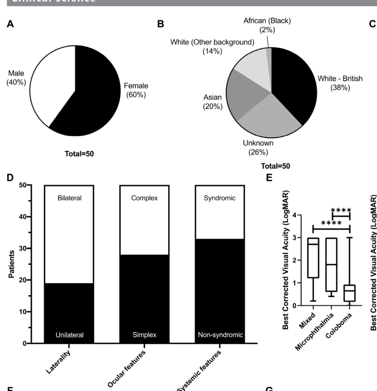
snippet: "Clinical analysis of 50 MAC patients (15 microphthalmia; 2 anophthalmia; 11 coloboma; and 22 mixed) from 44 unrelated families"
explanation: Microphthalmia is a defining and frequent feature of the MAC cohort.
- category: Ocular
name: Coloboma
description: >-
Missing ocular tissue resulting from incomplete optic-fissure closure;
may involve iris, ciliary body, retina/choroid, and/or optic nerve.
phenotype_term:
preferred_term: Coloboma
term:
id: HP:0000589
label: Coloboma
evidence:
- reference: PMID:39503780
reference_title: "Homozygosity for a hypomorphic mutation in frizzled class receptor 5 causes syndromic ocular coloboma with microcornea in humans."
supports: SUPPORT
evidence_source: HUMAN_CLINICAL
snippet: "Ocular coloboma (OC) is a congenital disorder caused by the incomplete closure of the embryonic ocular fissure."
explanation: Coloboma is a defining feature, mechanistically tied to fissure closure.
- category: Ocular
name: Iris coloboma
description: Keyhole-shaped iris defect from failure of fissure closure at the iris.
phenotype_term:
preferred_term: Iris coloboma
term:
id: HP:0000612
label: Iris coloboma
evidence:
- reference: PMID:20301552
reference_title: "Microphthalmia/Anophthalmia/Coloboma Spectrum – RETIRED CHAPTER, FOR HISTORICAL REFERENCE ONLY."
supports: SUPPORT
evidence_source: HUMAN_CLINICAL
snippet: "Iris coloboma causes the iris to appear keyhole-shaped."
explanation: GeneReviews baseline describes iris coloboma in the MAC spectrum.
- category: Ocular
name: Chorioretinal coloboma
description: Coloboma involving the retina and choroid.
phenotype_term:
preferred_term: Chorioretinal coloboma
term:
id: HP:0000567
label: Chorioretinal coloboma
evidence:
- reference: PMID:20301552
reference_title: "Microphthalmia/Anophthalmia/Coloboma Spectrum – RETIRED CHAPTER, FOR HISTORICAL REFERENCE ONLY."
supports: SUPPORT
evidence_source: HUMAN_CLINICAL
snippet: "Chorioretinal coloboma refers to coloboma of the retina and choroid."
explanation: GeneReviews baseline defines chorioretinal coloboma.
- category: Ocular
name: Anophthalmia
description: >-
Clinical absence of the globe with preserved ocular adnexa, at the severe
end of the MAC spectrum.
phenotype_term:
preferred_term: Anophthalmia
term:
id: HP:0000528
label: Anophthalmia
evidence:
- reference: PMID:20301552
reference_title: "Microphthalmia/Anophthalmia/Coloboma Spectrum – RETIRED CHAPTER, FOR HISTORICAL REFERENCE ONLY."
supports: SUPPORT
evidence_source: HUMAN_CLINICAL
snippet: "Anophthalmia refers to complete absence of the globe in the presence of ocular adnexa (eyelids, conjunctiva, and lacrimal apparatus)."
explanation: GeneReviews baseline defines anophthalmia within the spectrum.
- category: Ocular
name: Microcornea
description: Abnormally small cornea, reported with FZD5-related coloboma.
phenotype_term:
preferred_term: Microcornea
term:
id: HP:0000482
label: Microcornea
evidence:
- reference: PMID:39503780
reference_title: "Homozygosity for a hypomorphic mutation in frizzled class receptor 5 causes syndromic ocular coloboma with microcornea in humans."
supports: SUPPORT
evidence_source: HUMAN_CLINICAL
snippet: "we describe a case of syndromic bilateral OC with additional features such as microcornea, bone developmental anomalies, and mild intellectual disability"
explanation: Microcornea accompanies coloboma in the FZD5-related presentation.
- category: Ocular
name: Visual impairment
description: >-
Reduced vision is the principal functional consequence; MAC accounts for
~15–20% of severe childhood visual impairment and blindness worldwide.
phenotype_term:
preferred_term: Visual impairment
term:
id: HP:0000505
label: Visual impairment
evidence:
- reference: PMID:40038803
reference_title: "Management of anophthalmia, microphthalmia and coloboma in the newborn, shared care between neonatologist and ophthalmologist: a literature review."
supports: SUPPORT
evidence_source: HUMAN_CLINICAL
snippet: "Congenital ocular anomalies significantly contribute to global disability, with 15-20% of infant blindness attributed to these anomalies."
explanation: Supports visual impairment / blindness as the major MAC outcome.
- category: Neurologic
name: Developmental delay
description: >-
Intellectual/developmental delay is the most frequent systemic feature in
MAC patients with extraocular involvement.
phenotype_term:
preferred_term: Global developmental delay
term:
id: HP:0001263
label: Global developmental delay
evidence:
- reference: PMID:36192130
reference_title: "Real-world clinical and molecular management of 50 prospective patients with microphthalmia, anophthalmia and/or ocular coloboma."
supports: SUPPORT
evidence_source: HUMAN_CLINICAL
snippet: "34% had systemic involvement, most frequently intellectual/developmental delay (8/17)"
explanation: >-
Developmental delay is the leading systemic association in MAC patients
with extraocular features.
- category: Other
name: Sensorineural hearing loss
description: >-
Bilateral sensorineural hearing loss reported as a novel FOXE3-related
association in a MAC cohort.
phenotype_term:
preferred_term: Sensorineural hearing impairment
term:
id: HP:0000407
label: Sensorineural hearing impairment
evidence:
- reference: PMID:36192130
reference_title: "Real-world clinical and molecular management of 50 prospective patients with microphthalmia, anophthalmia and/or ocular coloboma."
supports: SUPPORT
evidence_source: HUMAN_CLINICAL
snippet: "New phenotypic associations were found for FOXE3 (bilateral sensorineural hearing loss) and MAB21L2 (unilateral microphthalmia)."
explanation: Documents the novel FOXE3 hearing-loss association.
- category: Ocular
name: Optic disc coloboma
subtype: IHH-Enhancer-MCOPCB13
description: >-
Colobomatous defect of the optic nerve head, frequently bilateral even where
microphthalmia is asymmetric, and the mildest end of the MCOPCB13 spectrum.
phenotype_term:
preferred_term: Optic nerve coloboma
term:
id: HP:0000588
label: Optic disc coloboma
evidence:
- reference: PMID:37580330
reference_title: "IHH enhancer variant within neighboring NHEJ1 intron causes microphthalmia anophthalmia and coloboma."
supports: SUPPORT
evidence_source: HUMAN_CLINICAL
snippet: "Optic nerve coloboma OU. Very high myopia OS>OD. Retinal atrophy OU. Nystagmus OU."
explanation: >-
Table 1 entry recording bilateral optic nerve coloboma in an affected
individual of pedigree 2.
- category: Ocular
name: High myopia
subtype: IHH-Enhancer-MCOPCB13
description: >-
High or very high myopia accompanies the structural malformation in several
affected individuals, in some with myopic chorioretinal degeneration and
retinal tears.
phenotype_term:
preferred_term: High myopia
term:
id: HP:0011003
label: High myopia
evidence:
- reference: PMID:37580330
reference_title: "IHH enhancer variant within neighboring NHEJ1 intron causes microphthalmia anophthalmia and coloboma."
supports: SUPPORT
evidence_source: HUMAN_CLINICAL
snippet: "High myopia with myopic chorioretinal degeneration and retinal tears OU."
explanation: >-
Table 1 entry recording bilateral high myopia with its chorioretinal
consequences.
- category: Ocular
name: Nystagmus
subtype: IHH-Enhancer-MCOPCB13
description: >-
Nystagmus is recorded in several affected individuals, in keeping with poor
visual input from early infancy.
phenotype_term:
preferred_term: Nystagmus
term:
id: HP:0000639
label: Nystagmus
evidence:
- reference: PMID:37580330
reference_title: "IHH enhancer variant within neighboring NHEJ1 intron causes microphthalmia anophthalmia and coloboma."
supports: SUPPORT
evidence_source: HUMAN_CLINICAL
snippet: "Strabismus (esotropia) OS. Nystagmus OU."
explanation: >-
Table 1 entry recording bilateral nystagmus alongside strabismus.
- category: Ocular
name: Strabismus
subtype: IHH-Enhancer-MCOPCB13
description: >-
Unilateral strabismus, esotropic where the deviation is specified, is
recorded in 2 of the 8 affected individuals tabulated in the index report,
in each case in the eye with the more severe structural malformation.
phenotype_term:
preferred_term: Strabismus
term:
id: HP:0000486
label: Strabismus
evidence:
- reference: PMID:37580330
reference_title: "IHH enhancer variant within neighboring NHEJ1 intron causes microphthalmia anophthalmia and coloboma."
supports: SUPPORT
evidence_source: HUMAN_CLINICAL
snippet: "Nuclear cataract OU. Strabismus OS, nystagmus OU."
explanation: >-
Table 1 entry for individual VI-7 of pedigree 1, recording left-sided
strabismus.
- reference: PMID:37580330
reference_title: "IHH enhancer variant within neighboring NHEJ1 intron causes microphthalmia anophthalmia and coloboma."
supports: SUPPORT
evidence_source: HUMAN_CLINICAL
snippet: "Strabismus (esotropia) OS. Nystagmus OU."
explanation: >-
A second tabulated individual with left esotropia, which is what makes
this a recurrent rather than a singleton feature of the subtype.
genetic:
- name: SOX2
subtype: SOX2-MAC
association: Causal dominant variant (often de novo)
notes: >-
SOX2 loss-of-function (often de novo) is the most common single-gene cause
of severe bilateral microphthalmia/anophthalmia in MAC, estimated at 10–15%
of cases.
gene_term:
preferred_term: SOX2
term:
id: hgnc:11195
label: SOX2
evidence:
- reference: PMID:40038803
reference_title: "Management of anophthalmia, microphthalmia and coloboma in the newborn, shared care between neonatologist and ophthalmologist: a literature review."
supports: SUPPORT
evidence_source: HUMAN_CLINICAL
snippet: "Chromosomal aberrations and mutations in genes such as PAX6, SOX2, OTX2, and CHD7 are contributors."
explanation: >-
Names SOX2 explicitly as a causal MAC gene; SOX2 is the leading monogenic
cause within the spectrum.
- name: OTX2
subtype: OTX2-MAC
association: Causal dominant variant (often de novo)
notes: >-
OTX2 loss-of-function variants (often de novo, dominant) cause severe
bilateral microphthalmia/anophthalmia and are estimated to explain roughly
2–5% of MAC cases.
gene_term:
preferred_term: OTX2
term:
id: hgnc:8522
label: OTX2
evidence:
- reference: PMID:40038803
reference_title: "Management of anophthalmia, microphthalmia and coloboma in the newborn, shared care between neonatologist and ophthalmologist: a literature review."
supports: SUPPORT
evidence_source: HUMAN_CLINICAL
snippet: "Chromosomal aberrations and mutations in genes such as PAX6, SOX2, OTX2, and CHD7 are contributors."
explanation: >-
Names OTX2 explicitly among the genes whose mutations contribute to the
MAC spectrum.
- name: PAX6
subtype: PAX6-MAC
association: Causal dominant haploinsufficiency
notes: >-
PAX6 haploinsufficiency is a classic contributor to the MAC spectrum,
producing colobomatous microphthalmia and a range of anterior-segment and
ocular malformations.
gene_term:
preferred_term: PAX6
term:
id: hgnc:8620
label: PAX6
evidence:
- reference: PMID:40038803
reference_title: "Management of anophthalmia, microphthalmia and coloboma in the newborn, shared care between neonatologist and ophthalmologist: a literature review."
supports: SUPPORT
evidence_source: HUMAN_CLINICAL
snippet: "Chromosomal aberrations and mutations in genes such as PAX6, SOX2, OTX2, and CHD7 are contributors."
explanation: >-
Names PAX6 explicitly among the genes whose mutations contribute to the
MAC spectrum.
- name: MAB21L2
subtype: MAB21L2-MAC
association: Causal coding and non-coding regulatory variant
notes: >-
Coding MAB21L2 variants and non-coding regulatory deletions upstream of
MAB21L2 cause microphthalmia and coloboma.
gene_term:
preferred_term: MAB21L2
term:
id: hgnc:6758
label: MAB21L2
evidence:
- reference: PMID:39455595
reference_title: "Deletion upstream of MAB21L2 highlights the importance of evolutionarily conserved non-coding sequences for eye development."
supports: SUPPORT
evidence_source: HUMAN_CLINICAL
snippet: "We report two families with variants in or near MAB21L2, a gene where genetic variants are known to cause AMC in humans and animal models."
explanation: Confirms MAB21L2 as an established MAC gene.
- name: FZD5
subtype: FZD5-Coloboma
association: Causal dominant-negative or recessive hypomorphic variant
notes: >-
FZD5 (Wnt receptor) variants cause isolated and syndromic coloboma via
disrupted Wnt signaling; dominant-negative and recessive hypomorphic alleles
are documented.
gene_term:
preferred_term: FZD5
term:
id: hgnc:4043
label: FZD5
evidence:
- reference: PMID:39503780
reference_title: "Homozygosity for a hypomorphic mutation in frizzled class receptor 5 causes syndromic ocular coloboma with microcornea in humans."
supports: SUPPORT
evidence_source: HUMAN_CLINICAL
snippet: "Mutations in the Wnt receptor FZD5, which is expressed throughout eye development, have been linked to both isolated and complex forms of coloboma."
explanation: Confirms FZD5 as a coloboma gene in isolated and complex forms.
- name: FOXE3
subtype: FOXE3-MAC
association: Causal variant
notes: >-
FOXE3 variants cause anterior-segment dysgenesis, microphthalmia, and
coloboma; bilateral sensorineural hearing loss is a reported association.
gene_term:
preferred_term: FOXE3
term:
id: hgnc:3808
label: FOXE3
evidence:
- reference: PMID:36192130
reference_title: "Real-world clinical and molecular management of 50 prospective patients with microphthalmia, anophthalmia and/or ocular coloboma."
supports: SUPPORT
evidence_source: HUMAN_CLINICAL
snippet: "novel variants in six known MAC genes (SOX2, KMT2D, MAB21L2, ALDH1A3, BCOR and FOXE3)"
explanation: FOXE3 is among the solved MAC genes in this prospective cohort.
- name: ALDH1A3
subtype: ALDH1A3-MAC
association: Causal biallelic variant
notes: >-
Recessive ALDH1A3 variants disrupt retinaldehyde dehydrogenase / retinoic
acid synthesis, causing autosomal recessive microphthalmia/anophthalmia.
gene_term:
preferred_term: ALDH1A3
term:
id: hgnc:409
label: ALDH1A3
evidence:
- reference: PMID:36192130
reference_title: "Real-world clinical and molecular management of 50 prospective patients with microphthalmia, anophthalmia and/or ocular coloboma."
supports: SUPPORT
evidence_source: HUMAN_CLINICAL
snippet: "novel variants in six known MAC genes (SOX2, KMT2D, MAB21L2, ALDH1A3, BCOR and FOXE3)"
explanation: ALDH1A3 is among the solved MAC genes; links to the retinoid mechanism.
- name: ABCB6
association: Causal dominant variant, ClinGen Limited
subtype: MCOPCB7
gene_term:
preferred_term: ABCB6
term:
id: hgnc:47
label: ABCB6
relationship_type: CAUSATIVE
notes: >-
ABCB6 is an ATP-binding cassette transporter expressed in the retina and the
retinal pigment epithelium. The dominant coloboma association rests on one
Chinese pedigree found by positional cloning, one further allele in three
sporadic microphthalmia-with-coloboma cases, and a zebrafish morpholino
knockdown rescued by wild-type but not mutant mRNA. ClinGen's General Inborn
Errors of Metabolism expert panel rates the relationship Limited, and that
rating is recorded here rather than argued with.
evidence:
- reference: PMID:22226084
reference_title: ABCB6 mutations cause ocular coloboma.
supports: SUPPORT
evidence_source: HUMAN_CLINICAL
quote_role: PRIMARY_RESULT
snippet: "Our results demonstrate that ABCB6 mutations cause ocular coloboma."
explanation: The discovery report's own causal conclusion.
- reference: PMID:22226084
reference_title: ABCB6 mutations cause ocular coloboma.
supports: SUPPORT
evidence_source: MODEL_ORGANISM
quote_role: PRIMARY_RESULT
snippet: "The knockdown phenotype can be corrected with coinjection of the wild-type, but not mutant, ABCB6 mRNA, suggesting that the phenotypes observed in zebrafish are due to insufficient abcb6 function."
explanation: >-
The rescue experiment behind the loss-of-function reading, and the only
functional support for the association.
- reference: CGGV:assertion_45ec3fba-48fd-4bf4-b865-2c2d7216e8eb-2024-04-26T180000.000Z
reference_title: "ABCB6 / microphthalmia, isolated, with coloboma 7 (Limited)"
supports: SUPPORT
evidence_source: OTHER
snippet: "ABCB6 | HGNC:47 | microphthalmia, isolated, with coloboma 7 | MONDO:0013783 | AD | Limited | SOP10 | General Inborn Errors of Metabolism Gene Curation Expert Panel | 2024-04-26T18:00:00.000Z"
explanation: The ClinGen classification and date quoted in the notes.
- name: TENM3
association: Causal recessive null variant in a single consanguineous family
subtype: MCOPCB9
gene_term:
preferred_term: TENM3
term:
id: hgnc:29944
label: TENM3
relationship_type: CAUSATIVE
notes: >-
TENM3 encodes a teneurin transmembrane protein, one of four vertebrate
orthologs of Drosophila odz. HGNC records ODZ3 as a previous symbol, and the
discovery report uses that name throughout. The association comes from exome
and autozygome analysis of one family with third-cousin parents and two
children with isolated colobomatous microphthalmia. The committed ClinGen
gene-validity snapshot carries no assertion for this gene under either
symbol.
evidence:
- reference: PMID:22766609
reference_title: Homozygous null mutation in ODZ3 causes microphthalmia in humans.
supports: SUPPORT
evidence_source: HUMAN_CLINICAL
quote_role: PRIMARY_RESULT
snippet: "Exome and autozygome analysis identified a null mutation in ODZ3, one of four vertebrate orthologs of odz in Drosophila."
explanation: The gene-discovery report, and the source of the ortholog statement in the notes.
- reference: PMID:22766609
reference_title: Homozygous null mutation in ODZ3 causes microphthalmia in humans.
supports: SUPPORT
evidence_source: HUMAN_CLINICAL
quote_role: PRIMARY_RESULT
snippet: "Our data highlight a role for ODZ3 in the early development of the human eye."
explanation: >-
The authors' own conclusion, which is a role claim rather than an
established gene-disease assertion.
- name: RBP4
association: Causal dominant-negative variant with incomplete penetrance and a maternal effect
subtype: MCOPCB10
gene_term:
preferred_term: RBP4
term:
id: hgnc:9922
label: RBP4
relationship_type: CAUSATIVE
notes: >-
RBP4 encodes serum retinol binding protein, which carries retinol from
hepatic stores to peripheral tissues including the placenta and the fetal
eye. The malformation alleles bind retinol poorly yet bind the cell-surface
receptor STRA6 with higher affinity, so they occupy the receptor without
delivering vitamin A; that is the dominant-negative step. A transmitting
mother's mutant protein acts at the placenta as well as in the fetus, so
maternal transmission raises the chance an allele-carrying fetus is affected,
and the authors present this as a mode of maternal inheritance distinct from
imprinting and from oocyte-derived mRNA. This is a distinct RBP4 phenotype
from the recessive one in which serum RBP4 is undetectable and the result is
a progressive retinal dystrophy, curated as
Progressive_Retinal_Dystrophy_Due_To_Retinol_Transport_Defect. The committed
ClinGen gene-validity snapshot carries no assertion for RBP4.
evidence:
- reference: PMID:25910211
reference_title: "Biochemical Basis for Dominant Inheritance, Variable Penetrance, and Maternal Effects in RBP4 Congenital Eye Disease."
supports: SUPPORT
evidence_source: HUMAN_CLINICAL
quote_role: PRIMARY_RESULT
snippet: "We identified missense mutations in RBP4, encoding serum retinol binding protein, in three families with eye malformations of differing severity, including bilateral anophthalmia."
explanation: The gene-discovery report and the range of severity this record covers.
- reference: PMID:25910211
reference_title: "Biochemical Basis for Dominant Inheritance, Variable Penetrance, and Maternal Effects in RBP4 Congenital Eye Disease."
supports: SUPPORT
evidence_source: IN_VITRO
quote_role: PRIMARY_RESULT
snippet: "The disease mutations greatly reduce retinol binding to RBP, yet paradoxically increase the affinity of RBP for its cell surface receptor, STRA6."
explanation: The two biochemical changes the dominant-negative mechanism in the notes rests on.
- reference: PMID:25910211
reference_title: "Biochemical Basis for Dominant Inheritance, Variable Penetrance, and Maternal Effects in RBP4 Congenital Eye Disease."
supports: SUPPORT
evidence_source: HUMAN_CLINICAL
quote_role: PRIMARY_RESULT
snippet: "These findings establish a previously uncharacterized mode of maternal inheritance, distinct from imprinting and oocyte-derived mRNA, and define a group of hereditary disorders plausibly modulated by dietary vitamin A."
explanation: >-
The authors' framing of the maternal effect, which the notes reports as
their claim rather than as settled nosology.
- name: IHH
subtype: IHH-Enhancer-MCOPCB13
association: Causal non-coding regulatory variant in an IHH enhancer hosted by an NHEJ1 intron
relationship_type: CAUSATIVE
variant_origin: GERMLINE
gene_term:
preferred_term: IHH
term:
id: hgnc:5956
label: IHH
notes: >-
MONDO, OMIM and the curation stub for this concept index MCOPCB13 under
NHEJ1, because the variant's coordinates fall inside an NHEJ1 intron. That
is a positional label, not a statement about NHEJ1 function: the primary
report states the allele is not predicted to affect NHEJ1, and the gene
whose expression is disturbed is IHH. NHEJ1's own established disease
association is a different one - ClinGen's gene-disease validity table
carries exactly one NHEJ1 assertion, Cernunnos-XLF deficiency (MONDO:0012650,
autosomal recessive, Definitive), and none for microphthalmia or coloboma;
the same table carries no IHH assertion at all, so neither the host gene nor
the regulatory target has a ClinGen assertion covering this phenotype.
NHEJ1 is recorded here as the host gene of the affected region rather than
as a causal gene binding.
affected_regions:
- name: IHH enhancer i8
regulatory_element_type: ENHANCER
chromosomal_region: 2q35
within_gene:
preferred_term: NHEJ1
term:
id: hgnc:25737
label: NHEJ1
description: >-
A conserved non-coding element of the IHH enhancer cluster, named i8 when
the cluster was characterised in limb development, lying within an intron
of NHEJ1 on the reference genome. The relationship recorded here locates
the element; it does not assert that NHEJ1 is regulated by it or that
NHEJ1 sequence function is disturbed.
variants:
- name: NHEJ1 NM_024782.2:c.588+18131A>G
variant_type: single nucleotide variant
genomic_contexts:
- intron
regulatory_category: mLOE
gene:
preferred_term: NHEJ1
term:
id: hgnc:25737
label: NHEJ1
regulatory_target_gene:
preferred_term: IHH
term:
id: hgnc:5956
label: IHH
description: >-
Homozygous founder substitution reported in two consanguineous Jewish
Iranian pedigrees, absent from gnomAD and from 87 ethnically matched
controls, and submitted to ClinVar as SCV002769719. The coordinates lie in
NHEJ1 intron sequence and the allele is not predicted to alter NHEJ1
splicing or protein. Expression loss is classified mLOE rather than LOE
because the affected individuals have no skeletal or cranial phenotype,
so IHH expression driven by the rest of the cluster is evidently retained
outside the eye.
functional_effects:
- regulatory_element_type: ENHANCER
regulatory_mechanism: >-
Loss of eye-specific enhancer activity; the substituted base sits at a
predicted transcription-factor binding motif and at the summit of the
chromatin-accessibility peak marking the element in developing retinal
pigment epithelium.
affected_developmental_stage: >-
Embryonic eye morphogenesis, before optic fissure closure (mouse E11.5;
chicken Hamburger-Hamilton stages 24-26).
description: >-
Wild-type i8 drives reporter expression in the developing eye of
transgenic mouse embryos and the patient allele does not.
evidence:
- reference: PMID:37580330
reference_title: "IHH enhancer variant within neighboring NHEJ1 intron causes microphthalmia anophthalmia and coloboma."
supports: SUPPORT
evidence_source: HUMAN_CLINICAL
snippet: "NHEJ1 (NM_024782.2): c.588+18131 A > G (NG_007880.1: g.37317 A > G), submitted to ClinVar (SCV002769719), has no reported frequency in gnomAD"
explanation: >-
Gives the variant's nomenclature, its ClinVar submission, and its absence
from population data.
- reference: PMID:37580330
reference_title: "IHH enhancer variant within neighboring NHEJ1 intron causes microphthalmia anophthalmia and coloboma."
supports: SUPPORT
evidence_source: HUMAN_CLINICAL
snippet: "RFLP screening of 87 ethnically matched control samples identified no other individuals heterozygous or homozygous for this variant"
explanation: >-
Ancestry-matched control screening supporting the allele's rarity in the
founder population.
- reference: PMID:37580330
reference_title: "IHH enhancer variant within neighboring NHEJ1 intron causes microphthalmia anophthalmia and coloboma."
supports: SUPPORT
evidence_source: MODEL_ORGANISM
snippet: "the summit of the observed i8 peak overlaps with the conserved PAX4 binding motif and the orthologous site of the c.588+18131A > G variant"
explanation: >-
Locates the substituted base at the accessibility summit and a predicted
transcription-factor motif, which is the proposed molecular mechanism.
evidence:
- reference: PMID:37580330
reference_title: "IHH enhancer variant within neighboring NHEJ1 intron causes microphthalmia anophthalmia and coloboma."
supports: SUPPORT
evidence_source: HUMAN_CLINICAL
snippet: "a MAC disease-associated 0.5 Mb genomic locus on chromosome 2q35 (maximal LOD score 6.8)"
explanation: >-
Linkage result establishing the 2q35 locus that contains the IHH enhancer
cluster and NHEJ1.
- reference: PMID:37580330
reference_title: "IHH enhancer variant within neighboring NHEJ1 intron causes microphthalmia anophthalmia and coloboma."
supports: SUPPORT
evidence_source: MODEL_ORGANISM
snippet: "this variant is within an Ihh enhancer that drives gene expression in the developing eye and that the identified variant affects this eye-specific enhancer activity"
explanation: >-
Functional result identifying IHH as the gene whose regulation is
disturbed, which is the basis for recording IHH rather than NHEJ1 here.
- reference: CGGV:assertion_765b118e-c60a-4696-b401-f63b3349d6d2-2021-05-20T151233.276Z
reference_title: "NHEJ1 / Cernunnos-XLF deficiency (Definitive)"
supports: NO_EVIDENCE
evidence_source: OTHER
snippet: "NHEJ1 | HGNC:25737 | Cernunnos-XLF deficiency | MONDO:0012650 | AR | Definitive"
explanation: >-
Cited because it is the assertion readers will find when they look NHEJ1
up, and it is about a different disease. ClinGen's only curated NHEJ1
gene-disease relationship is Cernunnos-XLF deficiency; it says nothing
either way about microphthalmia or coloboma, so it neither supports nor
refutes this record.
environmental:
- name: Retinoid pathway teratogens
exposure_term:
preferred_term: retinoid teratogen exposure
term:
id: ECTO:9001346
label: exposure to retinoid
influences_mechanisms:
- target: Retinoic acid signaling disruption
environmental_effect: TRIGGERS
causal_link_type: DIRECT
description: >-
Retinoid drugs act on the very signalling pathway this node names, so
the exposure perturbs retinoic acid signalling directly rather than
through an intermediate.
evidence:
- reference: PMID:3162101
reference_title: "Retinoic acid embryopathy."
supports: SUPPORT
evidence_source: HUMAN_CLINICAL
snippet: "The malformations included microtia/anotia (15 infants), micrognathia (6), cleft palate (3), conotruncal heart defects and aortic-arch abnormalities (8), thymic defects (7), retinal or optic-nerve abnormalities (4), and central nervous system malformations (18)."
explanation: >-
Isotretinoin is a retinoic acid analogue, and human pregnancies exposed
to it produced a characteristic malformation pattern that includes
ocular defects. Because retinoids act as ligands of the retinoic acid
receptors, the exposure perturbs the signalling this node names
directly rather than through an intermediate.
description: >-
Prenatal exposures that disrupt retinoid (vitamin A / retinoic acid) biology
— including isotretinoin/retinoic-acid embryopathy and maternal vitamin A
deficiency — are associated with microphthalmia and coloboma, acting as a
phenocopy of genetic retinoid-pathway lesions (e.g. ALDH1A3).
evidence:
- reference: PMID:39296666
reference_title: The first review on prenatal drug exposure and ocular malformation occurrence.
supports: SUPPORT
evidence_source: HUMAN_CLINICAL
snippet: "medications are specifically known to alter eye morphogenesis during embryonic development"
explanation: >-
Teratology review identifying medications (including retinoids) that alter
eye morphogenesis.
- name: Thalidomide and other ocular teratogens
influences_mechanisms:
- target: Failure of optic fissure closure
environmental_effect: PREDISPOSES
causal_link_type: INDIRECT_UNKNOWN_INTERMEDIATES
description: >-
Groups several unrelated environmental contributors, including
gestational infection and maternal vitamin A deficiency, whose common
feature is raising the chance that fissure closure fails rather than a
shared mechanism.
evidence:
- reference: PMID:40491727
reference_title: "Genetic and environmental factors contributing to anophthalmia and microphthalmia: Current understanding and future directions."
supports: SUPPORT
evidence_source: HUMAN_CLINICAL
snippet: "the contribution of environmental factors such as gestational-acquired infections, maternal vitamin A deficiency (VAD), exposure to X-rays, solvent misuse, and thalidomide exposure."
explanation: >-
Names gestational infections and maternal vitamin A deficiency among
environmental contributors to the malformation, a heterogeneous set of
risk factors rather than one mechanism.
description: >-
Thalidomide is a classic ocular teratogen, and additional environmental
contributors to anophthalmia/microphthalmia include gestational infections,
maternal vitamin A deficiency, X-ray exposure, and solvent misuse.
evidence:
- reference: PMID:40491727
reference_title: "Genetic and environmental factors contributing to anophthalmia and microphthalmia: Current understanding and future directions."
supports: SUPPORT
evidence_source: HUMAN_CLINICAL
snippet: "the contribution of environmental factors such as gestational-acquired infections, maternal vitamin A deficiency (VAD), exposure to X-rays, solvent misuse, and thalidomide exposure."
explanation: >-
Review enumerating environmental contributors (infections, vitamin A
deficiency, X-rays, solvents, thalidomide) to A/M.
treatments:
- name: Genetic counseling
action_category: COUNSELING_INFORMATIONAL
description: >-
Genetic counseling and diagnostic genetic testing (chromosomal microarray,
clinical exome or whole-genome sequencing) guide recurrence-risk assessment
and family planning in the genetically heterogeneous MAC spectrum.
treatment_term:
preferred_term: Genetic Counseling
term:
id: NCIT:C15240
label: Genetic Counseling
evidence:
- reference: PMID:40038803
reference_title: "Management of anophthalmia, microphthalmia and coloboma in the newborn, shared care between neonatologist and ophthalmologist: a literature review."
supports: SUPPORT
evidence_source: HUMAN_CLINICAL
snippet: "This work provides a literature review offering insights for effective management and genetic counseling in a pediatric context."
explanation: Genetic counseling is part of recommended MAC management.
- name: Ophthalmic surgical management
description: >-
Surgical interventions in colobomatous microphthalmia include cataract
surgery / lensectomy and vitreoretinal surgery for coloboma-associated
retinal detachment, with management individualized by visual potential.
therapeutic_modality: SURGERY
treatment_term:
preferred_term: surgical procedure
term:
id: NCIT:C15329
label: Surgical Procedure
target_mechanisms:
- target: Disrupted optic-cup and globe morphogenesis
description: >-
Addresses the structural consequences of the morphogenetic defect, and in
the anophthalmic socket supports orbital growth. It does not act on optic
fissure closure, which has already failed in utero.
evidence:
- reference: PMID:20301552
reference_title: "Microphthalmia/Anophthalmia/Coloboma Spectrum – RETIRED CHAPTER, FOR HISTORICAL REFERENCE ONLY."
supports: SUPPORT
evidence_source: HUMAN_CLINICAL
snippet: "An oculoplastic surgeon can help determine the most suitable options for surgical intervention after age six months"
explanation: >-
GeneReviews describes oculoplastic surgical intervention as part of MAC
spectrum management.
- name: Supportive and rehabilitative care
description: >-
Multidisciplinary supportive care includes prosthetic/conformer expansion of
the orbit for severe microphthalmia/anophthalmia, low-vision aids and vision
rehabilitation, and developmental/early-intervention support.
treatment_term:
preferred_term: supportive care
term:
id: NCIT:C15747
label: Supportive Care
target_mechanisms:
- target: Disrupted optic-cup and globe morphogenesis
description: >-
Addresses the visual impairment resulting from the malformed globe.
Purely supportive.
evidence:
- reference: PMID:20301552
reference_title: "Microphthalmia/Anophthalmia/Coloboma Spectrum – RETIRED CHAPTER, FOR HISTORICAL REFERENCE ONLY."
supports: SUPPORT
evidence_source: HUMAN_CLINICAL
snippet: "Prosthetic intervention is appropriate for those with severe microphthalmia and anophthalmia."
explanation: GeneReviews baseline supports prosthetic/supportive management.
clinical_trials:
- name: NCT01778543
status: RECRUITING
description: >-
NEI natural-history and genetics study deep-phenotyping individuals with
MAC and unaffected relatives to identify associated genes and build a
DNA/cell-line repository.
target_phenotypes:
- preferred_term: Coloboma
term:
id: HP:0000589
label: Coloboma
- preferred_term: Microphthalmia
term:
id: HP:0000568
label: Microphthalmia
evidence:
- reference: clinicaltrials:NCT01778543
reference_title: "Pathogenesis and Genetics of Microphthalmia, Anophthalmia and Uveal Coloboma ( MAC)"
supports: SUPPORT
snippet: "Uveal coloboma is part of a spectrum of developmental eye conditions that include anophthalmia and microphthalmia, typically referred to as \"MAC\""
explanation: Trial directly targets the MAC spectrum genetics.
- name: NCT04833361
status: COMPLETED
description: >-
NEI pilot study exploring maternal first-trimester exposures as potential
environmental causes of uveal coloboma, linking survey data to affected
children's clinical data.
target_phenotypes:
- preferred_term: Coloboma
term:
id: HP:0000589
label: Coloboma
evidence:
- reference: clinicaltrials:NCT04833361
reference_title: Pilot Evaluation of Potential Environmental Causes of Uveal Coloboma
supports: SUPPORT
snippet: "To explore maternal factors and exposures during the first trimester of pregnancy as potential causes of uveal coloboma"
explanation: Trial investigates environmental causes of coloboma in the spectrum.
animal_models:
- species: Dog
genotype: Homozygous 7.8 kb deletion in NHEJ1 intron 4 (Collie eye anomaly allele)
category: Naturally occurring
description: >-
Collie eye anomaly is a naturally occurring autosomal recessive canine
ocular malformation - choroidal hypoplasia with optic nerve head coloboma -
that maps to a homozygous 7.8 kb deletion inside an intron of the canine
NHEJ1 orthologue. The human MCOPCB13 report placed that canine deletion on
the human genome and found it removes a different member of the same IHH
enhancer cluster, i9, where the human variant disrupts i8. The dog is
therefore a natural experiment in the same enhancer cluster, at a
neighbouring element, with a lesion of a different class.
publication: PMID:17916641
modeled_mechanisms:
- target: Failure of optic fissure closure
relationship: PARTIALLY_RECAPITULATES
fidelity: MODERATE
model_scale: TISSUE
description: >-
The affected dog shows optic nerve head coloboma with choroidal
hypoplasia, so it reproduces the failure-of-optic-fissure-closure outcome
after a lesion in the same intronic enhancer cluster. It is partial
because the canine disease is confined to the posterior pole and does not
include the microphthalmia or anophthalmia seen in MCOPCB13.
limitations: >-
The canine lesion is a 7.8 kb deletion of i9, not the human single
nucleotide substitution in i8, so it does not model the human allele. The
canine deletion's own causal status is contested: it is discordant with
the phenotype in some breeds and populations, where it behaves as a linked
marker rather than the causal variant. IHH has not been shown to be the
canine target; the 2007 report describes the deleted sequence as a
conserved protein-binding domain without naming a regulated gene.
divergences:
- divergence_type: CAUSE_UNREPRESENTED
materiality: QUALIFYING
description: >-
The element disrupted is i9 in the dog and i8 in the human patients,
and the canine lesion is a 7.8 kb deletion rather than a single
nucleotide substitution. These are separate enhancers of the same
cluster, so the MCOPCB13 allele is not encoded in this model and the
dog does not report on the activity of the element that is lesioned in
MCOPCB13.
- divergence_type: SPECIES_MISMATCH
materiality: QUALIFYING
description: >-
Optic fissure closure is read out in the canine eye. Dogs have no
macula and a tapetal fundus, and the canine phenotype is choroidal
hypoplasia with optic nerve head coloboma rather than the microphthalmia
and anophthalmia that dominate the human pedigrees, so the severity and
distribution of the human malformation are not reproduced.
evidence:
- reference: PMID:17916641
reference_title: "Breed relationships facilitate fine-mapping studies: a 7.8-kb deletion cosegregates with Collie eye anomaly across multiple dog breeds."
supports: SUPPORT
evidence_source: MODEL_ORGANISM
snippet: "Sequence analysis revealed that all affected dogs share a homozygous deletion of 7.8 kb in the NHEJ1 gene. This intronic deletion spans a highly conserved binding domain to which several developmentally important proteins bind."
explanation: >-
Establishes the canine lesion as a deletion of conserved regulatory
sequence inside an NHEJ1 intron rather than an NHEJ1 coding lesion.
- reference: PMID:37580330
reference_title: "IHH enhancer variant within neighboring NHEJ1 intron causes microphthalmia anophthalmia and coloboma."
supports: SUPPORT
evidence_source: COMPUTATIONAL
snippet: "it encompasses another experimentally verified enhancer of Ihh (in mice), termed 'i9' by Will et al"
explanation: >-
The human report's own mapping of the canine deletion onto the IHH
enhancer cluster, which is what makes the dog informative here.
- reference: PMID:26732749
reference_title: "Discrepancy in compliance between the clinical and genetic diagnosis of choroidal hypoplasia in Danish Rough Collies and Shetland Sheepdogs."
supports: REFUTE
evidence_source: MODEL_ORGANISM
snippet: "the intronic deletion in NHEJ1 is not the causative mutation but, rather, a marker linked to the locus underlying the trait in some populations"
explanation: >-
Argues the canine deletion is a linked marker rather than the causal
variant in Danish Rough Collies, which limits how far this model can be
pushed as corroboration.
evidence:
- reference: PMID:28702949
reference_title: "Genetic analysis of optic nerve head coloboma in the Nova Scotia Duck Tolling Retriever identifies discordance with the NHEJ1 intronic deletion (collie eye anomaly mutation)."
supports: REFUTE
evidence_source: MODEL_ORGANISM
snippet: "Collection of ONH coloboma-affected NSDTR showed lack of concordance of the NHEJ1 intronic deletion with ONH coloboma"
explanation: >-
A second breed in which optic nerve head coloboma does not track with the
deletion, recorded here so the cross-species argument is not read as
stronger than it is.
discussions:
- discussion_id: mcopcb13_ihh_enhancer_human_evidence
kind: HUMAN_MODEL_MISMATCH
status: OPEN
prompt: >-
Is IHH expression actually reduced in the human developing eye in
microphthalmia/coloboma 13, or is the reduction inferred entirely from
mouse and chicken assays of the enhancer?
attaches_to:
- pathophysiology#IHH i8 enhancer disruption
- pathophysiology#Reduced IHH signaling to periocular mesenchyme and RPE
rationale: >-
Every step from the variant to reduced IHH output rests on non-human
systems: a transgenic mouse reporter assay at E11.5, chicken RPE ATAC-seq
and electroporation, and mouse genetics for the requirement of Ihh in RPE
and scleral mesenchyme. No measurement of IHH transcript or protein in
human ocular tissue from an affected individual has been reported, and the
variant has been described in two related consanguineous pedigrees from one
founder community, with no independent family reported since. The
translational question is sharpened rather than settled by the human
phenotype: the same enhancer cluster is required for IHH expression in limb
and skull, yet the affected individuals have no skeletal or cranial
abnormality, so either the element's eye and limb activities are separable
sub-modules or the human allele spares the limb-active part. Neither
possibility has been tested.
notes: >-
This is recorded as a model-to-human translation question rather than as an
absent mechanism. The enhancer's eye activity, the allele's effect on it,
and the dependence of periocular mesenchyme and RPE on Ihh are each
supported by experiments; what is missing is a human tissue measurement and
a second, unrelated family.
References & Deep Research
References
1Deep Research
1Deep research results are used as seeds for research; they do not undergo the same validation as the main records and may contain errors. How we use deep research.
Evaluations and curation notes (1)
Add MCOPCB13 (MONDO:0975809) as the IHH-Enhancer-MCOPCB13 subtype · 2026-09-24T17:45:00Z · View source
Curated microphthalmia/coloboma 13 (MONDO:0975809, OMIM 620968) after deciding it is a subtype of this entry rather than a new Disease entry. The stub named NHEJ1 (hgnc:25737) as the causal gene; the primary report (PMID:37580330, Wormser et al. 2023, npj Genomic Medicine, PMC full text) states the homozygous founder allele NM_024782.2:c.588+18131A>G is not predicted to affect NHEJ1 and lies inside 'i8', a conserved enhancer of the neighbouring gene IHH (hgnc:5956, verified against the HGNC REST API and cache/hgnc/terms.csv; the deep-research report offered HGNC:5384 for IHH, which is wrong and was not used). Recorded IHH as the causal gene with NHEJ1 as the host gene of the affected region, using the noncoding-variant-impact pattern: Variant.gene NHEJ1, regulatory_target_gene IHH, variant_type single nucleotide variant, genomic_contexts intron, regulatory_category mLOE, functional_effects regulatory_element_type ENHANCER, and affected_regions 'IHH enhancer i8' with within_gene NHEJ1. Added two pathophysiology nodes joining the existing 'Failure of optic fissure closure' and 'Disrupted optic-cup and globe morphogenesis' nodes, three subtype-scoped phenotypes from Table 1 of PMID:37580330, one animal model (canine Collie eye anomaly, PMID:17916641) carrying REFUTE evidence for its own limitations (PMID:26732749, PMID:28702949), and a HUMAN_MODEL_MISMATCH discussion recording that every step from variant to reduced IHH output rests on mouse and chicken assays with no human tissue measurement and no second unrelated family. Checked ClinGen directly by downloading the live gene-validity CSV on 2026-09-24: NHEJ1 has one assertion (Cernunnos-XLF deficiency, MONDO:0012650, AR, Definitive) and IHH has none, so no ClinGen assertion covers this phenotype; cited the NHEJ1 row as supports NO_EVIDENCE. Deep research: falcon requested, returned HTTP 402 (out of credits), claude_code produced the report under fallback; preflight-dr returned WARN because IHH is mentioned more often than NHEJ1, which is the expected signal for this disease rather than a wrong-disease report. Report reference validation 10/10 resolved, 0 off topic; term validation flagged UBERON:0000384 as non-existent and UBERON:0001789/UBERON:0001786 as mislabelled, none of which were bound. Validated under linkml-reference-validator 0.3.0rc1 in a dedicated venv: just validate (58/58 snippets, 36 titles, 0 issues), count-verified-snippets, validate-terms, check-duplicate-keys, check-entity-refs, check-causal-targets, check-qualifier-terms, check-enum-values, list-gene-term-mismatches, check-snippet-length, check-title-snippets, check-snippet-grading, check-folded-hyphens, check-environmental-evidence, check-coarse-phenotypes, check-case-collisions, pytest tests/test_data.py -k Microphthalmia, and validate-disorders.
Question: You are an expert researcher providing comprehensive, well-cited information.
Provide detailed information focusing on: 1. Key concepts and definitions with current understanding 2. Recent developments and latest research (prioritize 2023-2024 sources) 3. Current applications and real-world implementations 4. Expert opinions and analysis from authoritative sources 5. Relevant statistics and data from recent studies
Format as a comprehensive research report with proper citations. Include URLs and publication dates where available. Always prioritize recent, authoritative sources and provide specific citations for all major claims.
Disease Characteristics Research Template
Target Disease
- Disease Name: Microphthalmia with Coloboma
- MONDO ID: (if available)
- Category: Mendelian
Research Objectives
Please provide a comprehensive research report on Microphthalmia with Coloboma covering all of the disease characteristics listed below. This report will be used to populate a disease knowledge base entry. Be thorough and cite primary literature (PMID preferred) for all claims.
For each section, suggested databases/resources are listed. These are the first places you should search for information on each topic.
1. Disease Information
Search first: OMIM, Orphanet, ICD-10/ICD-11, MeSH, PubMed
- What is the disease? Provide a concise overview.
- What are the key identifiers? (OMIM, Orphanet, ICD-10/ICD-11, MeSH, Mondo)
- What are the common synonyms and alternative names?
- Is the information derived from individual patients (e.g., EHR) or aggregated disease-level resources?
2. Etiology
- Disease Causal Factors: What are the primary causes? (genetic, environmental, infectious, mechanistic)
- Risk Factors:
Search first: PubMed, Cochrane Library, UpToDate, clinical guidelines, ClinVar, ClinGen, GWAS Catalog, PheGenI, CTD, CDC, WHO, epidemiological databases
- Genetic risk factors (causal variants, susceptibility loci, modifier genes)
- Environmental risk factors (toxins, lifestyle, occupational exposures, age, sex, family history)
- Protective Factors:
Search first: PubMed, Cochrane Library, clinical trial databases, GWAS Catalog, gnomAD, WHO, CDC, nutrition databases
- Genetic protective factors (protective variants, modifier alleles)
- Environmental protective factors (diet, lifestyle, exposures that reduce risk)
- Gene-Environment Interactions: How do genetic and environmental factors interact to influence disease?
Search first: CTD, PubMed, PheGenI, GxE databases
3. Phenotypes
Search first: HPO (Human Phenotype Ontology), OMIM, Orphanet, PubMed, clinicaltrials.gov, MedDRA, SNOMED CT, DECIPHER, LOINC
For each phenotype, provide: - Phenotype type: symptoms, clinical signs, physical manifestations, behavioral changes, or laboratory abnormalities
For symptoms/signs: HPO, OMIM, Orphanet, PubMed For behavioral changes: HPO, DSM, RDoC (Research Domain Criteria), PubMed For laboratory abnormalities: LOINC, SNOMED CT, LabTests Online, PubMed - Phenotype characteristics: Search first: OMIM, Orphanet, HPO, PubMed - Age of symptom onset (neonatal, childhood, adult-onset, late-onset) - Symptom severity (mild, moderate, severe, variable) - Symptom progression (stable, progressive, episodic, fluctuating) - Frequency among affected individuals (percentage or qualitative) - Quality of life impact: Effects on daily functioning and well-being (per-phenotype when possible) Search first: EQ-5D database, SF-36, WHO QOL databases, PubMed - Suggest HPO (Human Phenotype Ontology) terms for each phenotype
4. Genetic/Molecular Information
- Causal Genes: Gene mutations or chromosomal abnormalities responsible for disease (gene symbols, OMIM IDs)
Search first: OMIM, ClinVar, HGMD, Ensembl, NCBI Gene
- Pathogenic Variants:
- Affected genes (gene symbols, HGNC IDs) > Search first: OMIM, NCBI Gene, Ensembl, HGNC, UniProt, GeneCards
- Variant classification (pathogenic, likely pathogenic, VUS per ACMG/AMP guidelines) > Search first: ClinVar, ClinGen, ACMG/AMP guidelines, VarSome
- Variant type/class (missense, frameshift, nonsense, splice-site, structural)
- Allele frequency in population databases > Search first: gnomAD, 1000 Genomes, ExAC, TOPMed, dbSNP
- Somatic vs germline origin > Search first: COSMIC (somatic), ClinVar, ICGC, TCGA
- Functional consequences (loss of function, gain of function, dominant negative)
- Modifier Genes: Genes that modify disease severity or expression
- Epigenetic Information: DNA methylation, histone modifications, chromatin changes affecting disease
Search first: ENCODE, Roadmap Epigenomics, MethBase, DiseaseMeth
- Chromosomal Abnormalities: Large-scale genetic changes (aneuploidy, translocations, inversions)
Search first: DECIPHER, ClinVar, ECARUCA, UCSC Genome Browser
5. Environmental Information
- Environmental Factors: Non-genetic contributing factors (toxins, radiation, pollution, occupational exposure)
Search first: CTD (Comparative Toxicogenomics Database), TOXNET, PubMed, EPA databases
- Lifestyle Factors: Behavioral factors (smoking, diet, exercise, alcohol consumption)
Search first: CDC databases, WHO, PubMed, NHANES
- Infectious Agents: If applicable, pathogens causing or triggering disease (bacteria, viruses, fungi, parasites)
Search first: NCBI Taxonomy, ViPR, BV-BRC, MicrobeDB, GIDEON
6. Mechanism / Pathophysiology
- Molecular Pathways: Specific signaling cascades or biochemical pathways involved (Wnt, MAPK, mTOR, PI3K-AKT, etc.)
Search first: KEGG, Reactome, WikiPathways, PathBank, BioCyc
- Cellular Processes: Cell-level mechanisms (apoptosis, autophagy, cell cycle dysregulation, inflammation, etc.)
Search first: Gene Ontology (GO), Reactome, KEGG, PubMed
- Protein Dysfunction: How protein structure or function is altered (misfolding, aggregation, loss of function, gain of function)
Search first: UniProt, PDB (Protein Data Bank), InterPro, Pfam, AlphaFold
- Metabolic Changes: Alterations in metabolic processes (energy metabolism, lipid metabolism, amino acid metabolism)
Search first: KEGG, BioCyc, HMDB (Human Metabolome Database), BRENDA
- Immune System Involvement: Role of immune response (autoimmunity, immunodeficiency, chronic inflammation)
Search first: ImmPort, Immunome Database, IEDB, Gene Ontology
- Tissue Damage Mechanisms: How tissues/ are injured (oxidative stress, ischemia, fibrosis, necrosis)
Search first: PubMed, Gene Ontology, Reactome
- Biochemical Abnormalities: Specific molecular defects (enzyme deficiencies, receptor dysfunction, ion channel defects)
Search first: BRENDA, UniProt, KEGG, OMIM, PubMed
- Epigenetic Changes: DNA methylation, histone modifications affecting gene expression in disease
Search first: ENCODE, Roadmap Epigenomics, MethBase, DiseaseMeth
- Molecular Profiling (if available):
- Transcriptomics/gene expression changes > Search first: GEO (Gene Expression Omnibus), ArrayExpress, GTEx, Human Cell Atlas, SRA
- Proteomics findings > Search first: PRIDE, ProteomeXchange, Human Protein Atlas, STRING, BioGRID
- Metabolomics signatures > Search first: MetaboLights, Metabolomics Workbench, HMDB, METLIN
- Lipidomics alterations > Search first: LIPID MAPS, SwissLipids, LipidHome, Metabolomics Workbench
- Genomic structural features > Search first: UCSC Genome Browser, Ensembl, NCBI, dbVar, DGV
- Advanced Technologies (if applicable):
- Single-cell analysis findings (cell-type specific mechanisms, cellular heterogeneity) > Search first: Human Cell Atlas, Single Cell Portal, GEO, CELLxGENE
- Spatial transcriptomics findings > Search first: GEO, Spatial Research, Vizgen, 10x Genomics data
- Multi-omics integration results > Search first: TCGA, ICGC, cBioPortal, LinkedOmics, PubMed
- Functional genomics screens (CRISPR, RNAi) > Search first: DepMap, GenomeRNAi, PubMed, BioGRID ORCS
For each mechanism, describe: - The causal chain from initial trigger to clinical manifestation - Which mechanisms are upstream vs downstream - What cell types and biological processes are involved - Suggest GO terms for biological processes and CL terms for cell types
7. Anatomical Structures Affected
- Organ Level:
- Primary organs directly affected
- Secondary organ involvement (complications, secondary effects)
- Body systems involved (cardiovascular, nervous, digestive, respiratory, endocrine, etc.)
Search first: Uberon, FMA (Foundational Model of Anatomy), OMIM, HPO, ICD-11, MeSH, SNOMED CT
- Tissue and Cell Level:
- Specific tissue types affected (epithelial, connective, muscle, nervous)
- Specific cell populations targeted (with Cell Ontology terms)
Search first: Uberon, Human Protein Atlas, Cell Ontology, Human Cell Atlas, CellMarker, PanglaoDB
- Subcellular Level:
- Cellular compartments involved (mitochondria, nucleus, ER, lysosomes) (with GO Cellular Component terms)
Search first: Gene Ontology (Cellular Component), UniProt, Human Protein Atlas
- Localization:
- Specific anatomical sites (with UBERON terms) > Search first: FMA, Uberon, NeuroNames (for brain), SNOMED CT
- Lateralization (unilateral, bilateral, asymmetric) > Search first: HPO, clinical literature, imaging databases
8. Temporal Development
- Onset:
- Typical age of onset (congenital, pediatric, adult, geriatric)
- Onset pattern (acute, subacute, chronic, insidious)
Search first: OMIM, Orphanet, HPO, PubMed
- Progression:
- Disease stages (early, intermediate, advanced, end-stage) > Search first: Cancer Staging Manual (AJCC), WHO classifications, PubMed
- Progression rate (rapid, slow, variable)
- Disease course pattern (episodic, relapsing-remitting, progressive, stable)
- Disease duration (self-limited, chronic lifelong)
Search first: Disease registries, longitudinal cohort databases, natural history studies, PubMed, Orphanet, OMIM
- Patterns:
- Remission patterns (spontaneous, treatment-induced) > Search first: Clinical trial databases, disease registries, PubMed
- Critical periods (time windows of vulnerability or opportunity for intervention) > Search first: PubMed, developmental biology databases, clinical guidelines
9. Inheritance and Population
- Epidemiology:
- Prevalence (cases per 100,000 at given time)
- Incidence (new cases per 100,000 per year)
Search first: Orphanet, CDC, WHO, GBD (Global Burden of Disease), national registries, SEER, disease registries
- For Genetic Etiology:
- Inheritance pattern (AD, AR, X-linked, mitochondrial, multifactorial, polygenic) > Search first: OMIM, Orphanet, ClinVar, GTR (Genetic Testing Registry)
- Penetrance (complete, incomplete, age-dependent) > Search first: ClinVar, OMIM, PubMed, ClinGen
- Expressivity (variable, consistent) > Search first: OMIM, ClinVar, PubMed
- Genetic anticipation (increasing severity in successive generations) > Search first: OMIM, PubMed (especially for repeat expansion disorders)
- Germline mosaicism > Search first: ClinVar, OMIM, genetic counseling literature, PubMed
- Founder effects (population-specific mutations) > Search first: gnomAD, population genetics databases, PubMed
- Consanguinity role > Search first: OMIM, population studies, genetic counseling resources
- Carrier frequency > Search first: gnomAD, carrier screening databases, GeneReviews, GTR
- Population Demographics:
- Affected populations (ethnic or demographic groups with higher prevalence) > Search first: gnomAD, 1000 Genomes, PAGE Study, PubMed, population registries
- Geographic distribution (endemic areas, regional variation) > Search first: WHO, CDC, GBD, Orphanet, geographic epidemiology databases
- Geographic distribution of specific variants
- Sex ratio (male:female) > Search first: Disease registries, OMIM, PubMed, epidemiological databases
- Age distribution of affected individuals > Search first: CDC, disease registries, SEER, Orphanet
10. Diagnostics
- Clinical Tests:
- Laboratory tests (blood, urine, tissue chemistry, specific enzyme assays) > Search first: LOINC, LabTests Online, PubMed
- Biomarkers (proteins, metabolites, genetic markers, circulating biomarkers) > Search first: FDA Biomarker List, BEST (Biomarkers, EndpointS, and other Tools), PubMed
- Imaging studies (X-ray, CT, MRI, PET, ultrasound) > Search first: RadLex, DICOM, Radiopaedia, imaging databases
- Functional tests (pulmonary function, cardiac stress tests) > Search first: LOINC, clinical guidelines, PubMed
- Electrophysiology (EEG, EMG, ECG, nerve conduction studies) > Search first: LOINC, clinical neurophysiology databases, PubMed
- Biopsy findings (histopathology, immunohistochemistry) > Search first: SNOMED CT, College of American Pathologists resources, PubMed
- Pathology findings (microscopic examination) > Search first: SNOMED CT, Digital Pathology databases, PubMed
- Genetic Testing:
Search first: GTR (Genetic Testing Registry), GeneReviews, ClinGen
- Overview of recommended genetic testing approach
- Whole genome sequencing (WGS) utility > Search first: GTR, ClinVar, GEL (Genomics England), gnomAD
- Whole exome sequencing (WES) utility > Search first: GTR, ClinVar, OMIM, GeneMatcher
- Gene panels (which panels, which genes) > Search first: GTR, ClinVar, laboratory-specific databases
- Single gene testing > Search first: GTR, ClinVar, OMIM, GeneReviews
- Chromosomal microarray (CMA) > Search first: DECIPHER, ClinVar, dbVar, ECARUCA
- Karyotyping > Search first: Chromosome Abnormality Database, ClinVar, cytogenetics resources
- FISH > Search first: ClinVar, cytogenetics databases, PubMed
- Mitochondrial DNA testing > Search first: MITOMAP, MSeqDR, ClinVar, GTR
- Repeat expansion testing > Search first: GTR, ClinVar, repeat expansion databases, PubMed
- Omics-Based Diagnostics (if applicable):
- RNA sequencing / transcriptomics > Search first: GEO, ArrayExpress, GTEx, RNA-seq databases
- Proteomics > Search first: PRIDE, ProteomeXchange, FDA Biomarker database
- Metabolomics > Search first: MetaboLights, Metabolomics Workbench, HMDB
- Epigenomics > Search first: GEO, ENCODE, Roadmap Epigenomics, MethBase
- Liquid biopsy > Search first: COSMIC, ClinVar, liquid biopsy databases, PubMed
- Clinical Criteria:
- Standardized diagnostic criteria (DSM, ICD, society guidelines) > Search first: DSM-5, ICD-11, clinical society guidelines, UpToDate
- Differential diagnosis (other conditions to rule out, with distinguishing features) > Search first: DynaMed, UpToDate, clinical decision support systems
- Screening:
- Screening methods for asymptomatic individuals (newborn screening, carrier screening, cascade screening) > Search first: ACMG recommendations, CDC newborn screening, GTR
11. Outcome/Prognosis
- Survival and Mortality:
- Survival rate (5-year, 10-year, overall) > Search first: SEER, cancer registries, disease-specific registries, PubMed
- Life expectancy (with and without treatment if applicable) > Search first: Orphanet, disease registries, actuarial databases, PubMed
- Mortality rate > Search first: CDC, WHO, GBD, national mortality databases
- Disease-specific mortality (deaths directly attributable to disease) > Search first: Disease registries, CDC Wonder, GBD, PubMed
- Morbidity and Function:
- Morbidity (disease-related disability and health impacts) > Search first: GBD, WHO, disability databases, PubMed
- Disability outcomes (long-term functional impairments) > Search first: ICF (International Classification of Functioning), disability registries
- Quality of life measures (EQ-5D, SF-36, PROMIS, disease-specific tools) > Search first: EQ-5D database, SF-36, PROMIS, PubMed
- Disease Course:
- Complications (secondary problems: infections, organ failure, etc.) > Search first: ICD codes, disease registries, clinical databases, PubMed
- Recovery potential (likelihood and extent of recovery, with vs without treatment) > Search first: Natural history studies, rehabilitation databases, PubMed
- Prediction:
- Prognostic factors (age, disease severity, biomarkers, treatment response) > Search first: Prognostic models databases, clinical calculators, PubMed
- Prognostic biomarkers (molecular markers predicting disease course) > Search first: FDA Biomarker database, PubMed, cancer prognostic databases
12. Treatment
- Pharmacotherapy:
- Pharmacological treatments (drug names, drug classes, mechanisms of action) > Search first: DrugBank, RxNorm, ATC classification, DailyMed, FDA databases
- Pharmacogenomics (how genetic variants affect drug metabolism, efficacy, toxicity) > Search first: PharmGKB, CPIC (Clinical Pharmacogenetics), FDA Table of PGx Biomarkers
- Advanced Therapeutics:
- Gene therapy (viral vectors, CRISPR, gene replacement, gene editing) > Search first: ClinicalTrials.gov, FDA gene therapy database, ASGCT resources
- Cell therapy (stem cell transplant, CAR-T, cellular therapeutics) > Search first: ClinicalTrials.gov, FDA cell therapy database, FACT standards
- RNA-based therapies (ASOs, siRNA, mRNA therapies) > Search first: ClinicalTrials.gov, FDA approvals, PubMed
- Targeted therapies (treatments directed at specific molecular targets) > Search first: My Cancer Genome, OncoKB, ClinicalTrials.gov, FDA approvals
- Immunotherapies (checkpoint inhibitors, monoclonal antibodies) > Search first: Cancer Immunotherapy Database, FDA approvals, ClinicalTrials.gov
- Surgical and Interventional:
- Surgical interventions (types of surgery, timing, outcomes) > Search first: CPT codes, surgical registries, clinical guidelines, PubMed
- Supportive and Rehabilitative:
- Supportive care (symptom management, pain control, nutrition) > Search first: Clinical guidelines, Cochrane Library, PubMed
- Rehabilitation (physical therapy, occupational therapy, speech therapy) > Search first: Rehabilitation medicine databases, clinical guidelines, PubMed
- Experimental:
- Experimental treatments in clinical trials (with NCT identifiers if available) > Search first: ClinicalTrials.gov, EU Clinical Trials Register, WHO ICTRP
- Treatment Outcomes:
- Treatment response rates > Search first: Clinical trial databases, FDA reviews, systematic reviews, PubMed
- Side effects and adverse events > Search first: FDA Adverse Event Reporting System (FAERS), MedWatch, PubMed
- Treatment Strategy:
- Treatment algorithms (clinical pathways, decision trees) > Search first: Clinical practice guidelines, NCCN Guidelines, UpToDate
- Combination therapies > Search first: ClinicalTrials.gov, treatment guidelines, PubMed
- Personalized medicine approaches (genotype-guided treatment) > Search first: My Cancer Genome, CIViC, PharmGKB, precision medicine databases
For each treatment, suggest MAXO (Medical Action Ontology) terms where applicable.
13. Prevention
- Prevention Levels:
- Primary prevention (preventing disease occurrence: vaccination, risk factor modification) > Search first: CDC, WHO, USPSTF recommendations, Cochrane Library
- Secondary prevention (early detection and treatment: screening programs, early intervention) > Search first: USPSTF, CDC screening guidelines, WHO
- Tertiary prevention (preventing complications in those with disease) > Search first: Clinical guidelines, disease management protocols, PubMed
- Immunization: Vaccine strategies (if applicable)
Search first: CDC vaccine schedules, WHO immunization, FDA vaccine database
- Screening and Early Detection:
- Screening programs (population-based: newborn screening, cancer screening) > Search first: CDC screening programs, USPSTF, cancer screening databases
- Genetic screening (carrier screening, preimplantation genetic diagnosis, prenatal testing) > Search first: ACMG recommendations, ACOG guidelines, GTR
- Risk stratification (identifying high-risk individuals for targeted prevention) > Search first: Risk prediction models, clinical calculators, PubMed
- Behavioral Interventions: Lifestyle modifications to reduce risk
Search first: CDC, WHO, behavioral intervention databases, Cochrane Library
- Counseling: Genetic counseling (risk assessment, family planning guidance)
Search first: NSGC resources, ACMG guidelines, GeneReviews
- Public Health:
- Public health interventions (sanitation, vector control, health education) > Search first: CDC, WHO, public health databases, PubMed
- Environmental interventions (reducing environmental risk factors) > Search first: EPA databases, WHO environmental health, PubMed
- Prophylaxis: Preventive medications or procedures
Search first: Clinical guidelines, FDA approvals, PubMed
14. Other Species / Natural Disease
- Taxonomy: Species affected (with NCBI Taxon identifiers)
Search first: NCBI Taxonomy
- Breed: Specific breeds affected (with VBO identifiers if applicable)
Search first: VBO (Vertebrate Breed Ontology)
- Gene: Orthologous genes in other species (with NCBI Gene IDs)
Search first: NCBI Gene
- Natural Disease:
- Naturally occurring disease in other species (companion animals, wildlife) > Search first: OMIA (Online Mendelian Inheritance in Animals), VetCompass, PubMed
- Veterinary relevance and importance in animal health > Search first: OMIA, veterinary databases, PubMed
- Comparative Biology:
- Comparative pathology (similarities and differences across species) > Search first: OMIA, comparative pathology databases, PubMed
- Evolutionary conservation of disease mechanisms > Search first: HomoloGene, OrthoMCL, Alliance of Genome Resources
- Transmission (if applicable):
- Zoonotic potential > Search first: CDC zoonotic diseases, WHO zoonoses, GIDEON
- Cross-species susceptibility > Search first: NCBI Taxonomy, veterinary databases, PubMed
15. Model Organisms
- Model Types:
- Model organism type (mammalian, invertebrate, cellular, in vitro) > Search first: Alliance of Genome Resources, model organism databases
- Specific model systems (mouse, rat, zebrafish, Drosophila, C. elegans, yeast, cell lines, organoids, iPSCs) > Search first: MGI, RGD, ZFIN, FlyBase, WormBase, SGD, ATCC, Cellosaurus
- Induced models (drug treatment, surgical intervention, environmental manipulation) > Search first: MGI, model organism databases, PubMed
- Genetic Models:
- Types available (knockout, knock-in, transgenic, conditional, humanized) > Search first: MGI, IMPC, KOMP, EuMMCR, IMSR
- Model Characteristics:
- Phenotype recapitulation (how well model reproduces human disease features) > Search first: Model organism databases, comparative studies, PubMed
- Model limitations (aspects of human disease not captured) > Search first: Model organism databases, PubMed, review articles
- Applications:
- Research applications (what aspects of disease can be studied) > Search first: Model organism databases, PubMed
- Resources:
- Model databases > Search first: MGI, RGD, ZFIN, FlyBase, WormBase, IMSR, EMMA, MMRRC
Citation Requirements
- Cite primary literature (PMID preferred) for all mechanistic and clinical claims
- Prioritize recent reviews and landmark papers
- Include direct quotes from abstracts where possible to support key statements
- Distinguish evidence source types: human clinical, model organism, in vitro, computational
Output Format
Structure your response as a comprehensive narrative organized by the sections above. For each section, provide: - Factual content with specific details (numbers, percentages, gene names, variant nomenclature) - Ontology term suggestions (HPO, GO, CL, UBERON, CHEBI, MAXO, MONDO) where applicable - Evidence citations with PMIDs - Direct quotes from abstracts to support key claims - Clear indication when information is not available or not applicable for this disease
This report will be used to populate a disease knowledge base entry with: - Pathophysiology descriptions with causal chains - Gene/protein annotations (HGNC, GO terms) - Phenotype associations (HP terms) with frequencies - Cell type involvement (CL terms) - Anatomical locations (UBERON terms) - Chemical entities (CHEBI terms) - Treatment annotations (MAXO terms) - Evidence items with PMIDs and exact abstract quotes - Epidemiology, prognosis, diagnostic, and prevention information - Animal model descriptions with phenotype recapitulation details
Microphthalmia with Coloboma (MAC/AMC Spectrum) — Comprehensive Disease Characteristics Report (Mendelian)
Executive summary
Microphthalmia with coloboma is typically curated as part of the microphthalmia–anophthalmia–coloboma (MAC/AMC) spectrum, a set of congenital structural eye malformations that are genetically heterogeneous and variably syndromic. Recent clinical cohorts (2023) and exome-based studies (2024) highlight: (i) frequent additional ocular findings and extraocular morbidity; (ii) modest molecular diagnostic yields with traditional targeted panels in unselected cohorts; and (iii) improved yields and new gene discoveries using clinical exome sequencing and non-coding/regulatory variant detection. (harding2023realworldclinicaland pages 1-1, kunisetty2024highclinicalexome pages 1-2, ceroni2024deletionupstreamof pages 1-2)
| Topic | Data | Source |
|---|---|---|
| Definition: Microphthalmia | Small eye; axial length/eye volume <2 SD below age-adjusted mean | (kunisetty2024highclinicalexome pages 1-2, harding2023realworldclinicaland pages 1-2) |
| Definition: Anophthalmia | Clinical absence of ocular tissue / no visible globe | (kunisetty2024highclinicalexome pages 1-2, harding2023realworldclinicaland pages 1-2) |
| Definition: Coloboma | Missing ocular tissue; typically inferonasal defects reflect incomplete optic fissure closure; may involve iris, ciliary body, retina/choroid/optic nerve and other ocular structures | (kunisetty2024highclinicalexome pages 1-2, harding2023realworldclinicaland pages 1-2) |
| Epidemiology: combined MAC/AMC | ~6–30 per 100,000 live births/children per year for the MAC spectrum | (ceroni2024deletionupstreamof pages 1-2, holt2023theimpactof pages 1-3) |
| Epidemiology: microphthalmia | 2–17 per 100,000 births | (kunisetty2024highclinicalexome pages 1-2, russo2025managementofanophthalmia pages 1-2) |
| Epidemiology: anophthalmia | 0.6–4.2 per 100,000 births | (kunisetty2024highclinicalexome pages 1-2, russo2025managementofanophthalmia pages 1-2) |
| Epidemiology: coloboma | 2–14 per 100,000 births; some reviews cite 2–19 per 100,000 live births | (kunisetty2024highclinicalexome pages 1-2, russo2025managementofanophthalmia pages 1-2) |
| Contribution to childhood visual impairment | MAC/AMC account for ~15–20%, sometimes stated as up to 20%, of severe childhood visual impairment/blindness | (kunisetty2024highclinicalexome pages 1-2, ceroni2024deletionupstreamof pages 1-2, holt2023theimpactof pages 1-3) |
| Harding et al. 2023 cohort | 50 patients / 44 unrelated families; phenotype mix: 15 microphthalmia, 2 anophthalmia, 11 coloboma, 22 mixed | (harding2023realworldclinicaland pages 1-1) |
| Harding et al. 2023: complex ocular features | 22/50 (44%) had additional ocular features | (harding2023realworldclinicaland pages 1-1) |
| Harding et al. 2023: systemic involvement | 17/50 (34%) had systemic manifestations | (harding2023realworldclinicaland pages 1-1) |
| Harding et al. 2023: developmental delay | Most frequent systemic feature; 8/17 (47%) of those with systemic involvement had intellectual/developmental delay | (harding2023realworldclinicaland pages 1-1) |
| Harding et al. 2023: retinal detachment | 7/80 affected eyes (9%), all associated with coloboma | (harding2023realworldclinicaland pages 7-8) |
| Harding et al. 2023: overall molecular diagnostic yield | Testing in 39 families identified a genetic association in 11/39 (28%); paper also reports an overall molecular diagnostic rate of ~33% in tested cohorts/patients | (harding2023realworldclinicaland pages 1-1, harding2023realworldclinicaland pages 10-11, harding2023realworldclinicaland pages 8-9) |
| Harding et al. 2023: by test modality | Family-level solve rates: WGS 4/17 (24%); targeted panel 3/18 (17%); aCGH 3/3 (100%); familial variant testing 1/1 | (harding2023realworldclinicaland pages 8-9) |
| Harding et al. 2023: genes solved | Pathogenic variants/CNVs in SOX2, PAX6, KMT2D, EPHA2, MAB21L2, ALDH1A3, BCOR, FOXE3, plus large deletions on chr10, chr11, X | (harding2023realworldclinicaland pages 8-9) |
| Recent discovery: MAB21L2 regulatory deletion | 2024; Ceroni et al.; DOI: https://doi.org/10.1038/s41467-024-53553-2; found an ~113.5 kb homozygous deletion 19.38 kb upstream of MAB21L2, removing conserved non-coding elements; zebrafish/Xenopus modeling caused small lens, coloboma, microphthalmia; supports non-coding regulatory variants as MAC causes | (ceroni2024deletionupstreamof pages 1-2) |
| Recent discovery: FZD5 hypomorphic recessive variant | 2024; Cortés-González et al.; DOI: https://doi.org/10.1007/s00439-024-02712-y; reported homozygous FZD5 missense variant causing syndromic bilateral ocular coloboma with microcornea; functional assays showed hypomorphic/partial loss-of-function in WNT signaling, expanding FZD5 disease mechanisms beyond dominant-negative alleles | (cortesgonzalez2024homozygosityfora pages 1-2) |
Table: This table condenses core disease definitions, epidemiology, major 2023 cohort statistics, and two high-impact 2024 genetic discoveries for microphthalmia with coloboma/MAC. It is useful as a quick reference for clinical, genetic, and knowledge-base curation work.
1. Disease information
1.1 Disease overview (current understanding)
Microphthalmia with coloboma refers to a congenital combination of (a) a reduced-size eye and (b) a colobomatous defect, most often involving inferonasal ocular structures due to incomplete embryonic fissure closure. In practice and in the genetics literature it is frequently discussed within the MAC/AMC spectrum (microphthalmia, anophthalmia, and coloboma), because patients often have mixed phenotypes and overlapping etiologies. (kunisetty2024highclinicalexome pages 1-2, harding2023realworldclinicaland pages 1-2)
Definitions (quoted/near-quoted from primary sources): - Microphthalmia: “reduced volume … axial length … less than two standard deviations below the … age-adjusted mean” (kunisetty2024highclinicalexome pages 1-2). - Anophthalmia: “clinical absence of ocular tissue” / “no visible … globe” (kunisetty2024highclinicalexome pages 1-2, harding2023realworldclinicaland pages 1-2). - Coloboma: “tissue is missing from … ocular structures” and typical inferonasal lesions are linked to incomplete fusion of the optic fissure (kunisetty2024highclinicalexome pages 1-2).
1.2 Key identifiers (ontology and clinical coding)
Available from retrieved sources in this run: - Genomics England PanelApp structural eye disorders panel used clinically for gene selection: https://panelapp.genomicsengland.co.uk/panels/509/ (referenced in a 2024 Nature Communications paper discussing AMC diagnostic panels) (ceroni2024deletionupstreamof pages 1-2).
Not retrievable with the current tool evidence: - MONDO ID, OMIM disease entry ID(s), Orphanet ID(s), ICD-10/ICD-11 code(s), and MeSH descriptor IDs were not directly present in the retrieved full-text excerpts and therefore cannot be asserted here without external database lookup beyond the current evidence set.
1.3 Synonyms / alternative names
Commonly used umbrella terms include: - MAC (microphthalmia, anophthalmia, coloboma) (holt2023theimpactof pages 1-3, kunisetty2024highclinicalexome pages 1-2) - AMC/AMC spectrum (anophthalmia, microphthalmia, coloboma) (ceroni2024deletionupstreamof pages 1-2) - “structural developmental eye anomalies” (context of MAC) (harding2023realworldclinicaland pages 1-1)
1.4 Evidence provenance
This report is derived from aggregated disease-level resources (reviews, trials registries) and cohort-based primary literature (e.g., prospective clinical cohorts, sequencing cohorts), not EHR-only summaries. (harding2023realworldclinicaland pages 1-1, kunisetty2024highclinicalexome pages 1-2, NCT01778543 chunk 1)
2. Etiology
2.1 Disease causal factors
MAC/AMC etiology is multifactorial, dominated by genetic causes but with recognized environmental/teratogenic contributors and likely gene–environment interactions. Harding et al. explicitly state: “The aetiology of MAC is complicated by a multitude of genetic and environmental factors.” (harding2023realworldclinicaland pages 9-10)
2.2 Genetic risk factors (causal genes)
Genetic heterogeneity is substantial: - A 2023 prospective MAC cohort identified pathogenic/likely pathogenic variants or CNVs in SOX2, PAX6, KMT2D, EPHA2, MAB21L2, ALDH1A3, BCOR, FOXE3, plus large deletions on chromosomes 10, 11 and X. (harding2023realworldclinicaland pages 8-9) - A 2024 clinical exome sequencing (cES) study in 189 individuals with nonisolated MAC concluded cES is effective and suggested additional low-penetrance MAC expansions in BRCA2, BRIP1, KAT6A, KAT6B, NSF, RAC1, SMARCA4, SMC1A, TUBA1A. (kunisetty2024highclinicalexome pages 1-2)
Abstract quote (cES yield): “We found the efficacy of cES in nonisolated MAC to be between 32.3% (61/189) and 48.1% (91/189).” (kunisetty2024highclinicalexome pages 1-2)
2.3 Environmental risk factors (including drugs) and infectious contributors
A 2024 scoping review emphasized that “prenatal medication exposure is recognized to be involved in fetal malformations” and that “several medications are specifically known to alter eye morphogenesis … leading to congenital eye defects” (dubucs2024thefirstreview pages 1-2). This review and other clinical summaries highlight exposures with reported association to ocular malformations including microphthalmia/coloboma: - Retinoid pathway disruption (vitamin A/retinoic acid dysregulation; isotretinoin/retinoic acid embryopathy) (dubucs2024thefirstreview pages 3-4, dubucs2024thefirstreview pages 11-12) - Thalidomide (classic ocular teratogen; review notes coloboma frequently reported in historical series) (dubucs2024thefirstreview pages 2-3) - Antiepileptics including valproate (coloboma associations in fetal valproate spectrum), carbamazepine, phenytoin/phenobarbital (dubucs2024thefirstreview pages 5-6) - Mycophenolate mofetil, methotrexate, warfarin/coumarins, methimazole (dubucs2024thefirstreview pages 5-6, dubucs2024thefirstreview pages 3-4) - Ionizing radiation/X-rays, solvents (e.g., trichloroethylene/toluene/xylene) and maternal hyperthermia/influenza in some reviews (goyal2025geneticandenvironmental pages 4-5, russo2025managementofanophthalmia pages 5-6) - TORCH and other congenital infections cited as causes of congenital ocular anomalies in general, including rubella/CMV/toxoplasmosis/HSV (dubucs2024thefirstreview pages 2-3)
2.4 Protective factors
Protective factors were not quantified in the retrieved primary evidence for MAC specifically. Mechanistically, maintenance of normal retinoic-acid signaling and avoidance of known teratogens during early gestation are implied prevention strategies (Sections 13–14 in Dubucs et al. emphasize modifiable exposures), but specific protective effect sizes were not available in retrieved excerpts. (dubucs2024thefirstreview pages 1-2)
2.5 Gene–environment interactions
The 2024 prenatal drug exposure review highlights genetic “phenocopies”: distinct insults (genetic or environmental/drug) that converge on shared pathways (e.g., retinoid biology) can produce similar ocular malformations. This supports a gene–environment interaction framework at the pathway level. (dubucs2024thefirstreview pages 11-12)
3. Phenotypes (with HPO suggestions)
3.1 Core ocular phenotypes (congenital)
- Microphthalmia (HP:0000568)
- Coloboma (general; HP:0000589), with possible subtypes:
- Iris coloboma (HP:0000612)
- Chorioretinal coloboma (HP:0001137)
- Optic disc coloboma (HP:0000588)
- Anophthalmia (HP:0000528) in severe spectrum cases
3.2 Additional ocular features and complications (frequency where available)
From a prospective 50-patient cohort (Moorfields Eye Hospital ocular genetics service; referrals 2017–2020): - “Complex” additional ocular features: 22/50 (44%) (harding2023realworldclinicaland pages 1-1) - Retinal detachment (RD): 7/80 affected eyes (9%), all associated with coloboma (harding2023realworldclinicaland pages 7-8) - Cataract occurred and lensectomy was performed in multiple patients in the cohort (with complications including secondary glaucoma reported after lensectomy in some eyes) (harding2023realworldclinicaland pages 7-8)
Suggested HPO terms: - Cataract (HP:0000518) - Retinal detachment (HP:0000541) - Glaucoma (HP:0000501) - Aphakia (HP:0000458)
3.3 Extraocular/systemic phenotypes (frequency where available)
In the same prospective cohort: - Systemic manifestations: 17/50 (34%) (harding2023realworldclinicaland pages 1-1) - Most frequent systemic feature: intellectual/developmental delay in 8/17 (47%) of those with systemic involvement (harding2023realworldclinicaland pages 1-1)
Suggested HPO terms: - Global developmental delay (HP:0001263) - Intellectual disability (HP:0001249) - Sensorineural hearing impairment (HP:0000407) (noted as a novel association for FOXE3 in the cohort) (harding2023realworldclinicaland pages 1-1)
3.4 Age of onset, severity, progression
- Onset: congenital/embryonic. Eye morphogenesis occurs early: the 2024 review notes “the eye develops very early with all its structures in place before the 8th week of gestation.” (dubucs2024thefirstreview pages 2-3)
- Course/progression: structural anomalies are typically stable, but complications (e.g., cataract, retinal detachment) can evolve and require monitoring/intervention. (harding2023realworldclinicaland pages 7-8)
3.5 Quality of life impact
Direct QoL statistics specific to microphthalmia with coloboma were not present in retrieved excerpts. However, MAC/AMC are consistently framed as contributing substantially to childhood severe visual impairment and blindness (15–20% / up to 20%), implying major functional and psychosocial impact. (kunisetty2024highclinicalexome pages 1-2, ceroni2024deletionupstreamof pages 1-2)
4. Genetic / molecular information
4.1 Causal genes (illustrative, not exhaustive)
High-confidence and/or cohort-supported genes in retrieved evidence include: - Transcription factor and developmental regulators: SOX2, OTX2, PAX6, FOXE3 (ceroni2024deletionupstreamof pages 1-2, harding2023realworldclinicaland pages 8-9) - Retinoid biology: ALDH1A3, STRA6 (harding2023realworldclinicaland pages 8-9, kunisetty2024highclinicalexome pages 1-2) - Other ocular developmental genes/regulators: MAB21L2, FZD5, BCOR, KMT2D, EPHA2 (harding2023realworldclinicaland pages 8-9, ceroni2024deletionupstreamof pages 1-2, cortesgonzalez2024homozygosityfora pages 1-2)
Gene contribution estimates reported in a 2024 Nature Communications paper: SOX2 and OTX2 are described as “the most common causes … explaining 10–15% and 2–5% of cases, respectively.” (ceroni2024deletionupstreamof pages 1-2)
4.2 Pathogenic variant classes and functional consequences
Observed variant classes in retrieved primary literature include: - Frameshift/nonsense/LoF (e.g., SOX2 c.867del; KMT2D c.6354del; MAB21L2 nonsense; BCOR frameshift; FOXE3 frameshift) (harding2023realworldclinicaland pages 8-9) - Missense (e.g., ALDH1A3 c.104T>C p.Phe35Ser reported as VUS in a compatible phenotype; MAB21L2 missense in 2024 report) (harding2023realworldclinicaland pages 8-9, ceroni2024deletionupstreamof pages 1-2) - Copy-number variants / structural variants including large deletions (chromosomes 10/11/X in the cohort; upstream MAB21L2 deletion) (harding2023realworldclinicaland pages 8-9, ceroni2024deletionupstreamof pages 1-2) - Non-coding regulatory deletions: ~113.5 kb homozygous deletion upstream of MAB21L2 deleting conserved regulatory elements bound by OTX2 (ceroni2024deletionupstreamof pages 1-2)
4.3 Inheritance patterns, penetrance/expressivity
Within the MAC spectrum, inheritance is variable (autosomal dominant, autosomal recessive, X-linked; often de novo in practice). In the 2024 FZD5 report, prior FZD5 coloboma mechanisms often involved dominant-negative effects, but the authors report a recessive/homozygous hypomorphic allele in a syndromic case, expanding inheritance/mechanism diversity. (cortesgonzalez2024homozygosityfora pages 1-2)
4.4 Diagnostic yield statistics (recent)
Prospective “real-world” cohort (Harding et al., British Journal of Ophthalmology; published Oct 2023; DOI URL https://doi.org/10.1136/bjo-2022-321991): - Genetic testing used in 39 families: WGS 17/39, targeted panel 18/39, aCGH 3/39, familial variant testing 1/39 (harding2023realworldclinicaland pages 7-8) - “Genetic association” identified in 11/39 (28%) families (harding2023realworldclinicaland pages 8-9) - Solve rates by test modality: WGS 4/17 (24%), targeted panel 3/18 (17%), aCGH 3/3 (100%) (harding2023realworldclinicaland pages 8-9)
Clinical exome sequencing (Kunisetty et al., IOVS; published Mar 19, 2024; DOI URL https://doi.org/10.1167/iovs.65.3.25): - Diagnostic efficacy 32.3%–48.1% in nonisolated MAC (kunisetty2024highclinicalexome pages 1-2) - Key conclusion (quote): cES “may identify putatively damaging variants that would be missed if only a clinically available ophthalmologic gene panel was obtained.” (kunisetty2024highclinicalexome pages 1-2)
5. Mechanism / pathophysiology
5.1 Causal chain (high-level)
1) Upstream trigger: germline coding variants, structural variants (CNVs), or non-coding regulatory variants in eye-development genes; or teratogenic/infectious exposures in early gestation (weeks ~3–8 are emphasized for vulnerability). (ceroni2024deletionupstreamof pages 1-2, dubucs2024thefirstreview pages 2-3) 2) Developmental disruption: altered gene regulatory networks and signaling (retinoic acid signaling, transcription factor networks; WNT signaling via FZD5), causing impaired optic vesicle/optic cup morphogenesis and optic fissure closure. (cortesgonzalez2024homozygosityfora pages 1-2, ceroni2024deletionupstreamof pages 1-2) 3) Anatomical malformations: microphthalmia (reduced axial length), coloboma (missing tissue), sometimes anophthalmia. (kunisetty2024highclinicalexome pages 1-2) 4) Downstream outcomes: visual impairment, cataract, retinal detachment, secondary glaucoma, and syndromic features depending on gene/system involvement. (harding2023realworldclinicaland pages 7-8, harding2023realworldclinicaland pages 1-1)
5.2 Key pathways and mechanisms (with evidence)
A. Non-coding regulatory mechanisms (2024 advance) - Ceroni et al. report a ~113.5 kb homozygous deletion upstream of MAB21L2; conservation analysis identified 15 conserved elements and ChIP-seq showed two bind Otx2; functional perturbation in zebrafish and Xenopus produced small lens, coloboma, microphthalmia**. This supports enhancer loss / dysregulated developmental transcription as a disease mechanism. (ceroni2024deletionupstreamof pages 1-2)
B. WNT signaling via FZD5 (2024 advance) - Cortés-González et al. describe FZD5 mutations as disrupting WNT signaling (dominant-negative alleles may sequester WNT ligands), and report a homozygous hypomorphic FZD5 missense** variant causing syndromic coloboma with microcornea, supported by zebrafish and TOPFlash functional assays. (cortesgonzalez2024homozygosityfora pages 1-2)
**C. Transcription factor networks and developmental regulators - Cohort data show frequent involvement of developmental regulators and transcription factors (SOX2, PAX6, FOXE3) and chromatin/gene expression regulators (KMT2D, BCOR) in solved cases, consistent with early developmental disruption as the proximate mechanism. (harding2023realworldclinicaland pages 9-10, harding2023realworldclinicaland pages 8-9)
5.3 Suggested ontology terms
GO Biological Process (suggestions): - eye development (GO:0001654) - camera-type eye morphogenesis (GO:0048592) - optic cup morphogenesis (GO:0003409) - retina development (GO:0060041) - Wnt signaling pathway (GO:0016055)
Cell types (CL suggestions): - neuroepithelial cell / retinal progenitor cell (broadly consistent with early optic cup neuroepithelium; exact CL IDs not asserted from retrieved text)
Anatomy (UBERON suggestions): - eye (UBERON:0000970) - retina (UBERON:0000966) - optic nerve (UBERON:0001130) - lens (UBERON:0000969)
6. Anatomical structures affected
Primary affected structures include the globe and internal ocular tissues (iris, retina/choroid, optic nerve) consistent with the coloboma definition (“tissue is missing from … eyelid, cornea, iris, lens, … retina, choroid, and/or optic nerve”). (kunisetty2024highclinicalexome pages 1-2)
Complication-relevant structures include lens (cataract/aphakia after surgery) and retina (retinal detachment in coloboma-associated eyes). (harding2023realworldclinicaland pages 7-8)
7. Temporal development
- Critical window: early embryogenesis; ocular structures established before week 8, and optic fissure closure occurs by week 7 (as summarized in the 2024 prenatal exposure review). (dubucs2024thefirstreview pages 2-3)
- Clinical course: congenital malformation with later complications (RD, cataract) and variable need for surgical rehabilitation and developmental supports. (harding2023realworldclinicaland pages 7-8, harding2023realworldclinicaland pages 9-10)
8. Inheritance and population
8.1 Epidemiology (statistics)
Across recent sources, reported birth prevalence ranges are: - Microphthalmia: 2–17 per 100,000 births (kunisetty2024highclinicalexome pages 1-2) - Anophthalmia: 0.6–4.2 per 100,000 births (kunisetty2024highclinicalexome pages 1-2) - Coloboma: 2–14 per 100,000 births (kunisetty2024highclinicalexome pages 1-2) - Combined MAC/AMC spectrum: approximately 6–30 per 100,000 (ceroni2024deletionupstreamof pages 1-2, holt2023theimpactof pages 1-3)
Contribution to visual impairment: - MAC account for “approximately 15% to 20% of severe visual impairment and blindness in children worldwide.” (kunisetty2024highclinicalexome pages 1-2) - Multiple sources state MAC/AMC can account for “up to 20% of childhood visual impairment.” (ceroni2024deletionupstreamof pages 1-2, holt2023theimpactof pages 1-3)
8.2 Population genetics notes
Founder variants, carrier frequencies, and variant geographic clustering were not extractable from the retrieved evidence set.
9. Diagnostics
9.1 Clinical and imaging evaluation
A 2023 “real-world” cohort recommends a multidisciplinary approach with “full phenotyping (with ophthalmic and systemic examination including parents, appropriate imaging and paediatric review for children)” and measurement of axial length by “ultrasound B-scan or orbital MRI.” (harding2023realworldclinicaland pages 9-10)
9.2 Genetic testing strategy (real-world implementation)
Evidence from 2023–2024 supports using a tiered strategy: - Chromosomal microarray (aCGH/CMA) to capture CNVs (high solve rate in the 2023 cohort’s small aCGH-tested subset, and recognition of large deletions in solved cases). (harding2023realworldclinicaland pages 8-9) - Clinical exome sequencing for nonisolated/syndromic MAC because many diagnoses may be missed by restricted ophthalmic panels; 2024 cES cohort emphasizes under-coverage of panel content. (kunisetty2024highclinicalexome pages 1-2) - Whole genome sequencing when available to detect a broader spectrum including structural/non-coding variants (and practical use in the 2023 cohort). (harding2023realworldclinicaland pages 8-9)
9.3 Differential diagnosis (examples)
Not comprehensively extractable from the retrieved excerpts, but syndromic entities repeatedly implicated include STRA6-related syndromic microphthalmia (Matthew–Wood syndrome) and other multisystem disorders where MAC is part of the phenotype. (kunisetty2024highclinicalexome pages 1-2)
9.4 Screening / prenatal diagnosis
The early timing of eye development implies that prevention/screening must target early pregnancy exposures, and prenatal genetic testing is feasible when familial pathogenic variants are known, but specific guideline statements were not present in retrieved excerpts. (dubucs2024thefirstreview pages 2-3)
10. Outcomes / prognosis
Quantitative survival/mortality is not applicable for most isolated MAC. Functional outcomes are driven by visual impairment severity and complications: - Retinal detachment occurred in 9% of affected eyes in a prospective cohort, with variable management and some eyes becoming phthisical when untreated/unsuitable for surgery. (harding2023realworldclinicaland pages 7-8) - Systemic/neurodevelopmental involvement is common (34% in the cohort), influencing long-term developmental outcomes and service needs. (harding2023realworldclinicaland pages 1-1)
11. Treatment (current applications and real-world implementation)
11.1 Ophthalmic interventions and supportive care
In the 2023 cohort, real-world care included: - Cataract management: lensectomy performed in several individuals; secondary glaucoma occurred in some eyes post-lensectomy. (harding2023realworldclinicaland pages 7-8) - Retinal detachment: vitreoretinal surgery attempted selectively based on visual potential; others managed conservatively due to extent or poor prognosis, with some eyes progressing to painless phthisis. (harding2023realworldclinicaland pages 5-7, harding2023realworldclinicaland pages 7-8) - Emphasis on multidisciplinary care and visual/aesthetic rehabilitation is stated as part of the recommended pathway. (harding2023realworldclinicaland pages 9-10)
MAXO suggestions (treatment actions): - Cataract extraction / lensectomy (MAXO term suggestion: cataract extraction) - Vitreoretinal surgery (MAXO suggestion: retinal detachment repair) - Genetic counseling (MAXO suggestion: genetic counseling) - Vision rehabilitation (MAXO suggestion: vision rehabilitation therapy)
11.2 Clinical trials / ongoing studies (real-world research implementations)
Key ClinicalTrials.gov studies directly relevant to MAC/AMC include: - NCT01778543 (NEI; recruiting; start 2013-01-08; enrollment 600): “Pathogenesis and Genetics of Microphthalmia, Anophthalmia and Uveal Coloboma (MAC)” — deep phenotyping + repository of DNA/cell lines to define ocular/systemic associations and risk factors. (NCT01778543 chunk 1) - NCT06293560 (Baylor; recruiting; start 2022-09-25; target enrollment 3,000): “Microphthalmia, Anophthalmia, and Coloboma Genetic Epidemiology in Children (MAGIC)” — staged phenotyping and genetic variant characterization, with optional deep phenotyping at NIH. (NCT06293560 chunk 1) - NCT04833361 (NEI; completed; enrollment 76): “Potential Environmental Causes of Uveal Coloboma” — maternal first-trimester exposures (e.g., hypothyroidism, alcohol) via phone survey + linkage to child clinical data to generate hypotheses. (NCT04833361 chunk 1) - NCT06408701 (Toulouse; recruiting; start 2024-11-05; target enrollment 20): “Modeling Ocular Developmental Diseases From 3D Cultures of Optic Vesicle Organoids …” — patient-derived hiPSC organoids for mechanistic studies and preclinical testing. (NCT06408701 chunk 1)
12. Prevention
The 2024 prenatal drug exposure review emphasizes modifiable exposures and calls for “high epidemiological vigilance,” noting that “medication exposures are potentially modifiable risk factors,” which creates prevention opportunities (avoid known teratogens; optimize pregnancy medication safety). (dubucs2024thefirstreview pages 1-2)
Practical prevention themes supported by recent reviews include: - Avoidance of known ocular teratogens during the early gestational window (e.g., isotretinoin/retinoic acid; thalidomide; certain antiepileptics; mycophenolate) (dubucs2024thefirstreview pages 3-4) - Management of maternal conditions and review of medications in pregnancy, given early eye development timing. (dubucs2024thefirstreview pages 2-3)
13. Other species / natural disease
Natural disease in non-human species was not captured in the retrieved evidence set.
14. Model organisms (key evidence)
- Zebrafish and Xenopus tropicalis were used to model non-coding regulatory deletion upstream of MAB21L2, recapitulating microphthalmia and coloboma phenotypes and supporting enhancer-level pathogenic mechanisms. (ceroni2024deletionupstreamof pages 1-2)
- Zebrafish + in vitro TOPFlash assays were used to characterize a recessive hypomorphic FZD5 allele affecting WNT signaling, linking pathway function to syndromic coloboma phenotypes. (cortesgonzalez2024homozygosityfora pages 1-2)
Visual evidence (figures/tables)
Harding et al. figure panels summarizing cohort demographics/ocular-systemic features and genetic testing yield by methodology were retrieved for visual support. (harding2023realworldclinicaland media 1f6eb4a9, harding2023realworldclinicaland media b580f419)
Expert opinion and analysis (authoritative interpretations)
- A translational perspective emphasizes that MAC genetic testing has major clinical impact but requires “careful and thorough phenotyping” and that diagnostic yield is higher in severely affected bilateral cases than unilateral/mild cases. (holt2023theimpactof pages 1-3)
- Recent high-impact primary research argues that a substantial fraction of severe AMC remains undiagnosed because many analyses focus on coding SNVs/CNVs and miss intronic/regulatory variants, reinforcing the need for genome-scale and regulatory-aware diagnostics. (ceroni2024deletionupstreamof pages 1-2)
Notes on citation format
Where available, the report provides DOI URLs and publication months/years (e.g., Oct 2023; Mar 2024; Oct/Nov 2024). PubMed IDs (PMIDs) were not present in the retrieved excerpts; therefore, PMID-specific citations could not be verified within the current evidence set.
References
-
(harding2023realworldclinicaland pages 1-1): Philippa Harding, Sri Gore, Samantha Malka, Jayashree Rajkumar, Ngozi Oluonye, and Mariya Moosajee. Real-world clinical and molecular management of 50 prospective patients with microphthalmia, anophthalmia and/or ocular coloboma. The British Journal of Ophthalmology, 107:1925-1935, Oct 2023. URL: https://doi.org/10.1136/bjo-2022-321991, doi:10.1136/bjo-2022-321991. This article has 26 citations.
-
(kunisetty2024highclinicalexome pages 1-2): Bhavana Kunisetty, Bailey A. Martin-Giacalone, Xiaonan Zhao, Pamela N. Luna, Brian P. Brooks, Robert B. Hufnagel, Chad A. Shaw, Jill A. Rosenfeld, A. J. Agopian, Philip J. Lupo, and Daryl A. Scott. High clinical exome sequencing diagnostic rates and novel phenotypic expansions for nonisolated microphthalmia, anophthalmia, and coloboma. Investigative Opthalmology & Visual Science, 65:25, Mar 2024. URL: https://doi.org/10.1167/iovs.65.3.25, doi:10.1167/iovs.65.3.25. This article has 7 citations.
-
(ceroni2024deletionupstreamof pages 1-2): Fabiola Ceroni, Munevver B. Cicekdal, Richard Holt, Elena Sorokina, Nicolas Chassaing, Samuel Clokie, Thomas Naert, Lidiya V. Talbot, Sanaa Muheisen, Dorine A. Bax, Yesim Kesim, Emma C. Kivuva, Catherine Vincent-Delorme, Soeren S. Lienkamp, Julie Plaisancié, Elfride De Baere, Patrick Calvas, Kris Vleminckx, Elena V. Semina, and Nicola K. Ragge. Deletion upstream of mab21l2 highlights the importance of evolutionarily conserved non-coding sequences for eye development. Nature Communications, Oct 2024. URL: https://doi.org/10.1038/s41467-024-53553-2, doi:10.1038/s41467-024-53553-2. This article has 7 citations and is from a highest quality peer-reviewed journal.
-
(harding2023realworldclinicaland pages 1-2): Philippa Harding, Sri Gore, Samantha Malka, Jayashree Rajkumar, Ngozi Oluonye, and Mariya Moosajee. Real-world clinical and molecular management of 50 prospective patients with microphthalmia, anophthalmia and/or ocular coloboma. The British Journal of Ophthalmology, 107:1925-1935, Oct 2023. URL: https://doi.org/10.1136/bjo-2022-321991, doi:10.1136/bjo-2022-321991. This article has 26 citations.
-
(holt2023theimpactof pages 1-3): Richard Holt. The impact of genetic testing for microphthalmia, anophthalmia, and coloboma: experiences from a translational approach. Expert Review of Ophthalmology, 18:351-353, Nov 2023. URL: https://doi.org/10.1080/17469899.2023.2281441, doi:10.1080/17469899.2023.2281441. This article has 0 citations.
-
(russo2025managementofanophthalmia pages 1-2): Monica Russo, Serena Palmeri, Alice Zucconi, Aldo Vagge, and Cesare Arioni. Management of anophthalmia, microphthalmia and coloboma in the newborn, shared care between neonatologist and ophthalmologist: a literature review. Italian Journal of Pediatrics, Mar 2025. URL: https://doi.org/10.1186/s13052-025-01882-3, doi:10.1186/s13052-025-01882-3. This article has 6 citations and is from a peer-reviewed journal.
-
(harding2023realworldclinicaland pages 7-8): Philippa Harding, Sri Gore, Samantha Malka, Jayashree Rajkumar, Ngozi Oluonye, and Mariya Moosajee. Real-world clinical and molecular management of 50 prospective patients with microphthalmia, anophthalmia and/or ocular coloboma. The British Journal of Ophthalmology, 107:1925-1935, Oct 2023. URL: https://doi.org/10.1136/bjo-2022-321991, doi:10.1136/bjo-2022-321991. This article has 26 citations.
-
(harding2023realworldclinicaland pages 10-11): Philippa Harding, Sri Gore, Samantha Malka, Jayashree Rajkumar, Ngozi Oluonye, and Mariya Moosajee. Real-world clinical and molecular management of 50 prospective patients with microphthalmia, anophthalmia and/or ocular coloboma. The British Journal of Ophthalmology, 107:1925-1935, Oct 2023. URL: https://doi.org/10.1136/bjo-2022-321991, doi:10.1136/bjo-2022-321991. This article has 26 citations.
-
(harding2023realworldclinicaland pages 8-9): Philippa Harding, Sri Gore, Samantha Malka, Jayashree Rajkumar, Ngozi Oluonye, and Mariya Moosajee. Real-world clinical and molecular management of 50 prospective patients with microphthalmia, anophthalmia and/or ocular coloboma. The British Journal of Ophthalmology, 107:1925-1935, Oct 2023. URL: https://doi.org/10.1136/bjo-2022-321991, doi:10.1136/bjo-2022-321991. This article has 26 citations.
-
(cortesgonzalez2024homozygosityfora pages 1-2): Vianney Cortés-González, Miguel Rodriguez-Morales, Paris Ataliotis, Claudine Mayer, Julie Plaisancié, Nicolas Chassaing, Hane Lee, Jean-Michel Rozet, Florencia Cavodeassi, and Lucas Fares Taie. Homozygosity for a hypomorphic mutation in frizzled class receptor 5 causes syndromic ocular coloboma with microcornea in humans. Human Genetics, 143:1509-1521, Nov 2024. URL: https://doi.org/10.1007/s00439-024-02712-y, doi:10.1007/s00439-024-02712-y. This article has 4 citations and is from a peer-reviewed journal.
-
(NCT01778543 chunk 1): Pathogenesis and Genetics of Microphthalmia, Anophthalmia and Uveal Coloboma (MAC). National Eye Institute (NEI). 2013. ClinicalTrials.gov Identifier: NCT01778543
-
(harding2023realworldclinicaland pages 9-10): Philippa Harding, Sri Gore, Samantha Malka, Jayashree Rajkumar, Ngozi Oluonye, and Mariya Moosajee. Real-world clinical and molecular management of 50 prospective patients with microphthalmia, anophthalmia and/or ocular coloboma. The British Journal of Ophthalmology, 107:1925-1935, Oct 2023. URL: https://doi.org/10.1136/bjo-2022-321991, doi:10.1136/bjo-2022-321991. This article has 26 citations.
-
(dubucs2024thefirstreview pages 1-2): Charlotte Dubucs, Julie Plaisancié, Monique Courtade-Saidi, and Christine Damase-Michel. The first review on prenatal drug exposure and ocular malformation occurrence. Frontiers in Pediatrics, Sep 2024. URL: https://doi.org/10.3389/fped.2024.1379875, doi:10.3389/fped.2024.1379875. This article has 6 citations.
-
(dubucs2024thefirstreview pages 3-4): Charlotte Dubucs, Julie Plaisancié, Monique Courtade-Saidi, and Christine Damase-Michel. The first review on prenatal drug exposure and ocular malformation occurrence. Frontiers in Pediatrics, Sep 2024. URL: https://doi.org/10.3389/fped.2024.1379875, doi:10.3389/fped.2024.1379875. This article has 6 citations.
-
(dubucs2024thefirstreview pages 11-12): Charlotte Dubucs, Julie Plaisancié, Monique Courtade-Saidi, and Christine Damase-Michel. The first review on prenatal drug exposure and ocular malformation occurrence. Frontiers in Pediatrics, Sep 2024. URL: https://doi.org/10.3389/fped.2024.1379875, doi:10.3389/fped.2024.1379875. This article has 6 citations.
-
(dubucs2024thefirstreview pages 2-3): Charlotte Dubucs, Julie Plaisancié, Monique Courtade-Saidi, and Christine Damase-Michel. The first review on prenatal drug exposure and ocular malformation occurrence. Frontiers in Pediatrics, Sep 2024. URL: https://doi.org/10.3389/fped.2024.1379875, doi:10.3389/fped.2024.1379875. This article has 6 citations.
-
(dubucs2024thefirstreview pages 5-6): Charlotte Dubucs, Julie Plaisancié, Monique Courtade-Saidi, and Christine Damase-Michel. The first review on prenatal drug exposure and ocular malformation occurrence. Frontiers in Pediatrics, Sep 2024. URL: https://doi.org/10.3389/fped.2024.1379875, doi:10.3389/fped.2024.1379875. This article has 6 citations.
-
(goyal2025geneticandenvironmental pages 4-5): Shiwali Goyal, Shailja Tibrewal, Ria Ratna, and Vanita Vanita. Genetic and environmental factors contributing to anophthalmia and microphthalmia: current understanding and future directions. World Journal of Clinical Pediatrics, Jun 2025. URL: https://doi.org/10.5409/wjcp.v14.i2.101982, doi:10.5409/wjcp.v14.i2.101982. This article has 10 citations.
-
(russo2025managementofanophthalmia pages 5-6): Monica Russo, Serena Palmeri, Alice Zucconi, Aldo Vagge, and Cesare Arioni. Management of anophthalmia, microphthalmia and coloboma in the newborn, shared care between neonatologist and ophthalmologist: a literature review. Italian Journal of Pediatrics, Mar 2025. URL: https://doi.org/10.1186/s13052-025-01882-3, doi:10.1186/s13052-025-01882-3. This article has 6 citations and is from a peer-reviewed journal.
-
(harding2023realworldclinicaland pages 5-7): Philippa Harding, Sri Gore, Samantha Malka, Jayashree Rajkumar, Ngozi Oluonye, and Mariya Moosajee. Real-world clinical and molecular management of 50 prospective patients with microphthalmia, anophthalmia and/or ocular coloboma. The British Journal of Ophthalmology, 107:1925-1935, Oct 2023. URL: https://doi.org/10.1136/bjo-2022-321991, doi:10.1136/bjo-2022-321991. This article has 26 citations.
-
(NCT06293560 chunk 1): Melissa Anne Richard. Microphthalmia, Anophthalmia, and Coloboma Genetic Epidemiology in Children. Baylor College of Medicine. 2022. ClinicalTrials.gov Identifier: NCT06293560
-
(NCT04833361 chunk 1): Potential Environmental Causes of Uveal Coloboma. National Eye Institute (NEI). 2023. ClinicalTrials.gov Identifier: NCT04833361
-
(NCT06408701 chunk 1): Modeling Ocular Developmental Diseases From 3D Cultures of Optic Vesicle Organoids Derived From hiPSCs of Patients With Ocular Malformations. University Hospital, Toulouse. 2024. ClinicalTrials.gov Identifier: NCT06408701
-
(harding2023realworldclinicaland media 1f6eb4a9): Philippa Harding, Sri Gore, Samantha Malka, Jayashree Rajkumar, Ngozi Oluonye, and Mariya Moosajee. Real-world clinical and molecular management of 50 prospective patients with microphthalmia, anophthalmia and/or ocular coloboma. The British Journal of Ophthalmology, 107:1925-1935, Oct 2023. URL: https://doi.org/10.1136/bjo-2022-321991, doi:10.1136/bjo-2022-321991. This article has 26 citations.
-
(harding2023realworldclinicaland media b580f419): Philippa Harding, Sri Gore, Samantha Malka, Jayashree Rajkumar, Ngozi Oluonye, and Mariya Moosajee. Real-world clinical and molecular management of 50 prospective patients with microphthalmia, anophthalmia and/or ocular coloboma. The British Journal of Ophthalmology, 107:1925-1935, Oct 2023. URL: https://doi.org/10.1136/bjo-2022-321991, doi:10.1136/bjo-2022-321991. This article has 26 citations.