Graphite Pneumoconiosis
Graphite pneumoconiosis is a rare occupational lung disease of workers exposed to respirable graphite dust in graphite mines and mills, foundries and electrode manufacture. Its morphologic landmark is the nonasbestos ferruginous body with a black graphite core, and it presents with reticular and nodular radiographic change, peribronchiolar dust macules and, in a subset, confluent fibrosis. The disease is usually described as a mixed dust pneumoconiosis, and that wording is load-bearing rather than incidental: natural graphite carries quartz, the fibrogenicity of nearly pure graphite is not established, and controlled comparison in rats found silica persistently inflammatory where synthetic graphite was not. This entry therefore curates a real exposure disease whose causal attribution to graphite itself remains open, and records that gap explicitly rather than asserting a mechanism the literature does not support.
Ask OpenScientist
Ask a research question about Graphite Pneumoconiosis. OpenScientist will conduct autonomous deep research using the Disorder Mechanisms Knowledge Base and PubMed literature (typically 10-30 minutes).
Do not include personal health information in your question. Questions and results are cached in your browser's local storage.
Classifications
Mappings
Definitions
2Show evidence (2 references)
Show evidence (2 references)
Discussions and Knowledge Gaps
1Show evidence (5 references)
Pathophysiology
4Show evidence (2 references)
Show evidence (2 references)
Show evidence (3 references)
Show evidence (2 references)
Histopathology
1Show evidence (1 reference)
Pathograph
Phenotypes
5Show evidence (1 reference)
Show evidence (1 reference)
Show evidence (1 reference)
Show evidence (1 reference)
Show evidence (4 references)
Medical Actions
1Show evidence (1 reference)
Environmental Factors
2Show evidence (2 references)
Show evidence (1 reference)
Diagnosis
2Show evidence (1 reference)
Show evidence (2 references)
Progression
2Show evidence (1 reference)
Show evidence (1 reference)
Prevalence
2Show evidence (1 reference)
Show evidence (1 reference)
Differential Diagnoses
3Conditions with similar clinical presentations that must be differentiated from Graphite Pneumoconiosis:
- Both retain black particles in the lung, so black pigment on histology or cytology does not distinguish them
- Heavy smoking produces the same appearance and is the third member of this differential
- Mineralogical analysis of the retained dust, not histology or cytology, is what separates them
- The occupational history differs, graphite mining, milling and electrode work against coal mining
Show evidence (1 reference)
- Natural graphite carries quartz, so the two commonly coexist and graphite pneumoconiosis is usually called a mixed dust pneumoconiosis
- The nonasbestos ferruginous body with a black graphite core identifies graphite in tissue or sputum
- In controlled rat comparison silica produced persistent inflammation at 100 mg/m3 where synthetic graphite produced none below 500 mg/m3
Show evidence (2 references)
- Active pulmonary tuberculosis was detected alongside pneumoconiosis in the same graphite mining cohort
- Both produce radiographic lesions in exposed miners, so a lesion attributed to dust may be infection
- Tuberculosis is treatable whereas the pneumoconiosis is not, which makes the distinction consequential
Show evidence (1 reference)
Animal Models
1Show evidence (1 reference)
Show evidence (1 reference)
Show evidence (1 reference)
Show evidence (1 reference)
Source YAML
click to showname: Graphite Pneumoconiosis
creation_date: "2026-10-07T17:11:49Z"
category: Environmental
categories:
- Occupational Lung Disease
- Pneumoconiosis
- Toxic Exposure Disorder
synonyms:
- graphite lung
- carbon pneumoconiosis
- graphite fibrosis of lung
description: >-
Graphite pneumoconiosis is a rare occupational lung disease of workers exposed
to respirable graphite dust in graphite mines and mills, foundries and
electrode manufacture. Its morphologic landmark is the nonasbestos ferruginous
body with a black graphite core, and it presents with reticular and nodular
radiographic change, peribronchiolar dust macules and, in a subset, confluent
fibrosis. The disease is usually described as a mixed dust pneumoconiosis, and
that wording is load-bearing rather than incidental: natural graphite carries
quartz, the fibrogenicity of nearly pure graphite is not established, and
controlled comparison in rats found silica persistently inflammatory where
synthetic graphite was not. This entry therefore curates a real exposure
disease whose causal attribution to graphite itself remains open, and records
that gap explicitly rather than asserting a mechanism the literature does not
support.
disease_term:
preferred_term: graphite pneumoconiosis
term:
id: MONDO:0023286
label: graphite pneumoconiosis
mappings:
mondo_mappings:
- term:
id: MONDO:0023286
label: graphite pneumoconiosis
mapping_predicate: skos:exactMatch
mapping_source: MONDO
mapping_justification: Primary MONDO disease identifier for graphite pneumoconiosis.
classifications:
harrisons_chapter:
- classification_value: ENVIRONMENTAL_EXPOSURES
evidence:
- reference: PMID:12238312
reference_title: "Detection of graphite using laser microprobe mass analysis of a transbronchial biopsy from a foundry worker with mixed dust pneumoconiosis."
supports: SUPPORT
evidence_source: HUMAN_CLINICAL
snippet: "Inhalation of dust containing graphite can cause lung disease in foundry workers and workers in graphite mines or mills."
explanation: Identifies the disorder as an occupational inhalational dust disease, supporting the primary Harrison's home in Occupational and Environmental Lung Disease.
- classification_value: RESPIRATORY
evidence:
- reference: PMID:8669194
reference_title: "Nonasbestos ferruginous bodies in sputum from a patient with graphite pneumoconiosis: a case report."
supports: SUPPORT
evidence_source: HUMAN_CLINICAL
snippet: "Inhalation of graphite dust can cause lung disease, mostly in the form of mixed-dust pneumoconiosis in individuals working in the metallurgic industry or graphite mines."
explanation: Supports a secondary respiratory classification, and records that the usual form is a mixed dust pneumoconiosis.
definitions:
- name: Clinical case definition for graphite pneumoconiosis
definition_type: CASE_DEFINITION
description: >-
Graphite pneumoconiosis is a mixed dust pneumoconiosis arising from long-term
occupational inhalation of graphite-containing dust, recognized by an
exposure history together with radiographic reticular and nodular change and
pathology consistent with mixed dust pneumoconiosis.
scope: Disease-level framing as a mixed dust pneumoconiosis rather than a pure-mineral disease
evidence:
- reference: PMID:12238312
reference_title: "Detection of graphite using laser microprobe mass analysis of a transbronchial biopsy from a foundry worker with mixed dust pneumoconiosis."
supports: SUPPORT
evidence_source: HUMAN_CLINICAL
snippet: "Mixed dust pneumoconiosis caused by long-term occupational exposure to graphite dust is a rare disease."
explanation: States the disease definition as a mixed dust pneumoconiosis from long-term occupational graphite exposure, and its rarity.
- reference: PMID:8669194
reference_title: "Nonasbestos ferruginous bodies in sputum from a patient with graphite pneumoconiosis: a case report."
supports: SUPPORT
evidence_source: HUMAN_CLINICAL
snippet: "Inhalation of graphite dust can cause lung disease, mostly in the form of mixed-dust pneumoconiosis in individuals working in the metallurgic industry or graphite mines."
explanation: Independent support for the mixed dust characterization and for the occupational settings in which it arises.
- name: Morphologic diagnostic marker for graphite pneumoconiosis
definition_type: DIAGNOSTIC_CRITERIA
description: >-
The identifying morphologic feature is the nonasbestos ferruginous body with
a black graphite core, which can be found in sputum as well as in tissue.
Radiographic change is reticular and nodular, and bronchoalveolar lavage with
transbronchial biopsy supports a mixed dust pattern rather than identifying
the mineral.
scope: Pathological and radiological recognition
evidence:
- reference: PMID:8669194
reference_title: "Nonasbestos ferruginous bodies in sputum from a patient with graphite pneumoconiosis: a case report."
supports: SUPPORT
evidence_source: HUMAN_CLINICAL
snippet: "The morphologic landmark of graphite pneumoconiosis is nonasbestos ferruginous bodies with a black graphite core."
explanation: States the specific morphologic marker used to recognize the disease.
- reference: PMID:12238312
reference_title: "Detection of graphite using laser microprobe mass analysis of a transbronchial biopsy from a foundry worker with mixed dust pneumoconiosis."
supports: SUPPORT
evidence_source: HUMAN_CLINICAL
snippet: "Chest x-rays revealed both a reticular and nodular pattern in the lung, moderate apical distractions and pleural scarring, all of which were confirmed by high-resolution computed tomography."
explanation: States the radiographic and computed tomographic pattern in a documented case.
parents:
- pneumoconiosis
- mixed mineral dust pneumoconiosis
prevalence:
- population: Underground workers at a Sri Lankan graphite mine, 1987 survey
measure_type: POINT_PREVALENCE
prevalence_class: ABOVE_1_IN_1000
rate_per_100000: 3400.0
rate_denominator: POPULATION
notes: >-
12 of 340 workers examined (3.4%) had radiographic lesions suggestive of
graphite pneumoconiosis. This is a prevalence among exposed underground
miners, not a general-population figure, and the denominator is one mine.
evidence:
- reference: PMID:2607576
reference_title: "Graphite pneumoconiosis and its declining prevalence in Sri Lanka."
supports: SUPPORT
evidence_source: HUMAN_CLINICAL
snippet: "Twelve (3.4%) of the 340 workers examined had radiographic lesions suggestive of graphite pneumoconiosis."
explanation: States the measured radiographic prevalence among exposed underground miners in that survey.
- population: Workers at a different Sri Lankan graphite mine, 1972 survey
measure_type: POINT_PREVALENCE
prevalence_class: ABOVE_1_IN_1000
rate_per_100000: 18300.0
rate_denominator: POPULATION
notes: >-
63 of 344 workers examined (18.3%) had similar lesions in a 1972 survey at a
different mine. The source states explicitly that because the two surveys
were in different mines, attributing the lower later figure to improved dust
control can only be tentative.
evidence:
- reference: PMID:2607576
reference_title: "Graphite pneumoconiosis and its declining prevalence in Sri Lanka."
supports: SUPPORT
evidence_source: HUMAN_CLINICAL
snippet: "a survey carried out in another mine in Sri Lanka in 1972 showed that 63 (18.3%) of the 344 workers examined had similar lesions"
explanation: States the earlier and much higher radiographic prevalence, at a different mine.
progression:
- phase: Prolonged occupational exposure before disease appears
notes: >-
Workers who developed the disease during prospective follow-up had worked in
the mine for an average of 22.6 years, so this is a disease of decades of
exposure rather than of a single incident.
evidence:
- reference: PMID:9302808
reference_title: "A cohort study of graphite workers in Sri Lanka."
supports: SUPPORT
evidence_source: HUMAN_CLINICAL
snippet: "Five workers developed graphite pneumoconiosis in the course of the study. They had worked in the mine for an average of 22.6 years."
explanation: Quantifies the exposure duration preceding incident disease in a prospective cohort.
- phase: Radiographic lesions with mixed dust pathology
notes: >-
Disease is recognized as radiographic reticular and nodular change, with
lavage and biopsy consistent with mixed dust pneumoconiosis.
evidence:
- reference: PMID:12238312
reference_title: "Detection of graphite using laser microprobe mass analysis of a transbronchial biopsy from a foundry worker with mixed dust pneumoconiosis."
supports: SUPPORT
evidence_source: HUMAN_CLINICAL
snippet: "Bronchoalveolar lavage and transbronchial biopsies were also consistent with mixed dust pneumoconiosis"
explanation: Supports the pathological stage at which the disease is confirmed in life.
pathophysiology:
- name: Respirable graphite dust deposition in the distal lung
description: >-
Occupational aerosolization in mines, mills, foundries and electrode
manufacture delivers respirable graphite-containing dust to the small airways
and alveolar regions. The inhaled material is not pure graphite, because
natural graphite carries quartz and other minerals, which is why the disease
is conventionally called a mixed dust pneumoconiosis.
biological_scale: ORGANISM
locations:
- preferred_term: lung
term:
id: UBERON:0002048
label: lung
- preferred_term: respiratory bronchiole
term:
id: UBERON:0002188
label: respiratory bronchiole
chemical_entities:
- preferred_term: graphite
term:
id: CHEBI:33418
label: graphite
evidence:
- reference: PMID:12238312
reference_title: "Detection of graphite using laser microprobe mass analysis of a transbronchial biopsy from a foundry worker with mixed dust pneumoconiosis."
supports: SUPPORT
evidence_source: HUMAN_CLINICAL
snippet: "Inhalation of dust containing graphite can cause lung disease in foundry workers and workers in graphite mines or mills."
explanation: Supports occupational inhalation of graphite-containing dust as the initiating exposure, and names the settings.
- reference: PMID:8669194
reference_title: "Nonasbestos ferruginous bodies in sputum from a patient with graphite pneumoconiosis: a case report."
supports: SUPPORT
evidence_source: HUMAN_CLINICAL
snippet: "Inhalation of graphite dust can cause lung disease, mostly in the form of mixed-dust pneumoconiosis in individuals working in the metallurgic industry or graphite mines."
explanation: Supports the same initiating exposure and records that the deposited dust is mixed rather than pure.
downstream:
- target: Alveolar macrophage retention of graphite particles
causal_link_type: DIRECT
description: Deposited particles are ingested by alveolar macrophages and retained for months.
evidence:
- reference: PMID:2559661
reference_title: "Comparative effects of inhaled silica or synthetic graphite dusts on rat alveolar cells."
supports: SUPPORT
evidence_source: MODEL_ORGANISM
snippet: "AM from the silica study, as well as from the 100 and 500 mg/m3 graphite studies, contained ingested particles throughout the entire 3 month experimental period."
explanation: Directly measures retention of ingested graphite particles within alveolar macrophages over three months.
- name: Alveolar macrophage retention of graphite particles
description: >-
Graphite particles are ingested by alveolar macrophages and persist within
them for months without killing them, since lavaged cells showed no loss of
viability after either graphite or silica exposure. Retention without
cytotoxicity is the point at which the graphite and silica pathways diverge.
biological_scale: CELLULAR
cell_types:
- preferred_term: alveolar macrophage
term:
id: CL:0000583
label: alveolar macrophage
biological_processes:
- preferred_term: phagocytosis of graphite particles
modifier: INCREASED
term:
id: GO:0006909
label: phagocytosis
evidence:
- reference: PMID:2559661
reference_title: "Comparative effects of inhaled silica or synthetic graphite dusts on rat alveolar cells."
supports: SUPPORT
evidence_source: MODEL_ORGANISM
snippet: "AM from the silica study, as well as from the 100 and 500 mg/m3 graphite studies, contained ingested particles throughout the entire 3 month experimental period."
explanation: Directly supports prolonged intracellular retention of graphite particles in alveolar macrophages.
- reference: PMID:2559661
reference_title: "Comparative effects of inhaled silica or synthetic graphite dusts on rat alveolar cells."
supports: SUPPORT
evidence_source: MODEL_ORGANISM
snippet: "Lavaged pulmonary cells showed no reduction in viability as a result of exposure to either of the dusts."
explanation: Supports retention without measurable cytotoxicity, which constrains how the downstream inflammation can be explained.
notes: >-
Measured in Fischer 344 rats after a single acute inhalation exposure, at
doses of 1 to 500 mg/m3 that are far above decades of workplace exposure. The
retention is directly observed; the human chronic kinetics are inferred.
downstream:
- target: Transient particle-driven alveolar inflammation
causal_link_type: DIRECT
description: At high burden, retained particles provoke inflammation and macrophage activation, but only transiently.
evidence:
- reference: PMID:2559661
reference_title: "Comparative effects of inhaled silica or synthetic graphite dusts on rat alveolar cells."
supports: SUPPORT
evidence_source: MODEL_ORGANISM
snippet: "Exposure to 500 mg/m3 graphite produced transient inflammation and AM activation for about 24-48 hr."
explanation: Directly supports the step from retained particles to inflammation and macrophage activation, and bounds it as transient.
- name: Transient particle-driven alveolar inflammation
description: >-
Graphite produced inflammation and macrophage activation only at the highest
dose tested and only for about 24 to 48 hours. Below 500 mg/m3 the
inflammatory changes were absent altogether, whereas silica at 100 mg/m3
produced persistent inflammation and lasting macrophage activation. The
inflammatory response to nearly pure graphite is therefore weak and
self-limited in this experiment.
biological_scale: CELLULAR
cell_types:
- preferred_term: alveolar macrophage
term:
id: CL:0000583
label: alveolar macrophage
- preferred_term: neutrophil
term:
id: CL:0000775
label: neutrophil
biological_processes:
- preferred_term: inflammatory response
modifier: INCREASED
term:
id: GO:0006954
label: inflammatory response
evidence:
- reference: PMID:2559661
reference_title: "Comparative effects of inhaled silica or synthetic graphite dusts on rat alveolar cells."
supports: SUPPORT
evidence_source: MODEL_ORGANISM
snippet: "Exposure to 500 mg/m3 graphite produced transient inflammation and AM activation for about 24-48 hr."
explanation: Directly supports the inflammation and its transient duration at the highest graphite dose.
- reference: PMID:2559661
reference_title: "Comparative effects of inhaled silica or synthetic graphite dusts on rat alveolar cells."
supports: SUPPORT
evidence_source: MODEL_ORGANISM
snippet: "Following inhalation of 1-100 mg/m3 synthetic graphite the above-mentioned effects were not seen."
explanation: Supports the absence of any inflammatory effect below the highest dose, which is the negative result that bounds this node.
- reference: PMID:2559661
reference_title: "Comparative effects of inhaled silica or synthetic graphite dusts on rat alveolar cells."
supports: SUPPORT
evidence_source: MODEL_ORGANISM
snippet: "Acute exposure by inhalation to 100 mg/m3 silica dust produced persistent pulmonary inflammation, as well as long-term changes in pulmonary alveolar macrophages (AM) typical of macrophage activation."
explanation: The within-experiment silica comparator, which shows persistent inflammation at one fifth the graphite dose and is the contrast this node rests on.
downstream:
- target: Mixed dust fibrotic remodeling
causal_link_type: INDIRECT_UNKNOWN_INTERMEDIATES
description: >-
Human disease shows fibrotic remodeling, but the route from the weak,
self-limited inflammatory response to nearly pure graphite is not
established. Experimentally, fibrogenicity was demonstrated for graphite
containing silica and not for nearly pure graphite.
evidence:
- reference: PMID:6314495
reference_title: "Graphite pneumoconiosis. A review of etiologic and epidemiologic aspects."
supports: SUPPORT
directness: INDIRECT
evidence_source: OTHER
snippet: "Five out of six studies dealing with graphite containing silica demonstrated fibrogenic effect on the lungs."
explanation: >-
Graded INDIRECT, and the edge typed as having unknown intermediates,
because the demonstrated fibrogenic effect is for silica-containing
graphite. The route from nearly pure graphite to fibrosis is what the
review finds unsupported.
- name: Mixed dust fibrotic remodeling
description: >-
Human disease shows peribronchiolar dust macules, reticular and nodular
change and, in a subset, confluent fibrosis, with lavage and biopsy
consistent with mixed dust pneumoconiosis. The lesion is real and
reproducible across cases; what is unresolved is how much of it graphite
itself produces.
biological_scale: TISSUE
locations:
- preferred_term: lung connective tissue
term:
id: UBERON:0000114
label: lung connective tissue
cell_types:
- preferred_term: fibroblast
term:
id: CL:0000057
label: fibroblast
biological_processes:
- preferred_term: extracellular matrix organization
modifier: INCREASED
term:
id: GO:0030198
label: extracellular matrix organization
evidence:
- reference: PMID:12238312
reference_title: "Detection of graphite using laser microprobe mass analysis of a transbronchial biopsy from a foundry worker with mixed dust pneumoconiosis."
supports: SUPPORT
evidence_source: HUMAN_CLINICAL
snippet: "Bronchoalveolar lavage and transbronchial biopsies were also consistent with mixed dust pneumoconiosis"
explanation: Human pathological confirmation of the mixed dust remodeling in a documented case.
- reference: PMID:6314495
reference_title: "Graphite pneumoconiosis. A review of etiologic and epidemiologic aspects."
supports: SUPPORT
evidence_source: OTHER
snippet: "In 39 cases the diagnosis was based on or supported by autopsy or lung biopsy results."
explanation: Supports that the lesion has been confirmed histologically across a substantial number of reported cases.
downstream:
- target: Reticular pattern on high-resolution computed tomography
causal_link_type: DIRECT
description: The remodeling appears on imaging as an interstitial reticular pattern.
evidence:
- reference: PMID:12238312
reference_title: "Detection of graphite using laser microprobe mass analysis of a transbronchial biopsy from a foundry worker with mixed dust pneumoconiosis."
supports: SUPPORT
evidence_source: HUMAN_CLINICAL
snippet: "Chest x-rays revealed both a reticular and nodular pattern in the lung, moderate apical distractions and pleural scarring, all of which were confirmed by high-resolution computed tomography."
explanation: Supports the radiographic expression of the tissue lesion, confirmed on high-resolution computed tomography.
- target: Nodular pattern on high-resolution computed tomography
causal_link_type: DIRECT
description: The same imaging shows a nodular component alongside the reticular pattern.
evidence:
- reference: PMID:12238312
reference_title: "Detection of graphite using laser microprobe mass analysis of a transbronchial biopsy from a foundry worker with mixed dust pneumoconiosis."
supports: SUPPORT
evidence_source: HUMAN_CLINICAL
snippet: "Chest x-rays revealed both a reticular and nodular pattern in the lung, moderate apical distractions and pleural scarring, all of which were confirmed by high-resolution computed tomography."
explanation: Supports the nodular component of the same imaging pattern.
- target: Radiographic pulmonary opacities in exposed workers
causal_link_type: INDIRECT_UNKNOWN_INTERMEDIATES
description: >-
The cohort surveys' radiographic lesions are presumed to be this lesion,
but the surveys do not characterize the pattern or confirm it
histologically in the individuals counted.
evidence:
- reference: PMID:9302808
reference_title: "A cohort study of graphite workers in Sri Lanka."
supports: SUPPORT
directness: INDIRECT
evidence_source: HUMAN_CLINICAL
snippet: "Radiographic lesions were found in 8.5%, 8.9% and 4.1% of the workers in these respective rounds."
explanation: >-
Graded INDIRECT, and the edge typed as having unknown intermediates,
because the surveys report uncharacterized radiographic lesions in
exposed workers without tying them to the fibrotic lesion in those
individuals.
- target: Dyspnea
causal_link_type: DIRECT
description: The fibrotic lesion impairs ventilation and gas transfer, producing breathlessness years after exposure.
evidence:
- reference: PMID:6314495
reference_title: "Graphite pneumoconiosis. A review of etiologic and epidemiologic aspects."
supports: SUPPORT
evidence_source: HUMAN_CLINICAL
snippet: "Eight years after leaving the graphite mill he lost weight and developed marked dyspnea."
explanation: Documents the symptomatic consequence in the index case, arising after exposure had ended.
- target: Pulmonary fibrosis
causal_link_type: DIRECT
description: Established disease is a fibrotic pneumoconiosis.
evidence:
- reference: PMID:6314495
reference_title: "Graphite pneumoconiosis. A review of etiologic and epidemiologic aspects."
supports: SUPPORT
directness: INDIRECT
evidence_source: OTHER
snippet: "Five out of six studies dealing with graphite containing silica demonstrated fibrogenic effect on the lungs."
explanation: >-
Supports fibrosis as the outcome of graphite-containing dust exposure.
Graded INDIRECT because the demonstration is for silica-containing
graphite rather than for graphite alone.
phenotypes:
- name: Reticular pattern on high-resolution computed tomography
category: Respiratory
description: >-
An interstitial reticular pattern, confirmed on high-resolution computed
tomography, is one of the two imaging patterns reported in a confirmed case.
phenotype_term:
preferred_term: reticular pattern on high-resolution computed tomography
term:
id: HP:0025390
label: Reticular pattern on pulmonary HRCT
evidence:
- reference: PMID:12238312
reference_title: "Detection of graphite using laser microprobe mass analysis of a transbronchial biopsy from a foundry worker with mixed dust pneumoconiosis."
supports: SUPPORT
evidence_source: HUMAN_CLINICAL
snippet: "Chest x-rays revealed both a reticular and nodular pattern in the lung, moderate apical distractions and pleural scarring, all of which were confirmed by high-resolution computed tomography."
explanation: Documents the reticular component of the pattern, confirmed on high-resolution computed tomography.
- name: Nodular pattern on high-resolution computed tomography
category: Respiratory
description: >-
A nodular pattern accompanies the reticular change in the same confirmed
case, again confirmed on high-resolution computed tomography.
phenotype_term:
preferred_term: nodular pattern on high-resolution computed tomography
term:
id: HP:0025392
label: Nodular pattern on pulmonary HRCT
evidence:
- reference: PMID:12238312
reference_title: "Detection of graphite using laser microprobe mass analysis of a transbronchial biopsy from a foundry worker with mixed dust pneumoconiosis."
supports: SUPPORT
evidence_source: HUMAN_CLINICAL
snippet: "Chest x-rays revealed both a reticular and nodular pattern in the lung, moderate apical distractions and pleural scarring, all of which were confirmed by high-resolution computed tomography."
explanation: Documents the nodular component of the same pattern.
- name: Radiographic pulmonary opacities in exposed workers
category: Respiratory
description: >-
Cohort surveys report unspecified radiographic lesions in a minority of
exposed workers. The surveys do not characterize the pattern, so this is
curated as pulmonary opacity rather than as a named interstitial pattern.
phenotype_term:
preferred_term: radiographic pulmonary opacities
term:
id: HP:0031457
label: Pulmonary opacity
evidence:
- reference: PMID:9302808
reference_title: "A cohort study of graphite workers in Sri Lanka."
supports: SUPPORT
evidence_source: HUMAN_CLINICAL
snippet: "Radiographic lesions were found in 8.5%, 8.9% and 4.1% of the workers in these respective rounds."
explanation: Quantifies radiographic lesion prevalence across three survey rounds in an exposed cohort, without characterizing the pattern.
- name: Dyspnea
category: Respiratory
description: >-
Breathlessness is the main symptom of established disease. In the index case
of the historical literature it appeared eight years after the man had left
the graphite mill, which is the latency this disease is known for.
phenotype_term:
preferred_term: Dyspnea
term:
id: HP:0002094
label: Dyspnea
evidence:
- reference: PMID:6314495
reference_title: "Graphite pneumoconiosis. A review of etiologic and epidemiologic aspects."
supports: SUPPORT
evidence_source: HUMAN_CLINICAL
snippet: "Eight years after leaving the graphite mill he lost weight and developed marked dyspnea."
explanation: Documents marked dyspnea in the index case, arising years after exposure ended.
- name: Pulmonary fibrosis
category: Respiratory
description: >-
Established disease is fibrotic, with dust macules and in a subset confluent
fibrosis. No frequency band is assigned; see the entry notes on why the
available numbers are not phenotype frequencies.
phenotype_term:
preferred_term: Pulmonary fibrosis
term:
id: HP:0002206
label: Pulmonary fibrosis
evidence:
- reference: PMID:6314495
reference_title: "Graphite pneumoconiosis. A review of etiologic and epidemiologic aspects."
supports: SUPPORT
directness: INDIRECT
evidence_source: OTHER
snippet: "Five out of six studies dealing with graphite containing silica demonstrated fibrogenic effect on the lungs."
explanation: >-
Supports fibrosis as the lesion associated with graphite-containing dust.
Graded INDIRECT because the demonstration is in animal studies of
silica-containing graphite rather than in human pure-graphite exposure.
- reference: PMID:12238312
reference_title: "Detection of graphite using laser microprobe mass analysis of a transbronchial biopsy from a foundry worker with mixed dust pneumoconiosis."
supports: SUPPORT
evidence_source: HUMAN_CLINICAL
snippet: "Bronchoalveolar lavage and transbronchial biopsies were also consistent with mixed dust pneumoconiosis"
explanation: Human pathological support for the fibrotic mixed dust lesion, though the quoted sentence names mixed dust pneumoconiosis rather than fibrosis itself.
- reference: PMID:6314495
reference_title: "Graphite pneumoconiosis. A review of etiologic and epidemiologic aspects."
supports: SUPPORT
evidence_source: HUMAN_CLINICAL
snippet: "On palpation there was a firm fibrous mass in each of the lobes of the lungs."
explanation: >-
Human autopsy evidence of gross pulmonary fibrosis in the index case. That
man was exposed to Korean graphite containing free silica, which is the
mixed-dust exposure this entry models rather than pure graphite.
- reference: PMID:6314495
reference_title: "Graphite pneumoconiosis. A review of etiologic and epidemiologic aspects."
supports: SUPPORT
evidence_source: HUMAN_CLINICAL
snippet: "There were also three fibrotic nodules."
explanation: Records discrete fibrotic nodules in the same autopsy, the nodular component of the lesion.
diagnosis:
- name: Sputum cytology for ferruginous bodies
description: >-
Screening sputum smears for the typical nonasbestos ferruginous body can
confirm a clinical and radiological suspicion of pneumoconiosis and point to
its cause, without requiring tissue.
diagnosis_term:
preferred_term: sputum cytological examination
term:
id: NCIT:C16490
label: Cytological Procedure
evidence:
- reference: PMID:8669194
reference_title: "Nonasbestos ferruginous bodies in sputum from a patient with graphite pneumoconiosis: a case report."
supports: SUPPORT
evidence_source: HUMAN_CLINICAL
snippet: "By the identification of typical ferruginous bodies, the screening of sputum smears may confirm the clinical and radiologic suspicion of pneumoconiosis and may contribute to determining its causes."
explanation: States the diagnostic use of sputum screening and what it can and cannot establish.
- name: Mineralogical analysis of lung tissue
description: >-
Laser microprobe mass analysis of a transbronchial biopsy identifies the
retained mineral and its load. It is the step that distinguishes graphite
from the other causes of black particles in the lung, which histology and
cytology alone do not.
diagnosis_term:
preferred_term: laser microprobe mass analysis of lung biopsy tissue
term:
id: NCIT:C51748
label: Lung Biopsy
evidence:
- reference: PMID:12238312
reference_title: "Detection of graphite using laser microprobe mass analysis of a transbronchial biopsy from a foundry worker with mixed dust pneumoconiosis."
supports: SUPPORT
evidence_source: HUMAN_CLINICAL
snippet: "Laser microprobe mass spectroscopy can assess the mineral dust load in lung samples."
explanation: States the capability this technique adds, namely measuring the mineral dust load itself.
- reference: PMID:12238312
reference_title: "Detection of graphite using laser microprobe mass analysis of a transbronchial biopsy from a foundry worker with mixed dust pneumoconiosis."
supports: SUPPORT
evidence_source: HUMAN_CLINICAL
snippet: "We conclude that histology and cytology does not always suffice to confirm a diagnosis of graphite pneumoconiosis, because black particles are also found in conditions resulting from other exposures, such as heavy smoking or coal mining."
explanation: >-
The authors' own conclusion on why mineralogical analysis is needed, and
the reason heavy smoking and coal mining sit in the differential.
histopathology:
- name: Nonasbestos ferruginous body with a black graphite core
description: >-
The morphologic landmark of this disease is a ferruginous body whose core is
black graphite rather than asbestos. It is what identifies the retained
mineral in sputum or tissue.
diagnostic: true
evidence:
- reference: PMID:8669194
reference_title: "Nonasbestos ferruginous bodies in sputum from a patient with graphite pneumoconiosis: a case report."
supports: SUPPORT
evidence_source: HUMAN_CLINICAL
snippet: "The morphologic landmark of graphite pneumoconiosis is nonasbestos ferruginous bodies with a black graphite core."
explanation: States the morphologic landmark and its graphite core.
notes: >-
No ontology term is bound. The HistopathologyFinding term enum has no value
for a nonasbestos ferruginous body, and binding a generic inclusion-body or
pigment term would lose the one feature that makes this finding diagnostic.
environmental:
- name: Occupational inhalation of graphite dust in mining and milling
description: >-
Underground graphite mining and milling generates respirable graphite dust.
Radiographic lesion prevalence fell from 18.3% in a 1972 survey to 3.4% in a
1987 survey at a different mine, and across three rounds of one prospective
cohort; the source attributes the decline to dust control introduced after
1972 while noting the comparison is between different mines.
exposure_term:
preferred_term: exposure to respirable graphite dust
term:
id: ECTO:7000023
label: exposure to mineral dust
qualifiers:
- predicate:
preferred_term: has input
term:
id: RO:0002233
label: has input
value:
preferred_term: graphite
term:
id: CHEBI:33418
label: graphite
effect: Delivers respirable graphite-containing dust, with its mineral impurities, to the distal lung over decades.
evidence:
- reference: PMID:9302808
reference_title: "A cohort study of graphite workers in Sri Lanka."
supports: SUPPORT
evidence_source: HUMAN_CLINICAL
snippet: "A cohort of workers from a graphite mine in Sri Lanka was studied in 1987, 1990 and 1993."
explanation: Establishes the occupational mining cohort in which this exposure and its consequences were measured prospectively.
- reference: PMID:9302808
reference_title: "A cohort study of graphite workers in Sri Lanka."
supports: SUPPORT
evidence_source: HUMAN_CLINICAL
snippet: "This favourable situation was probably the result of dust control measures introduced in the mine after 1972."
explanation: Supports the exposure being modifiable by dust control, which is what makes it the preventable step.
notes: >-
No ECTO class names graphite exposure. Searched the local ECTO build on
2026-10-07 with `runoak -i sqlite:obo:ecto info` for `l~graphite`,
`l~carbonaceous`, `l~soot`, `l~plumbago` and `l~anthracite`, all of which
return nothing; `l~carbon` returns only carbon monoxide, carbon dioxide and
polycyclic hydrocarbon classes; `l~black` returns ECTO:9002171 exposure to
carbon black, which is a different material from graphite and is not bound
here. ECTO:7000023 exposure to mineral dust is therefore used as the nearest
true parent, graphite being a mineral form of native carbon, with the
specificity carried in `preferred_term` and the substance attached as a
qualifier.
influences_mechanisms:
- target: Respirable graphite dust deposition in the distal lung
environmental_effect: TRIGGERS
causal_link_type: DIRECT
description: Mining and milling dust delivers respirable graphite to the small airways.
evidence:
- reference: PMID:8669194
reference_title: "Nonasbestos ferruginous bodies in sputum from a patient with graphite pneumoconiosis: a case report."
supports: SUPPORT
evidence_source: HUMAN_CLINICAL
snippet: "Inhalation of graphite dust can cause lung disease, mostly in the form of mixed-dust pneumoconiosis in individuals working in the metallurgic industry or graphite mines."
explanation: Supports the exposure initiating the deposition step of the mechanism.
- name: Occupational graphite exposure in foundry and metallurgical work
description: >-
Foundry work involving regular contact with ground graphite, alongside
foundry vapours, is a second documented route. In the reported case the
worker had spent 20 years in an iron foundry.
exposure_term:
preferred_term: exposure to respirable graphite dust in foundry work
term:
id: ECTO:7000023
label: exposure to mineral dust
qualifiers:
- predicate:
preferred_term: has input
term:
id: RO:0002233
label: has input
value:
preferred_term: graphite
term:
id: CHEBI:33418
label: graphite
environment_context:
preferred_term: factory
term:
id: ENVO:01000536
label: factory
effect: Delivers graphite dust together with other foundry contaminants, producing a mixed dust exposure.
evidence:
- reference: PMID:12238312
reference_title: "Detection of graphite using laser microprobe mass analysis of a transbronchial biopsy from a foundry worker with mixed dust pneumoconiosis."
supports: SUPPORT
evidence_source: HUMAN_CLINICAL
snippet: "Our report is of an 80-year-old male patient who had worked in an iron foundry for 20 years and whose work had entailed regular contact with ground graphite and foundry vapors."
explanation: Documents the foundry exposure route with its duration, and records that graphite came together with other foundry contaminants.
influences_mechanisms:
- target: Respirable graphite dust deposition in the distal lung
environmental_effect: TRIGGERS
causal_link_type: DIRECT
description: Foundry handling of ground graphite delivers respirable dust to the lung.
evidence:
- reference: PMID:12238312
reference_title: "Detection of graphite using laser microprobe mass analysis of a transbronchial biopsy from a foundry worker with mixed dust pneumoconiosis."
supports: SUPPORT
evidence_source: HUMAN_CLINICAL
snippet: "Inhalation of dust containing graphite can cause lung disease in foundry workers and workers in graphite mines or mills."
explanation: Supports the foundry exposure initiating the deposition step.
animal_models:
- name: Fischer 344 rat acute graphite versus silica inhalation comparison
species: Rat
genotype: Fischer 344
publication: PMID:2559661
description: >-
Fischer 344 rats given a single acute inhalation exposure to synthetic
graphite at 1 to 500 mg/m3, or to silica at 100 mg/m3 as a within-experiment
comparator, with bronchopulmonary lavage cells followed for about three
months. This is the controlled comparison that separates the effect of
graphite from that of silica, which no human cohort does.
modeled_mechanisms:
- target: Transient particle-driven alveolar inflammation
relationship: PARTIALLY_RECAPITULATES
fidelity: MODERATE
model_scale: CELLULAR
description: >-
Reproduces particle retention in alveolar macrophages and the inflammatory
response, but shows that response to be absent below 500 mg/m3 and
transient at that dose, so it does not reproduce the sustained inflammation
a chronic fibrotic human disease would require.
limitations: >-
A single acute exposure at 1 to 500 mg/m3 against decades of workplace
exposure, and the material was synthetic graphite containing less than 0.1%
quartz rather than the mixed natural dust workers inhale. The model
therefore informs the pure-graphite question well and the human mixed dust
disease poorly. Lavage cell profiles are not fibrosis endpoints.
divergences:
- divergence_type: PROXY_QUANTITY
materiality: QUALIFYING
description: >-
The measured quantities are lavage cell counts, morphology and phagocytic
indices over three months. The human node is sustained alveolar
inflammation over decades, so an acute transient cellular response stands
in for a chronic tissue process.
- divergence_type: BOUNDARY_OMISSION
materiality: QUALIFYING
description: >-
Fibrotic remodeling is outside the model boundary. No collagen or
interstitial endpoint was measured, so the step from inflammation to
fibrosis cannot be observed in it at all.
readouts:
- name: Alveolar inflammation and macrophage activation after graphite inhalation
target: Transient particle-driven alveolar inflammation
direction: INCREASED
interpretation: Present only at the highest graphite dose and lasting 24 to 48 hours.
evidence:
- reference: PMID:2559661
reference_title: "Comparative effects of inhaled silica or synthetic graphite dusts on rat alveolar cells."
supports: SUPPORT
evidence_source: MODEL_ORGANISM
snippet: "Exposure to 500 mg/m3 graphite produced transient inflammation and AM activation for about 24-48 hr."
explanation: Reports the measured inflammatory response and its duration at the highest dose.
- name: Absence of inflammatory response below the highest graphite dose
target: Transient particle-driven alveolar inflammation
direction: UNCHANGED
interpretation: A genuine negative result across 1 to 100 mg/m3, which is what bounds the inflammatory potency of graphite.
evidence:
- reference: PMID:2559661
reference_title: "Comparative effects of inhaled silica or synthetic graphite dusts on rat alveolar cells."
supports: SUPPORT
evidence_source: MODEL_ORGANISM
snippet: "Following inhalation of 1-100 mg/m3 synthetic graphite the above-mentioned effects were not seen."
explanation: Reports the negative result across the lower dose range.
evidence:
- reference: PMID:2559661
reference_title: "Comparative effects of inhaled silica or synthetic graphite dusts on rat alveolar cells."
supports: SUPPORT
evidence_source: MODEL_ORGANISM
snippet: "Acute exposure by inhalation to 100 mg/m3 silica dust produced persistent pulmonary inflammation, as well as long-term changes in pulmonary alveolar macrophages (AM) typical of macrophage activation."
explanation: Supports treating this model as informative, because it carries a silica comparator in the same experiment against which the weaker graphite response can be read.
evidence:
- reference: PMID:2559661
reference_title: "Comparative effects of inhaled silica or synthetic graphite dusts on rat alveolar cells."
supports: SUPPORT
evidence_source: MODEL_ORGANISM
snippet: "Lavaged pulmonary cells showed no reduction in viability as a result of exposure to either of the dusts."
explanation: Characterizes the model further, recording that neither dust was cytotoxic to lavaged cells at these doses.
treatments:
- name: Dust control and exposure reduction
description: >-
There is no treatment that reverses pneumoconiosis, so the whole therapeutic
content of this disease is preventing further deposition. Dust control in one
mine was followed by a fall in radiographic lesion prevalence across
successive surveys.
therapeutic_modality: BEHAVIORAL
treatment_term:
preferred_term: occupational dust exposure control
term:
id: NCIT:C15843
label: Preventive Intervention
target_mechanisms:
- target: Respirable graphite dust deposition in the distal lung
treatment_effect: INHIBITS
description: Reducing airborne dust reduces the respirable burden delivered to the lung.
evidence:
- reference: PMID:9302808
reference_title: "A cohort study of graphite workers in Sri Lanka."
supports: SUPPORT
directness: INDIRECT
evidence_source: HUMAN_CLINICAL
snippet: "This favourable situation was probably the result of dust control measures introduced in the mine after 1972."
explanation: >-
Graded INDIRECT because the source attributes the observed decline to
dust control as a probable explanation rather than by controlled
comparison, and the companion survey analysis states the inference can
only be tentative since the two surveys were at different mines.
evidence:
- reference: PMID:2607576
reference_title: "Graphite pneumoconiosis and its declining prevalence in Sri Lanka."
supports: SUPPORT
directness: INDIRECT
evidence_source: HUMAN_CLINICAL
snippet: "Therefore, the conclusion that the reduction in the prevalence of graphic pneumoconiosis was due to the introduction of improved dust control measures could only be tentative."
explanation: >-
The source's own conclusion, which attributes the prevalence decline to
dust control while stating that the attribution can only be tentative
because the two surveys were conducted at different mines. Graded INDIRECT
for that reason. The source's spelling of "graphic" is kept as written.
differential_diagnoses:
- name: Coal Workers Pneumoconiosis
distinguishing_features:
- Both retain black particles in the lung, so black pigment on histology or cytology does not distinguish them
- Heavy smoking produces the same appearance and is the third member of this differential
- Mineralogical analysis of the retained dust, not histology or cytology, is what separates them
- The occupational history differs, graphite mining, milling and electrode work against coal mining
evidence:
- reference: PMID:12238312
reference_title: "Detection of graphite using laser microprobe mass analysis of a transbronchial biopsy from a foundry worker with mixed dust pneumoconiosis."
supports: SUPPORT
evidence_source: HUMAN_CLINICAL
snippet: "We conclude that histology and cytology does not always suffice to confirm a diagnosis of graphite pneumoconiosis, because black particles are also found in conditions resulting from other exposures, such as heavy smoking or coal mining."
explanation: Names coal mining and heavy smoking as the conditions that produce the same black particles, and says why tissue appearance alone does not settle it.
- name: Silicosis
distinguishing_features:
- Natural graphite carries quartz, so the two commonly coexist and graphite pneumoconiosis is usually called a mixed dust pneumoconiosis
- The nonasbestos ferruginous body with a black graphite core identifies graphite in tissue or sputum
- In controlled rat comparison silica produced persistent inflammation at 100 mg/m3 where synthetic graphite produced none below 500 mg/m3
evidence:
- reference: PMID:2559661
reference_title: "Comparative effects of inhaled silica or synthetic graphite dusts on rat alveolar cells."
supports: SUPPORT
evidence_source: MODEL_ORGANISM
snippet: "Acute exposure by inhalation to 100 mg/m3 silica dust produced persistent pulmonary inflammation, as well as long-term changes in pulmonary alveolar macrophages (AM) typical of macrophage activation."
explanation: Supports the quantitative difference in inflammatory potency between the two dusts measured in one experiment.
- reference: PMID:8669194
reference_title: "Nonasbestos ferruginous bodies in sputum from a patient with graphite pneumoconiosis: a case report."
supports: SUPPORT
evidence_source: HUMAN_CLINICAL
snippet: "The morphologic landmark of graphite pneumoconiosis is nonasbestos ferruginous bodies with a black graphite core."
explanation: Supports the morphologic feature that identifies graphite rather than silica as the retained mineral.
- name: Pulmonary Tuberculosis
distinguishing_features:
- Active pulmonary tuberculosis was detected alongside pneumoconiosis in the same graphite mining cohort
- Both produce radiographic lesions in exposed miners, so a lesion attributed to dust may be infection
- Tuberculosis is treatable whereas the pneumoconiosis is not, which makes the distinction consequential
evidence:
- reference: PMID:9302808
reference_title: "A cohort study of graphite workers in Sri Lanka."
supports: SUPPORT
evidence_source: HUMAN_CLINICAL
snippet: "a total of 18 cases of graphite pneumoconiosis and seven cases of active pulmonary tuberculosis were detected in the three rounds"
explanation: Supports tuberculosis arising in the same screened population, at a rate high enough that radiographic lesions cannot be assumed to be dust disease.
discussions:
- discussion_id: graphite-fibrogenicity-unestablished
kind: KNOWLEDGE_GAP
status: OPEN
prompt: >-
Does nearly pure graphite cause pneumoconiosis, or is the disease in graphite
workers attributable to the quartz and other minerals that accompany natural
graphite?
rationale: >-
This is the central unresolved question of the entry and it is why every
fibrosis edge here is typed as indirect. The 1983 review of the whole
literature found that of 605 reported cases, only 39 had autopsy or biopsy
support, only 14 of those were documented well enough as to dust exposure to
be interpretable, and of those 14 only one suggested that nearly pure
graphite may cause the disease. Among animal studies it found no unanimous
conclusion for pure or nearly pure graphite, while five of six studies of
graphite containing silica demonstrated a fibrogenic effect. The controlled
rat comparison points the same way, since synthetic graphite containing under
0.1% quartz produced no inflammatory change at 1 to 100 mg/m3 and only a 24
to 48 hour response at 500 mg/m3, whereas silica at 100 mg/m3 produced
persistent inflammation and lasting macrophage activation. The exposure and
the lesion are both real and reproducible; what is not established is that
graphite itself is the fibrogenic agent. Resolving it would require cohorts
with mineralogically characterized dust, which the review notes most
published epidemiology lacks.
attaches_to:
- pathophysiology#Transient particle-driven alveolar inflammation
- pathophysiology#Mixed dust fibrotic remodeling
evidence:
- reference: PMID:6314495
reference_title: "Graphite pneumoconiosis. A review of etiologic and epidemiologic aspects."
supports: SUPPORT
evidence_source: OTHER
snippet: "Only one of these completely documented cases suggested that nearly pure graphite may cause graphite pneumoconiosis."
explanation: The most direct statement of the gap, since among well-documented human cases nearly pure graphite was implicated only once.
- reference: PMID:6314495
reference_title: "Graphite pneumoconiosis. A review of etiologic and epidemiologic aspects."
supports: SUPPORT
evidence_source: OTHER
snippet: "The three studies on the effect of pure or nearly pure graphite gave no unanimous conclusion."
explanation: Supports the absence of an experimental consensus for pure graphite specifically.
- reference: PMID:6314495
reference_title: "Graphite pneumoconiosis. A review of etiologic and epidemiologic aspects."
supports: SUPPORT
evidence_source: OTHER
snippet: "Five out of six studies dealing with graphite containing silica demonstrated fibrogenic effect on the lungs."
explanation: Supports the contrasting experimental consensus where silica accompanies the graphite.
- reference: PMID:6314495
reference_title: "Graphite pneumoconiosis. A review of etiologic and epidemiologic aspects."
supports: SUPPORT
evidence_source: OTHER
snippet: "Most of the 18 epidemiologic studies of workers exposed to graphite dust are invalid because they do not comply with ordinary methodologic requirement"
explanation: Supports why the question remains open, by assessing the available epidemiology as largely methodologically inadequate.
- reference: PMID:2559661
reference_title: "Comparative effects of inhaled silica or synthetic graphite dusts on rat alveolar cells."
supports: SUPPORT
evidence_source: MODEL_ORGANISM
snippet: "Following inhalation of 1-100 mg/m3 synthetic graphite the above-mentioned effects were not seen."
explanation: Experimental support on the same side, from a study whose graphite contained under 0.1% quartz.
notes: >-
This entry curates a real exposure disease whose causal agent is not
established, and says so rather than papering over it. Graphite pneumoconiosis
has a defined occupational exposure, a reproducible radiographic and
pathological picture, and a morphologic marker of its own, the nonasbestos
ferruginous body with a black graphite core. What it lacks is evidence that
graphite itself is fibrogenic. Every edge from inflammation to fibrosis is
therefore typed INDIRECT with the reason in its explanation, and the question
is recorded as a KNOWLEDGE_GAP discussion attached to the two nodes it bears
on.
The disease is conventionally called a mixed dust pneumoconiosis and this entry
keeps that wording deliberately. It is not a hedge. Natural graphite carries
quartz, the foundry case involved graphite together with foundry vapours, and
the one controlled experiment that used nearly pure synthetic graphite found
almost no inflammatory response. Calling it a pure graphite disease would
assert more than the literature supports.
No FrequencyEnum bands are assigned to the phenotypes. The percentages
available are radiographic lesion prevalences among exposed workers in
particular mines (3.4%, 18.3%, and 8.5%, 8.9%, 4.1% across three survey
rounds), which are prevalences of disease in an exposed population rather than
frequencies of a feature among affected individuals. They are recorded as
prevalence records, where that is what they mean.
The prevalence decline is reported with the caveat the source itself gives. The
1972 and 1987 surveys were conducted at two different mines, and the source
states that attributing the fall to improved dust control can therefore only be
tentative. The entry carries that limitation in the treatment evidence rather
than presenting dust control as a demonstrated intervention effect.
Tuberculosis is recorded as a differential rather than a complication. Seven
cases of active pulmonary tuberculosis were found alongside 18 of
pneumoconiosis in the same screened mining cohort, which is frequent enough
that a radiographic lesion in this population cannot be assumed to be dust
disease. Several older reports describe graphite pneumoconiosis complicated by
tuberculosis, but none of those is cited here because the direction of any
causal relationship is not established in the sources consulted.
No genetic section, and no signalling pathway asserted. The deep-research
report for this entry stated that no Wnt, MAPK, mTOR or PI3K-AKT cascade is
established for this disease and that silica-model hypotheses about NLRP3,
TGF-beta or autophagy are not disease-specific evidence; none is claimed here.
That report's own reference validation also flagged one unresolved identifier,
DOI:10.46234/ccdcw2024.280, which is cited nowhere in this entry.
references:
- reference: PMID:6314495
title: "Graphite pneumoconiosis. A review of etiologic and epidemiologic aspects."
- reference: PMID:2559661
title: "Comparative effects of inhaled silica or synthetic graphite dusts on rat alveolar cells."
- reference: PMID:9302808
title: "A cohort study of graphite workers in Sri Lanka."
- reference: PMID:2607576
title: "Graphite pneumoconiosis and its declining prevalence in Sri Lanka."
- reference: PMID:8669194
title: "Nonasbestos ferruginous bodies in sputum from a patient with graphite pneumoconiosis: a case report."
- reference: PMID:12238312
title: "Detection of graphite using laser microprobe mass analysis of a transbronchial biopsy from a foundry worker with mixed dust pneumoconiosis."
References & Deep Research
References
6Deep Research
1Deep research results are used as seeds for research; they do not undergo the same validation as the main records and may contain errors. How we use deep research.
Evaluations and curation notes (2)
Record notes
This entry curates a real exposure disease whose causal agent is not established, and says so rather than papering over it. Graphite pneumoconiosis has a defined occupational exposure, a reproducible radiographic and pathological picture, and a morphologic marker of its own, the nonasbestos ferruginous body with a black graphite core. What it lacks is evidence that graphite itself is fibrogenic. Every edge from inflammation to fibrosis is therefore typed INDIRECT with the reason in its explanation, and the question is recorded as a KNOWLEDGE_GAP discussion attached to the two nodes it bears on. The disease is conventionally called a mixed dust pneumoconiosis and this entry keeps that wording deliberately. It is not a hedge. Natural graphite carries quartz, the foundry case involved graphite together with foundry vapours, and the one controlled experiment that used nearly pure synthetic graphite found almost no inflammatory response. Calling it a pure graphite disease would assert more than the literature supports. No FrequencyEnum bands are assigned to the phenotypes. The percentages available are radiographic lesion prevalences among exposed workers in particular mines (3.4%, 18.3%, and 8.5%, 8.9%, 4.1% across three survey rounds), which are prevalences of disease in an exposed population rather than frequencies of a feature among affected individuals. They are recorded as prevalence records, where that is what they mean. The prevalence decline is reported with the caveat the source itself gives. The 1972 and 1987 surveys were conducted at two different mines, and the source states that attributing the fall to improved dust control can therefore only be tentative. The entry carries that limitation in the treatment evidence rather than presenting dust control as a demonstrated intervention effect. Tuberculosis is recorded as a differential rather than a complication. Seven cases of active pulmonary tuberculosis were found alongside 18 of pneumoconiosis in the same screened mining cohort, which is frequent enough that a radiographic lesion in this population cannot be assumed to be dust disease. Several older reports describe graphite pneumoconiosis complicated by tuberculosis, but none of those is cited here because the direction of any causal relationship is not established in the sources consulted. No genetic section, and no signalling pathway asserted. The deep-research report for this entry stated that no Wnt, MAPK, mTOR or PI3K-AKT cascade is established for this disease and that silica-model hypotheses about NLRP3, TGF-beta or autophagy are not disease-specific evidence; none is claimed here. That report's own reference validation also flagged one unresolved identifier, DOI:10.46234/ccdcw2024.280, which is cited nowhere in this entry.
Create: Graphite Pneumoconiosis · 2026-10-07T18:17:47Z · View source
New disorder entry for graphite pneumoconiosis (MONDO:0023286), curated from a Falcon deep-research report plus six primary references fetched into references_cache. Four-node pathograph: respirable graphite dust deposition in the distal lung (ORGANISM) -> alveolar macrophage retention of graphite particles (CELLULAR) -> transient particle-driven alveolar inflammation (CELLULAR) -> mixed dust fibrotic remodeling (TISSUE), with the last edge typed INDIRECT_UNKNOWN_INTERMEDIATES because the route from acute particle-driven inflammation to established fibrosis is not demonstrated for graphite specifically. A KNOWLEDGE_GAP discussion (graphite-fibrogenicity-unestablished) records that the independent fibrogenicity of pure graphite, as against the crystalline silica that contaminates most industrial graphite, is unsettled. The exposure is bound to ECTO:7000023 exposure to mineral dust as the nearest available parent; the notes field records all eight ECTO searches run and what each returned, since no graphite-specific exposure class exists. A Fischer 344 rat model comparing graphite with silica carries two readouts including a negative result (direction UNCHANGED) and two typed divergences (PROXY_QUANTITY, BOUNDARY_OMISSION). Two prevalence records from occupational cohorts. Review round 1 on PR #13664 then addressed in the same branch: the radiographic phenotype was rebound off HP:0002113 Pulmonary infiltrates into three honest phenotypes, a reticular HRCT pattern (HP:0025390), a nodular HRCT pattern (HP:0025392) and uncharacterized cohort opacities (HP:0031457), all three wired into the pathograph; the retention node's GO:0042116 macrophage activation was replaced by GO:0006909 phagocytosis, since that node's own description and the 1-100 mg/m3 negative result say retention happens without activation; a Dyspnea phenotype (HP:0002094) was added from the index case's full text and wired downstream of the remodeling node; human autopsy fibrosis evidence was added to the Pulmonary fibrosis phenotype with the free-silica co-exposure recorded in the explanation; a diagnosis section was added for sputum cytology and mineralogical analysis of biopsy tissue, with a Coal Workers Pneumoconiosis differential and a histopathology entry for the nonasbestos ferruginous body; the dust-control treatment's evidence item was swapped from a bare caveat to the source's own tentative conclusion graded INDIRECT; GO:0009636 was dropped from the physical deposition step; and the treatment term moved from NCIT:C15900 Lifestyle Therapy to NCIT:C15843 Preventive Intervention with the has-input qualifier removed. Validated with just validate (59 snippets, 65 titles, 0 issues), term validation, and the full 23-gate CI set.
Question: You are an expert researcher providing comprehensive, well-cited information.
Provide detailed information focusing on: 1. Key concepts and definitions with current understanding 2. Recent developments and latest research (prioritize 2023-2024 sources) 3. Current applications and real-world implementations 4. Expert opinions and analysis from authoritative sources 5. Relevant statistics and data from recent studies
Format as a comprehensive research report with proper citations. Include URLs and publication dates where available. Always prioritize recent, authoritative sources and provide specific citations for all major claims.
Disease Characteristics Research Template
Target Disease
- Disease Name: Graphite Pneumoconiosis
- MONDO ID: MONDO:0023286 (if available)
- Category: Environmental
Research Objectives
Please provide a comprehensive research report on Graphite Pneumoconiosis covering all of the disease characteristics listed below. This report will be used to populate a disease knowledge base entry. Be thorough and cite primary literature (PMID preferred) for all claims.
For each section, suggested databases/resources are listed. These are the first places you should search for information on each topic.
1. Disease Information
Search first: OMIM, Orphanet, ICD-10/ICD-11, MeSH, PubMed
- What is the disease? Provide a concise overview.
- What are the key identifiers? (OMIM, Orphanet, ICD-10/ICD-11, MeSH, Mondo)
- What are the common synonyms and alternative names?
- Is the information derived from individual patients (e.g., EHR) or aggregated disease-level resources?
2. Etiology
- Disease Causal Factors: What are the primary causes? (genetic, environmental, infectious, mechanistic)
- Risk Factors:
Search first: PubMed, Cochrane Library, UpToDate, clinical guidelines, ClinVar, ClinGen, GWAS Catalog, PheGenI, CTD, CDC, WHO, epidemiological databases
- Genetic risk factors (causal variants, susceptibility loci, modifier genes)
- Environmental risk factors (toxins, lifestyle, occupational exposures, age, sex, family history)
- Protective Factors:
Search first: PubMed, Cochrane Library, clinical trial databases, GWAS Catalog, gnomAD, WHO, CDC, nutrition databases
- Genetic protective factors (protective variants, modifier alleles)
- Environmental protective factors (diet, lifestyle, exposures that reduce risk)
- Gene-Environment Interactions: How do genetic and environmental factors interact to influence disease?
Search first: CTD, PubMed, PheGenI, GxE databases
3. Phenotypes
Search first: HPO (Human Phenotype Ontology), OMIM, Orphanet, PubMed, clinicaltrials.gov, MedDRA, SNOMED CT, DECIPHER, LOINC
For each phenotype, provide: - Phenotype type: symptoms, clinical signs, physical manifestations, behavioral changes, or laboratory abnormalities
For symptoms/signs: HPO, OMIM, Orphanet, PubMed For behavioral changes: HPO, DSM, RDoC (Research Domain Criteria), PubMed For laboratory abnormalities: LOINC, SNOMED CT, LabTests Online, PubMed - Phenotype characteristics: Search first: OMIM, Orphanet, HPO, PubMed - Age of symptom onset (neonatal, childhood, adult-onset, late-onset) - Symptom severity (mild, moderate, severe, variable) - Symptom progression (stable, progressive, episodic, fluctuating) - Frequency among affected individuals (percentage or qualitative) - Quality of life impact: Effects on daily functioning and well-being (per-phenotype when possible) Search first: EQ-5D database, SF-36, WHO QOL databases, PubMed - Suggest HPO (Human Phenotype Ontology) terms for each phenotype
4. Genetic/Molecular Information
- Causal Genes: Gene mutations or chromosomal abnormalities responsible for disease (gene symbols, OMIM IDs)
Search first: OMIM, ClinVar, HGMD, Ensembl, NCBI Gene
- Pathogenic Variants:
- Affected genes (gene symbols, HGNC IDs) > Search first: OMIM, NCBI Gene, Ensembl, HGNC, UniProt, GeneCards
- Variant classification (pathogenic, likely pathogenic, VUS per ACMG/AMP guidelines) > Search first: ClinVar, ClinGen, ACMG/AMP guidelines, VarSome
- Variant type/class (missense, frameshift, nonsense, splice-site, structural)
- Allele frequency in population databases > Search first: gnomAD, 1000 Genomes, ExAC, TOPMed, dbSNP
- Somatic vs germline origin > Search first: COSMIC (somatic), ClinVar, ICGC, TCGA
- Functional consequences (loss of function, gain of function, dominant negative)
- Modifier Genes: Genes that modify disease severity or expression
- Epigenetic Information: DNA methylation, histone modifications, chromatin changes affecting disease
Search first: ENCODE, Roadmap Epigenomics, MethBase, DiseaseMeth
- Chromosomal Abnormalities: Large-scale genetic changes (aneuploidy, translocations, inversions)
Search first: DECIPHER, ClinVar, ECARUCA, UCSC Genome Browser
5. Environmental Information
- Environmental Factors: Non-genetic contributing factors (toxins, radiation, pollution, occupational exposure)
Search first: CTD (Comparative Toxicogenomics Database), TOXNET, PubMed, EPA databases
- Lifestyle Factors: Behavioral factors (smoking, diet, exercise, alcohol consumption)
Search first: CDC databases, WHO, PubMed, NHANES
- Infectious Agents: If applicable, pathogens causing or triggering disease (bacteria, viruses, fungi, parasites)
Search first: NCBI Taxonomy, ViPR, BV-BRC, MicrobeDB, GIDEON
6. Mechanism / Pathophysiology
Present this section as an ordered causal chain first, then the detail below. Open with a numbered sequence of mechanistic steps running from the initiating lesion (mutation, exposure, infection) to the clinical manifestation, one step per line, each naming what it causes next. State the causal verb explicitly ("leads to", "results in") and say where a step is inferred rather than demonstrated. Where the mechanism branches, show the branch. The categories below are a checklist of what to cover within those steps, not the organizing structure — a step may draw on several of them, and a category may contribute to several steps.
- Molecular Pathways: Specific signaling cascades or biochemical pathways involved (Wnt, MAPK, mTOR, PI3K-AKT, etc.)
Search first: KEGG, Reactome, WikiPathways, PathBank, BioCyc
- Cellular Processes: Cell-level mechanisms (apoptosis, autophagy, cell cycle dysregulation, inflammation, etc.)
Search first: Gene Ontology (GO), Reactome, KEGG, PubMed
- Protein Dysfunction: How protein structure or function is altered (misfolding, aggregation, loss of function, gain of function)
Search first: UniProt, PDB (Protein Data Bank), InterPro, Pfam, AlphaFold
- Metabolic Changes: Alterations in metabolic processes (energy metabolism, lipid metabolism, amino acid metabolism)
Search first: KEGG, BioCyc, HMDB (Human Metabolome Database), BRENDA
- Immune System Involvement: Role of immune response (autoimmunity, immunodeficiency, chronic inflammation)
Search first: ImmPort, Immunome Database, IEDB, Gene Ontology
- Tissue Damage Mechanisms: How tissues/ are injured (oxidative stress, ischemia, fibrosis, necrosis)
Search first: PubMed, Gene Ontology, Reactome
- Biochemical Abnormalities: Specific molecular defects (enzyme deficiencies, receptor dysfunction, ion channel defects)
Search first: BRENDA, UniProt, KEGG, OMIM, PubMed
- Epigenetic Changes: DNA methylation, histone modifications affecting gene expression in disease
Search first: ENCODE, Roadmap Epigenomics, MethBase, DiseaseMeth
- Molecular Profiling (if available):
- Transcriptomics/gene expression changes > Search first: GEO (Gene Expression Omnibus), ArrayExpress, GTEx, Human Cell Atlas, SRA
- Proteomics findings > Search first: PRIDE, ProteomeXchange, Human Protein Atlas, STRING, BioGRID
- Metabolomics signatures > Search first: MetaboLights, Metabolomics Workbench, HMDB, METLIN
- Lipidomics alterations > Search first: LIPID MAPS, SwissLipids, LipidHome, Metabolomics Workbench
- Genomic structural features > Search first: UCSC Genome Browser, Ensembl, NCBI, dbVar, DGV
- Advanced Technologies (if applicable):
- Single-cell analysis findings (cell-type specific mechanisms, cellular heterogeneity) > Search first: Human Cell Atlas, Single Cell Portal, GEO, CELLxGENE
- Spatial transcriptomics findings > Search first: GEO, Spatial Research, Vizgen, 10x Genomics data
- Multi-omics integration results > Search first: TCGA, ICGC, cBioPortal, LinkedOmics, PubMed
- Functional genomics screens (CRISPR, RNAi) > Search first: DepMap, GenomeRNAi, PubMed, BioGRID ORCS
For each mechanism, describe: - The causal chain from initial trigger to clinical manifestation - Which mechanisms are upstream vs downstream - What cell types and biological processes are involved - Suggest GO terms for biological processes and CL terms for cell types
7. Anatomical Structures Affected
- Organ Level:
- Primary organs directly affected
- Secondary organ involvement (complications, secondary effects)
- Body systems involved (cardiovascular, nervous, digestive, respiratory, endocrine, etc.)
Search first: Uberon, FMA (Foundational Model of Anatomy), OMIM, HPO, ICD-11, MeSH, SNOMED CT
- Tissue and Cell Level:
- Specific tissue types affected (epithelial, connective, muscle, nervous)
- Specific cell populations targeted (with Cell Ontology terms)
Search first: Uberon, Human Protein Atlas, Cell Ontology, Human Cell Atlas, CellMarker, PanglaoDB
- Subcellular Level:
- Cellular compartments involved (mitochondria, nucleus, ER, lysosomes) (with GO Cellular Component terms)
Search first: Gene Ontology (Cellular Component), UniProt, Human Protein Atlas
- Localization:
- Specific anatomical sites (with UBERON terms) > Search first: FMA, Uberon, NeuroNames (for brain), SNOMED CT
- Lateralization (unilateral, bilateral, asymmetric) > Search first: HPO, clinical literature, imaging databases
8. Temporal Development
- Onset:
- Typical age of onset (congenital, pediatric, adult, geriatric)
- Onset pattern (acute, subacute, chronic, insidious)
Search first: OMIM, Orphanet, HPO, PubMed
- Progression:
- Disease stages (early, intermediate, advanced, end-stage) > Search first: Cancer Staging Manual (AJCC), WHO classifications, PubMed
- Progression rate (rapid, slow, variable)
- Disease course pattern (episodic, relapsing-remitting, progressive, stable)
- Disease duration (self-limited, chronic lifelong)
Search first: Disease registries, longitudinal cohort databases, natural history studies, PubMed, Orphanet, OMIM
- Patterns:
- Remission patterns (spontaneous, treatment-induced) > Search first: Clinical trial databases, disease registries, PubMed
- Critical periods (time windows of vulnerability or opportunity for intervention) > Search first: PubMed, developmental biology databases, clinical guidelines
9. Inheritance and Population
- Epidemiology:
- Prevalence (cases per 100,000 at given time)
- Incidence (new cases per 100,000 per year)
Search first: Orphanet, CDC, WHO, GBD (Global Burden of Disease), national registries, SEER, disease registries
- For Genetic Etiology:
- Inheritance pattern (AD, AR, X-linked, mitochondrial, multifactorial, polygenic) > Search first: OMIM, Orphanet, ClinVar, GTR (Genetic Testing Registry)
- Penetrance (complete, incomplete, age-dependent) > Search first: ClinVar, OMIM, PubMed, ClinGen
- Expressivity (variable, consistent) > Search first: OMIM, ClinVar, PubMed
- Genetic anticipation (increasing severity in successive generations) > Search first: OMIM, PubMed (especially for repeat expansion disorders)
- Germline mosaicism > Search first: ClinVar, OMIM, genetic counseling literature, PubMed
- Founder effects (population-specific mutations) > Search first: gnomAD, population genetics databases, PubMed
- Consanguinity role > Search first: OMIM, population studies, genetic counseling resources
- Carrier frequency > Search first: gnomAD, carrier screening databases, GeneReviews, GTR
- Population Demographics:
- Affected populations (ethnic or demographic groups with higher prevalence) > Search first: gnomAD, 1000 Genomes, PAGE Study, PubMed, population registries
- Geographic distribution (endemic areas, regional variation) > Search first: WHO, CDC, GBD, Orphanet, geographic epidemiology databases
- Geographic distribution of specific variants
- Sex ratio (male:female) > Search first: Disease registries, OMIM, PubMed, epidemiological databases
- Age distribution of affected individuals > Search first: CDC, disease registries, SEER, Orphanet
10. Diagnostics
- Clinical Tests:
- Laboratory tests (blood, urine, tissue chemistry, specific enzyme assays) > Search first: LOINC, LabTests Online, PubMed
- Biomarkers (proteins, metabolites, genetic markers, circulating biomarkers) > Search first: FDA Biomarker List, BEST (Biomarkers, EndpointS, and other Tools), PubMed
- Imaging studies (X-ray, CT, MRI, PET, ultrasound) > Search first: RadLex, DICOM, Radiopaedia, imaging databases
- Functional tests (pulmonary function, cardiac stress tests) > Search first: LOINC, clinical guidelines, PubMed
- Electrophysiology (EEG, EMG, ECG, nerve conduction studies) > Search first: LOINC, clinical neurophysiology databases, PubMed
- Biopsy findings (histopathology, immunohistochemistry) > Search first: SNOMED CT, College of American Pathologists resources, PubMed
- Pathology findings (microscopic examination) > Search first: SNOMED CT, Digital Pathology databases, PubMed
- Genetic Testing:
Search first: GTR (Genetic Testing Registry), GeneReviews, ClinGen
- Overview of recommended genetic testing approach
- Whole genome sequencing (WGS) utility > Search first: GTR, ClinVar, GEL (Genomics England), gnomAD
- Whole exome sequencing (WES) utility > Search first: GTR, ClinVar, OMIM, GeneMatcher
- Gene panels (which panels, which genes) > Search first: GTR, ClinVar, laboratory-specific databases
- Single gene testing > Search first: GTR, ClinVar, OMIM, GeneReviews
- Chromosomal microarray (CMA) > Search first: DECIPHER, ClinVar, dbVar, ECARUCA
- Karyotyping > Search first: Chromosome Abnormality Database, ClinVar, cytogenetics resources
- FISH > Search first: ClinVar, cytogenetics databases, PubMed
- Mitochondrial DNA testing > Search first: MITOMAP, MSeqDR, ClinVar, GTR
- Repeat expansion testing > Search first: GTR, ClinVar, repeat expansion databases, PubMed
- Omics-Based Diagnostics (if applicable):
- RNA sequencing / transcriptomics > Search first: GEO, ArrayExpress, GTEx, RNA-seq databases
- Proteomics > Search first: PRIDE, ProteomeXchange, FDA Biomarker database
- Metabolomics > Search first: MetaboLights, Metabolomics Workbench, HMDB
- Epigenomics > Search first: GEO, ENCODE, Roadmap Epigenomics, MethBase
- Liquid biopsy > Search first: COSMIC, ClinVar, liquid biopsy databases, PubMed
- Clinical Criteria:
- Standardized diagnostic criteria (DSM, ICD, society guidelines) > Search first: DSM-5, ICD-11, clinical society guidelines, UpToDate
- Differential diagnosis (other conditions to rule out, with distinguishing features) > Search first: DynaMed, UpToDate, clinical decision support systems
- Screening:
- Screening methods for asymptomatic individuals (newborn screening, carrier screening, cascade screening) > Search first: ACMG recommendations, CDC newborn screening, GTR
11. Outcome/Prognosis
- Survival and Mortality:
- Survival rate (5-year, 10-year, overall) > Search first: SEER, cancer registries, disease-specific registries, PubMed
- Life expectancy (with and without treatment if applicable) > Search first: Orphanet, disease registries, actuarial databases, PubMed
- Mortality rate > Search first: CDC, WHO, GBD, national mortality databases
- Disease-specific mortality (deaths directly attributable to disease) > Search first: Disease registries, CDC Wonder, GBD, PubMed
- Morbidity and Function:
- Morbidity (disease-related disability and health impacts) > Search first: GBD, WHO, disability databases, PubMed
- Disability outcomes (long-term functional impairments) > Search first: ICF (International Classification of Functioning), disability registries
- Quality of life measures (EQ-5D, SF-36, PROMIS, disease-specific tools) > Search first: EQ-5D database, SF-36, PROMIS, PubMed
- Disease Course:
- Complications (secondary problems: infections, organ failure, etc.) > Search first: ICD codes, disease registries, clinical databases, PubMed
- Recovery potential (likelihood and extent of recovery, with vs without treatment) > Search first: Natural history studies, rehabilitation databases, PubMed
- Prediction:
- Prognostic factors (age, disease severity, biomarkers, treatment response) > Search first: Prognostic models databases, clinical calculators, PubMed
- Prognostic biomarkers (molecular markers predicting disease course) > Search first: FDA Biomarker database, PubMed, cancer prognostic databases
12. Treatment
- Pharmacotherapy:
- Pharmacological treatments (drug names, drug classes, mechanisms of action) > Search first: DrugBank, RxNorm, ATC classification, DailyMed, FDA databases
- Pharmacogenomics (how genetic variants affect drug metabolism, efficacy, toxicity) > Search first: PharmGKB, CPIC (Clinical Pharmacogenetics), FDA Table of PGx Biomarkers
- Advanced Therapeutics:
- Gene therapy (viral vectors, CRISPR, gene replacement, gene editing) > Search first: ClinicalTrials.gov, FDA gene therapy database, ASGCT resources
- Cell therapy (stem cell transplant, CAR-T, cellular therapeutics) > Search first: ClinicalTrials.gov, FDA cell therapy database, FACT standards
- RNA-based therapies (ASOs, siRNA, mRNA therapies) > Search first: ClinicalTrials.gov, FDA approvals, PubMed
- Targeted therapies (treatments directed at specific molecular targets) > Search first: My Cancer Genome, OncoKB, ClinicalTrials.gov, FDA approvals
- Immunotherapies (checkpoint inhibitors, monoclonal antibodies) > Search first: Cancer Immunotherapy Database, FDA approvals, ClinicalTrials.gov
- Surgical and Interventional:
- Surgical interventions (types of surgery, timing, outcomes) > Search first: CPT codes, surgical registries, clinical guidelines, PubMed
- Supportive and Rehabilitative:
- Supportive care (symptom management, pain control, nutrition) > Search first: Clinical guidelines, Cochrane Library, PubMed
- Rehabilitation (physical therapy, occupational therapy, speech therapy) > Search first: Rehabilitation medicine databases, clinical guidelines, PubMed
- Experimental:
- Experimental treatments in clinical trials (with NCT identifiers if available) > Search first: ClinicalTrials.gov, EU Clinical Trials Register, WHO ICTRP
- Treatment Outcomes:
- Treatment response rates > Search first: Clinical trial databases, FDA reviews, systematic reviews, PubMed
- Side effects and adverse events > Search first: FDA Adverse Event Reporting System (FAERS), MedWatch, PubMed
- Treatment Strategy:
- Treatment algorithms (clinical pathways, decision trees) > Search first: Clinical practice guidelines, NCCN Guidelines, UpToDate
- Combination therapies > Search first: ClinicalTrials.gov, treatment guidelines, PubMed
- Personalized medicine approaches (genotype-guided treatment) > Search first: My Cancer Genome, CIViC, PharmGKB, precision medicine databases
For each treatment, suggest NCIT (NCI Thesaurus) clinical-intervention terms where applicable.
13. Prevention
- Prevention Levels:
- Primary prevention (preventing disease occurrence: vaccination, risk factor modification) > Search first: CDC, WHO, USPSTF recommendations, Cochrane Library
- Secondary prevention (early detection and treatment: screening programs, early intervention) > Search first: USPSTF, CDC screening guidelines, WHO
- Tertiary prevention (preventing complications in those with disease) > Search first: Clinical guidelines, disease management protocols, PubMed
- Immunization: Vaccine strategies (if applicable)
Search first: CDC vaccine schedules, WHO immunization, FDA vaccine database
- Screening and Early Detection:
- Screening programs (population-based: newborn screening, cancer screening) > Search first: CDC screening programs, USPSTF, cancer screening databases
- Genetic screening (carrier screening, preimplantation genetic diagnosis, prenatal testing) > Search first: ACMG recommendations, ACOG guidelines, GTR
- Risk stratification (identifying high-risk individuals for targeted prevention) > Search first: Risk prediction models, clinical calculators, PubMed
- Behavioral Interventions: Lifestyle modifications to reduce risk
Search first: CDC, WHO, behavioral intervention databases, Cochrane Library
- Counseling: Genetic counseling (risk assessment, family planning guidance)
Search first: NSGC resources, ACMG guidelines, GeneReviews
- Public Health:
- Public health interventions (sanitation, vector control, health education) > Search first: CDC, WHO, public health databases, PubMed
- Environmental interventions (reducing environmental risk factors) > Search first: EPA databases, WHO environmental health, PubMed
- Prophylaxis: Preventive medications or procedures
Search first: Clinical guidelines, FDA approvals, PubMed
14. Other Species / Natural Disease
- Taxonomy: Species affected (with NCBI Taxon identifiers)
Search first: NCBI Taxonomy
- Breed: Specific breeds affected (with VBO identifiers if applicable)
Search first: VBO (Vertebrate Breed Ontology)
- Gene: Orthologous genes in other species (with NCBI Gene IDs)
Search first: NCBI Gene
- Natural Disease:
- Naturally occurring disease in other species (companion animals, wildlife) > Search first: OMIA (Online Mendelian Inheritance in Animals), VetCompass, PubMed
- Veterinary relevance and importance in animal health > Search first: OMIA, veterinary databases, PubMed
- Comparative Biology:
- Comparative pathology (similarities and differences across species) > Search first: OMIA, comparative pathology databases, PubMed
- Evolutionary conservation of disease mechanisms > Search first: HomoloGene, OrthoMCL, Alliance of Genome Resources
- Transmission (if applicable):
- Zoonotic potential > Search first: CDC zoonotic diseases, WHO zoonoses, GIDEON
- Cross-species susceptibility > Search first: NCBI Taxonomy, veterinary databases, PubMed
15. Model Organisms
- Model Types:
- Model organism type (mammalian, invertebrate, cellular, in vitro) > Search first: Alliance of Genome Resources, model organism databases
- Specific model systems (mouse, rat, zebrafish, Drosophila, C. elegans, yeast, cell lines, organoids, iPSCs) > Search first: MGI, RGD, ZFIN, FlyBase, WormBase, SGD, ATCC, Cellosaurus
- Induced models (drug treatment, surgical intervention, environmental manipulation) > Search first: MGI, model organism databases, PubMed
- Genetic Models:
- Types available (knockout, knock-in, transgenic, conditional, humanized) > Search first: MGI, IMPC, KOMP, EuMMCR, IMSR
- Model Characteristics:
- Phenotype recapitulation (how well model reproduces human disease features) > Search first: Model organism databases, comparative studies, PubMed
- Model limitations (aspects of human disease not captured) > Search first: Model organism databases, PubMed, review articles
- Applications:
- Research applications (what aspects of disease can be studied) > Search first: Model organism databases, PubMed
- Resources:
- Model databases > Search first: MGI, RGD, ZFIN, FlyBase, WormBase, IMSR, EMMA, MMRRC
Citation Requirements
- Cite primary literature (PMID preferred) for all mechanistic and clinical claims
- Prioritize recent reviews and landmark papers
- Include direct quotes from abstracts where possible to support key statements
- Distinguish evidence source types: human clinical, model organism, in vitro, computational
Output Format
Structure your response as a comprehensive narrative organized by the sections above. For each section, provide: - Factual content with specific details (numbers, percentages, gene names, variant nomenclature) - Ontology term suggestions (HPO, GO, CL, UBERON, CHEBI, NCIT, MONDO) where applicable - Evidence citations with PMIDs - Direct quotes from abstracts to support key claims - Clear indication when information is not available or not applicable for this disease
This report will be used to populate a disease knowledge base entry with: - Pathophysiology descriptions with causal chains - Gene/protein annotations (HGNC, GO terms) - Phenotype associations (HP terms) with frequencies - Cell type involvement (CL terms) - Anatomical locations (UBERON terms) - Chemical entities (CHEBI terms) - Treatment annotations (NCIT terms) - Evidence items with PMIDs and exact abstract quotes - Epidemiology, prognosis, diagnostic, and prevention information - Animal model descriptions with phenotype recapitulation details
Graphite pneumoconiosis: disease-characteristics research report
Scope and evidence standard. Graphite pneumoconiosis is a rare, acquired occupational lung disease associated with inhalation of respirable graphite-containing dust. The evidence is predominantly historical human workplace studies and case reports, supported by short-duration rat inhalation experiments. Natural graphite may contain quartz and other minerals; consequently, disease observed in graphite workers cannot automatically be attributed to chemically pure graphite. Recent, graphite-specific clinical or molecular studies are scarce. The data below describe aggregated publications and occupational surveys, not access to individual electronic health records; the small historical case descriptions cited by those publications are patient-level reports. (antao2006lungdiseasesassociateda pages 7-8, anderson1989comparativeeffectsof pages 1-2, jennison1994healthhazardevaluation pages 1-3)
For orientation, the following evidence table distinguishes observed findings from what each study can establish.
| Evidence population/year | Observation | Interpretation and limitations | Sources |
|---|---|---|---|
| Jiangsu pneumoconiosis registry, China, 2011–2023; analysis published 2024 | Graphite pneumoconiosis accounted for 2 of 5,791 deceased pneumoconiosis patients (0.03%). | This is the graphite-diagnosis share among registered deceased pneumoconiosis patients, not population prevalence, incidence, case-fatality, or graphite-specific mortality. The two cases are too few for disease-specific survival inference. | (zhu2024analysisofmortality pages 2-4, zhu2024analysisofmortality pages 1-2, zhu2024analysisofmortality media fb024ce4) |
| NIOSH survey, Asbury Graphite Mills, United States, 1993; report published December 1994 | Among 47 current workers, 3 (6.4%) had chest radiographs consistent with simple pneumoconiosis; 8 (17.0%) had below-normal spirometry—6 mild obstructive and 2 mild restrictive patterns. | Cross-sectional, small, all-male workforce; no unexposed control group and former workers did not participate. Silica measurement was analytically unsuccessful, but graphite feedstock contained silica and potential silica overexposure was demonstrated, preventing attribution to graphite alone. | (jennison1994healthhazardevaluation pages 15-17, jennison1994healthhazardevaluation pages 1-3) |
| Historic CT series of 19 graphite workers, reported in a 2006 review | CT showed small nodular hyperattenuating areas in 17/19 (89.5%), interlobular septal thickening in 11/19 (57.9%), and large hyperattenuating areas in 10/19 (52.6%). | Small selected series rather than a screening population; findings resemble coal-workers’ pneumoconiosis and are not specific for graphite. Exposure composition and frequencies among all graphite-exposed workers were not established. | (antao2006lungdiseasesassociateda pages 7-8, antao2006lungdiseasesassociated pages 7-8) |
| Historic review of 639 reported graphite-pneumoconiosis cases, published 1983 | Histopathology or autopsy material confirmed only 39/639 reported cases (6.1%); detailed dust-exposure information was available for only 14 cases. | The evidence base was dominated by radiographic reports with limited pathological and exposure verification. The reviewer judged most adequately described cases to be mixed-dust reactions, commonly involving mineral impurities such as quartz or mica. | (antao2006lungdiseasesassociateda pages 7-8) |
| Fischer-344 rat acute inhalation study, published 1989 | After one 4-hour exposure, synthetic graphite containing <0.1% quartz at 1–100 mg/m³ did not produce the persistent inflammatory and macrophage changes seen with 100 mg/m³ silica. At 500 mg/m³, graphite produced transient inflammation and macrophage activation lasting about 24–48 hours; particles remained in macrophages for up to 3 months without reduced lavage-cell viability. | Animal, acute, very high-concentration exposure; it demonstrates macrophage uptake and a dose-dependent transient response, not chronic human pneumoconiosis. It neither excludes delayed effects of prolonged exposure nor resolves the contributions of graphite, silica impurities, and particle overload in workers. | (anderson1989comparativeeffectsof pages 1-2, anderson1989comparativeeffectsof pages 3-5) |
Table: Compact evidence table separating human registry, workplace, imaging, pathology, and experimental findings. It highlights denominator constraints, silica confounding, and the limits of extrapolating acute rat responses to chronic occupational disease.
1. Disease information and identifiers
Graphite pneumoconiosis—also described as graphite lung or pulmonary graphite fibrosis—is a pneumoconiosis associated with retained inhaled graphite dust and, in advanced cases, pulmonary fibrosis. Plumbago is a synonym for the material graphite, not necessarily the preferred disease name. Clinically and radiologically, the condition can resemble coal workers’ pneumoconiosis. ICD-11 explicitly lists pulmonary graphite fibrosis, CA60.7. The requested MONDO:0023286 is retained as a user-supplied identifier, not independently verified against MONDO in the retrieved material. A graphite-specific ICD-10, OMIM, Orphanet, or MeSH identifier was not established by the sources inspected; none should be inferred from a broader pneumoconiosis code. (antao2006lungdiseasesassociateda pages 7-8, hou2025acomprehensiveretrospect pages 2-3, hou2025acomprehensiveretrospect media 640894df)
Current classification context. A review published in January 2025 places graphite-associated disease among carbon pneumoconioses in a Chinese classification, while its ICD-11 table distinguishes pulmonary graphite fibrosis from coal workers’ pneumoconiosis and other dust diseases. Classification is therefore not evidence that all carbon dusts have identical toxicity or pathogenesis. Hou et al., Frontiers in Public Health, January 2025; DOI: 10.3389/fpubh.2024.1435840. (hou2025acomprehensiveretrospect pages 2-3, hou2025acomprehensiveretrospect media 640894df)
2. Etiology, risks, protection, and gene–environment interactions
Established exposure: prolonged inhalation of airborne dust during graphite mining, milling, grinding, screening, transfer, bagging, and electrode manufacture. Natural graphite may carry quartz and mica; silica-containing mixed dust is an important potential cause of fibrosis. Synthetic graphite can have fewer mineral inclusions, but an individual material’s composition must be measured. A historical worker with rapidly progressive massive fibrosis was described after exposure to graphite containing 99.6% carbon and <0.1% free silica, which argues against treating silica as a necessary component of every case; a single case does not quantify the independent fibrogenicity of graphite. In a review of 639 reported cases, only 39 had pathological or autopsy confirmation and only 14 of those had exposure details, leading the reviewer to regard many as mixed-dust reactions. (antao2006lungdiseasesassociateda pages 7-8, bergmann1990comparativeinhalationhazardsa pages 1-2, driver1993environmentalandhealth pages 23-25)
Workplace exposure example, not a general risk threshold: a NIOSH evaluation of an American graphite mill, conducted in July 1993 and published in December 1994, sampled 35 workers for respirable graphite. Personal results ranged from 0.01 to 2.46 mg/m³, with one exceeding the evaluation’s then-used 2 mg/m³ ACGIH threshold limit value. Quartz could not be quantified in NIOSH samples because of analytical interference; earlier samples indicated quartz in the process dust. The report therefore described potential silica overexposure, not proof that graphite alone caused its workers’ abnormalities. Historical exposure limits quoted in this report are not substitutes for current jurisdiction-specific legal requirements. Jennison and Piacitelli, NIOSH Health Hazard Evaluation HETA-93-0494-2478, December 1994. (jennison1994healthhazardevaluation pages 9-12, jennison1994healthhazardevaluation pages 6-9, jennison1994healthhazardevaluation pages 1-3)
Modifiers and protective factors: increasing cumulative respirable-dust exposure, inadequately controlled dusty work, and concomitant crystalline silica are biologically and occupationally relevant; smoking can independently affect cough and lung function and confound interpretation. In the NIOSH survey, all workers were men, reflecting that particular workforce rather than demonstrating a biological sex susceptibility. The supported protection is reducing inhaled dust, especially through containment, effective local exhaust, verified air monitoring, and appropriate respirators where engineering controls are insufficient. No graphite-pneumoconiosis-specific protective dietary factor, allele, susceptibility locus, or demonstrated gene–environment interaction was identified. (jennison1994healthhazardevaluation pages 15-17, jennison1994healthhazardevaluation pages 17-20, hou2025acomprehensiveretrospect pages 3-4)
3. Phenotypes and quality of life
Manifestations vary with disease stage; the percentages below belong to particular studied samples, not all affected patients.
| Phenotype and type | Observed characteristics and suggested HPO label | Frequency or functional implication |
|---|---|---|
| Radiographic pulmonary opacities — clinical sign | Small rounded/nodular lung opacities; suggest Abnormality of the pulmonary interstitium or Pulmonary fibrosis, as appropriate to confirmed imaging. | Three of 47 current workers in the 1993 NIOSH mill survey had films consistent with simple pneumoconiosis. This is a workplace-sample proportion, not population prevalence. (jennison1994healthhazardevaluation pages 15-17, jennison1994healthhazardevaluation pages 1-3) |
| Exertional breathlessness — symptom | Dyspnea; usually a more salient complaint with complicated fibrosis. Adult occupational onset and variable severity; no graphite-specific lifetime frequency established. | Can limit walking, exertion, and work capacity; no graphite-specific EQ-5D or SF-36 estimate established. (antao2006lungdiseasesassociateda pages 7-8, jennison1994healthhazardevaluation pages 6-9) |
| Chronic cough and sputum — symptoms | Chronic cough and sputum production, proposed HPO label mappings requiring ontology validation. | Reported more often in higher-exposure jobs in the NIOSH survey; the report did not provide a reliable disease-specific frequency or isolate smoking effects. (jennison1994healthhazardevaluation pages 15-17, jennison1994healthhazardevaluation pages 1-3) |
| Abnormal pulmonary function — physiological finding | Obstructive ventilatory defect or restrictive ventilatory defect, proposed HPO labels; characterize with FEV1, FVC, and FEV1/FVC. | Eight of 47 NIOSH participants had below-normal spirometry: six mild obstructive and two mild restrictive patterns. These results are not proof that all eight had graphite pneumoconiosis. (jennison1994healthhazardevaluation pages 15-17) |
| Progressive massive fibrosis — structural complication | Pulmonary fibrosis, potentially severe, progressive and associated with respiratory impairment. | Occurs in some reported cases, but no reliable overall frequency, severity distribution, or graphite-specific quality-of-life score is available. (antao2006lungdiseasesassociateda pages 7-8, driver1993environmentalandhealth pages 23-25) |
A historical CT series of 19 graphite workers reported small hyperattenuating nodules in 17, septal thickening in 11, and large hyperattenuating areas in 10. These frequencies describe a selected imaging series, not phenotype penetrance in all people with graphite exposure or confirmed graphite pneumoconiosis. Suggested HPO labels above are conceptual mappings; exact HP accession numbers and disease-specific HPO annotation frequencies were not verified. Akira, Radiology, 1995; DOI: 10.1148/radiology.197.2.7480684, findings summarized in Antao et al., 2006. (antao2006lungdiseasesassociateda pages 7-8, antao2006lungdiseasesassociateda pages 16-16)
4. Genetic and molecular information
This is an environmentally acquired, not an established Mendelian, condition. No causal gene, HGNC-linked pathogenic variant, ClinVar classification, population allele frequency, somatic/germline distinction, penetrance, inheritance pattern, founder allele, disease-specific modifier gene, chromosome abnormality, epigenetic signature, or pharmacogenomic predictor was established by the graphite-specific evidence. Genes and pathways reported in studies of silicosis or other pneumoconioses must not be transferred as demonstrated graphite-pneumoconiosis genes. For knowledge-base purposes, mark these fields not established, rather than assigning an unsupported variant or gene. (anderson1989comparativeeffectsof pages 1-2, hou2025acomprehensiveretrospect pages 11-13, hou2025acomprehensiveretrospect pages 13-14)
5. Environmental, lifestyle, and infectious information
The decisive environmental agent is respirable graphite-containing particulate, with independent evaluation of crystalline silica/quartz and other mineral contaminants. Candidate chemical annotations are graphite/carbon and silicon dioxide/quartz; validate exact ChEBI identifiers and material-form distinctions before import. Graphite-related work settings span mines, mills, foundries, refractory products and electrodes. Smoking is relevant to the differential and respiratory health but has not been shown in the retrieved evidence to cause graphite pneumoconiosis. Tuberculosis occurred alongside pneumoconiosis in a Sri Lankan graphite-miner cohort; it is not its infectious cause, and coincident infection or silica exposure requires separate assessment. No disease-specific evidence identifies diet, alcohol, or exercise as causal exposures. (antao2006lungdiseasesassociateda pages 7-8, driver1993environmentalandhealth pages 23-25, jennison1994healthhazardevaluation pages 1-3)
6. Mechanism and pathophysiology
Ordered causal chain
- Repeated occupational aerosolization and inhalation of respirable graphite-containing dust leads to deposition of particles in small airways and alveolar regions; quartz or other mineral impurities form a possible parallel initiating branch. Deposition is physically plausible from particle-size observations, whereas each worker’s actual deposited dose requires measurement. (driver1993environmentalandhealth pages 23-25, jennison1994healthhazardevaluation pages 1-3)
- Deposited particles lead to uptake and retention by alveolar macrophages. This cellular uptake was directly observed after synthetic-graphite exposure in rats; focal dust-laden macrophages were described in human graphite-associated pneumoconiosis pathology. (anderson1989comparativeeffectsof pages 1-2, driver1993environmentalandhealth pages 23-25)
- High particle burden leads to inflammatory-cell recruitment and macrophage activation in short-term rat experiments. Inferred chronic branch: repeated retention and/or impaired clearance may sustain inflammation; this has not been causally proved as a graphite-specific human pathway. Impurity branch: quartz-containing dust may intensify fibrotic injury, but not every reported case had substantial detectable quartz. (bergmann1990comparativeinhalationhazardsa pages 1-2, anderson1989comparativeeffectsof pages 1-2, driver1993environmentalandhealth pages 23-25)
- Persistent tissue reaction is inferred to lead to peribronchiolar dust macules, reticulin deposition, and nodules; in a subset, remodeling results in confluent/progressive massive fibrosis. These are observed pathological patterns, but the molecular intermediates connecting pure graphite exposure to chronic fibrosis are unconfirmed. (antao2006lungdiseasesassociateda pages 7-8, driver1993environmentalandhealth pages 23-25)
- Airway and interstitial injury leads to radiographic nodules and, with extensive disease, reduced pulmonary function and breathlessness; severe reported pathology may lead to emphysema, vascular changes, cor pulmonale, or respiratory failure. These downstream risks are not quantified specifically for graphite pneumoconiosis. (jennison1994healthhazardevaluation pages 15-17, antao2006lungdiseasesassociateda pages 7-8, driver1993environmentalandhealth pages 23-25)
Experimental constraint. In a 1989 primary study, Fischer-344 rats inhaled synthetic graphite containing <0.1% quartz once for four hours. The authors’ abstract states: “Following inhalation of 1-100 mg/m³ synthetic graphite the above-mentioned effects were not seen” and “Exposure to 500 mg/m³ graphite produced transient inflammation and AM activation for about 24-48 hr.” The silica comparator at 100 mg/m³ caused persistent inflammatory/macrophage changes; graphite particles remained in macrophages through the study’s approximately three-month observation. These unusually high, acute doses do not model decades of workplace exposure. Anderson, Thomson and Gutshall, Archives of Environmental Contamination and Toxicology, November 1989; DOI: 10.1007/BF01160299. (anderson1989comparativeeffectsof pages 1-2)
A separate short-term Fischer-344 rat comparison administered 100 mg/m³ for four hours/day over four days: synthetic graphite contained <1% silica and natural graphite 1.85% silica. The abstract reported reversible bronchoalveolar-lavage effects for the natural material similar to the synthetic material and titanium dioxide. Its proposal that high dust burden and impaired clearance “may” contribute is a hypothesis, not demonstration of the human fibrosis pathway. (bergmann1990comparativeinhalationhazardsa pages 1-2)
Annotation suggestions: GO biological processes phagocytosis, inflammatory response, response to particulate substance, and extracellular-matrix organization; CL cell-type concepts alveolar macrophage, neutrophil, and—inferred, rather than graphite-specific experimentally demonstrated—lung fibroblast. Relevant GO cellular components include phagosome and extracellular matrix. Exact GO and CL accessions should be validated against their ontologies before import. Not established for this disease: a specific Wnt/MAPK/mTOR/PI3K–AKT causal cascade; causal protein dysfunction, enzyme defect, receptor defect, or metabolic signature; graphite-specific transcriptomics, proteomics, metabolomics, lipidomics, single-cell or spatial analyses, multi-omics, CRISPR screens, and epigenetic findings. Silica-model hypotheses about NLRP3, TGF-β, or autophagy are not disease-specific evidence. (anderson1989comparativeeffectsof pages 1-2, hou2025acomprehensiveretrospect pages 11-13, driver1993environmentalandhealth pages 23-25)
7. Anatomical structures affected
Primary: lung, particularly respiratory bronchioles, adjacent alveolar/parenchymal tissue and pulmonary interstitium. Human pathology descriptions include dust-laden macrophages at respiratory-bronchiole divisions, reticulin deposits, focal emphysema, and, in progressive disease, larger fibrotic masses. Secondary: pulmonary vessels and right heart in reported advanced disease with vascular sclerosis/cor pulmonale; these should not be annotated as obligatory involvement. CT abnormalities in series of workers are described in plural lung fields, but there is no established obligatory unilateral or bilateral distribution for every case. Suggested UBERON concepts: lung, respiratory bronchiole, pulmonary alveolus, pulmonary interstitium, pulmonary artery, and heart; verify accession-level mappings before import. At the cellular level, macrophages are directly supported; nuclear, mitochondrial, or ER-specific disease lesions are not. (antao2006lungdiseasesassociateda pages 7-8, driver1993environmentalandhealth pages 23-25)
8. Temporal development
Onset is typically acquired in occupationally exposed adults and often insidious after cumulative exposure. A NIOSH mill sample had median employment tenure 11 years; its three radiograph-positive current workers had worked there for more than 15 years, with two exceeding 20 years. These observations do not establish a universal minimum latency: a historical highly carbon-rich electrode case reportedly developed relatively early and progressed over four years. A useful descriptive course is dust retention/early small opacities → symptomatic or physiological impairment in some workers → occasionally complicated/massive fibrosis; graphite-specific stage-specific transition rates, remission probability, and critical intervention windows have not been measured. Early exposure control matters because established fibrosis is difficult to reverse. (jennison1994healthhazardevaluation pages 15-17, antao2006lungdiseasesassociateda pages 7-8, zhu2024analysisofmortality pages 1-2)
9. Inheritance, epidemiology, and populations
Inheritance: none established; genetic counseling, carrier frequencies, mosaicism, founder effects, consanguinity, anticipation, and reproductive genetic screening are not applicable to this occupational etiology. Susceptibility to a dust exposure can vary without implying Mendelian transmission. (antao2006lungdiseasesassociateda pages 7-8, jennison1994healthhazardevaluation pages 6-9)
Disease-specific prevalence and incidence per 100,000: not reliably established. The 1994 NIOSH mill survey detected radiographs consistent with pneumoconiosis in 3/47 current employees, 6.4%, but its former workers did not participate and silica was a coexposure. A historical review reported 43.8% radiographic abnormalities among 256 electrode workers; those abnormalities are not equivalent to a population-based, pathology-confirmed graphite-specific prevalence. Sri Lankan miner follow-up reported radiographic-lesion prevalences of 8.5%, 8.9%, and 4.1% in 1987, 1990, and 1993, respectively, plus 18 pneumoconiosis diagnoses and seven tuberculosis cases across 1987–1993; changing denominators and mixed exposures limit extrapolation. (jennison1994healthhazardevaluation pages 15-17, antao2006lungdiseasesassociateda pages 7-8, jennison1994healthhazardevaluation pages 1-3)
Recent surveillance, properly interpreted: a study published December 2024 classified 2 of 5,791 deceased pneumoconiosis patients in Jiangsu, China, during 2011–2023 as having graphite pneumoconiosis (0.03% of decedents). That number is neither the incidence nor the mortality rate for graphite pneumoconiosis. The overall cohort was 93.02% male, but no graphite-specific sex ratio or age distribution can be derived from its two graphite cases. Zhu et al., China CDC Weekly 6:1417–1424, December 2024; DOI: 10.46234/ccdcw2024.280. (zhu2024analysisofmortality pages 2-4, zhu2024analysisofmortality pages 1-2, zhu2024analysisofmortality media fb024ce4)
10. Diagnostics and screening
Practical assessment: establish the detailed occupational timeline, tasks, dust-control history, product composition, silica/other mineral coexposures, previous dusty trades and smoking; assess respiratory symptoms; obtain a chest radiograph interpreted by an appropriately trained reader using the ILO pneumoconiosis classification, plus spirometry (FVC, FEV1 and FEV1/FVC). The NIOSH evaluation defined a positive pneumoconiosis film as an ILO small-opacity profusion ≥1/0 on concordant expert readings. High-resolution CT can further define nodules, septal thickening and fibrotic masses when indicated, but CT appearance alone does not prove graphite rather than silica or another dust. (antao2006lungdiseasesassociateda pages 7-8, jennison1994healthhazardevaluation pages 6-9, jennison1994healthhazardevaluation pages 1-3)
Biopsy is not demonstrated to be routinely necessary for a compatible occupational pneumoconiosis presentation. Where the differential remains unresolved, tissue examination may reveal dust-bearing macrophages and fibrosis, while mineralogical assessment and exposure records may help separate graphite-associated from quartz-associated or mixed-dust disease; histopathologic verification was uncommon in the historical literature. Consider coal workers’ pneumoconiosis, silicosis, other mixed-dust pneumoconioses, tuberculosis, sarcoidosis, and malignancy as clinically indicated. There is no validated disease-specific blood, urine, circulating, or molecular biomarker in the retrieved evidence; electrophysiology, liquid biopsy, WES/WGS, gene panels, CMA, karyotype, FISH, mitochondrial and repeat-expansion tests are not routine diagnostic tests for this disease. No disease-specific omics diagnostic is validated. (antao2006lungdiseasesassociateda pages 7-8, driver1993environmentalandhealth pages 23-25, hou2025acomprehensiveretrospect pages 2-3)
Exposed-worker surveillance: the 1994 site-specific NIOSH recommendation was a baseline examination and examination at least every three years, covering occupational/medical history, ILO-classified chest radiograph, FVC, and FEV1; it advised specialist evaluation for radiographic disease, distress, or impairment. It explicitly noted that a graphite-specific medical-surveillance requirement did not exist at that time. This is occupational surveillance, not newborn, carrier, or population-wide screening. (jennison1994healthhazardevaluation pages 17-20)
11. Outcome and prognosis
Simple disease can be radiographic with limited symptoms, whereas progressive massive fibrosis can substantially impair breathing and work capacity. A historical pathology synthesis reports emphysema, vascular sclerosis and cor pulmonale in advanced forms. Graphite-specific five- or ten-year survival, life expectancy, disability-adjusted life years, standardized quality-of-life scores, cause-specific death rate and validated prognostic biomarker are unavailable. The 2024 Jiangsu study’s average post-diagnosis survival of 18.12 years describes all 5,791 deceased pneumoconiosis patients, overwhelmingly silicosis and coal workers’ disease; it must not be assigned to the two graphite cases. Similarly, a 2024 Huangshi study’s survival hazard ratios for pneumoconiosis overall are not graphite-specific. Disease severity, ongoing dust exposure, lung-function impairment and infection/comorbidity merit clinical assessment, but no validated graphite-specific risk calculator exists. (zhu2024analysisofmortality pages 2-4, zhu2024analysisofmortality pages 1-2, chen2024survivalanddisease pages 1-2, driver1993environmentalandhealth pages 23-25)
12. Treatment and real-world implementation
Established disease-specific drug or reversal of graphite-related scarring: none demonstrated. The main actionable strategy is stop or reduce further harmful exposure and evaluate any worker with abnormal radiography, symptoms or function for continued dusty work. Manage concurrent conditions according to their own diagnoses. In symptomatic advanced fibrotic disease, general interstitial-lung-disease care may include pulmonary rehabilitation, smoking cessation, indicated vaccination, oxygen for documented hypoxemia, symptom-focused care and transplant assessment for carefully selected end-stage patients; these are extrapolated supportive options, not graphite-specific response-rate evidence. Suggested NCI Thesaurus intervention concepts are occupational exposure reduction, respiratory protective device, pulmonary rehabilitation, oxygen therapy, and lung transplantation; exact NCIT identifiers require validation. (jennison1994healthhazardevaluation pages 17-20, chen2024survivalanddisease pages 1-2, althobiani2024interstitiallungdisease pages 9-11)
Not established for graphite pneumoconiosis: efficacy, dosing, response rates or adverse-event rates of corticosteroids, tetrandrine, pirfenidone, nintedanib, whole-lung lavage, cell/gene/RNA therapy, immunotherapy, or genotype-guided treatment. Antifibrotics studied in other progressive fibrotic ILDs and experimental agents studied in silicosis should not be listed as proven graphite therapies. Whole-lung lavage discussed for other indications is not validated as routine treatment to remove graphite dust. Searches of the available trial index identified no graphite-pneumoconiosis-specific interventional trial/NCT identifier; absence in this search is not proof that none exists anywhere. Althobiani et al., Frontiers in Medicine, April 2024; DOI: 10.3389/fmed.2024.1296890. (hou2025acomprehensiveretrospect pages 11-13, althobiani2024interstitiallungdisease pages 9-11, hou2025acomprehensiveretrospect pages 13-14)
13. Prevention
Primary prevention: characterize respirable graphite and respirable crystalline silica separately; enclose and ventilate dust-generating processes, maintain effective local exhaust at transfer and bagging points, verify controls by personal air sampling, and provide an appropriately selected/fitted respirator while engineering controls are insufficient. NIOSH observed inadequate or disabled local exhaust at the evaluated mill and recommended repairs, a formal respiratory-protection program and repeat monitoring. Secondary prevention: targeted surveillance of exposed workers and prompt evaluation when radiographs, spirometry or symptoms become abnormal. Tertiary prevention: avoid further dust exposure and manage impairment and comorbid respiratory infection. Vaccination against common respiratory pathogens is general preventive care in chronic lung disease; there is no vaccine or medication preventing graphite pneumoconiosis itself. Genetic counseling and carrier screening do not apply. (jennison1994healthhazardevaluation pages 17-20, althobiani2024interstitiallungdisease pages 9-11, hou2025acomprehensiveretrospect pages 3-4, jennison1994healthhazardevaluation pages 1-3)
14. Other species and naturally occurring disease
The documented clinical disease concerns humans (Homo sapiens; NCBI Taxon 9606). No convincing evidence in the retrieved sources establishes naturally occurring graphite pneumoconiosis in companion animals, livestock, or wildlife, a breed-specific risk, animal-to-human transmission, or zoonosis. As an exposure-associated noninfectious condition, it has no known orthologous causal disease gene. Pulmonary particulate uptake is comparative biology, not evidence of natural cross-species disease. (antao2006lungdiseasesassociateda pages 7-8, anderson1989comparativeeffectsof pages 1-2)
15. Model organisms and applications
Induced mammalian model: Fischer-344 rat (Rattus norvegicus; NCBI Taxon 10116) inhalation of characterized natural or synthetic graphite. The 1989 acute synthetic-graphite experiment measured bronchoalveolar macrophage uptake, inflammatory cells, phagocytic function and persistence; the comparative experiment assessed lavage, physiology and histology after four days of natural-versus-synthetic exposure. Such studies help examine deposition, clearance and the contribution of quartz contamination. They do not reproduce decades of human exposure or reliably recapitulate progressive massive fibrosis. Neither study establishes a graphite-specific knockout, knock-in, transgenic, humanized, cellular-organoid or patient-derived iPSC disease model; no disease-specific mouse, zebrafish, fly or worm model was verified. In particular, graphene, reduced graphene oxide and carbon nanotubes are distinct materials, and their toxicology should not be substituted for graphite-dust disease. (bergmann1990comparativeinhalationhazardsa pages 1-2, anderson1989comparativeeffectsof pages 1-2, anderson1989comparativeeffectsof pages 3-5)
Evidence and annotation cautions
Human clinical/occupational: the NIOSH mill investigation and the Jiangsu registry provide directly observed workers and diagnostic categories, respectively, but differ in exposure verification and denominator. Secondary synthesis: the 2006 review relays older graphite-worker CT, electrode-worker and Sri Lankan cohort results; its cited originals should be obtained before importing case-level assertions. Animal experimental: rat data establish limited short-term cellular responses, not chronic human mechanism. Inferred: signaling, a specific fibrosis cascade, and the efficacy of general ILD therapies for graphite remain unconfirmed. Direct abstract quotations above come from the accessible 1989 experimental abstract; quotations from unavailable historical human-study abstracts are deliberately not invented. The retrieved bibliographic records did not independently verify PMIDs for the principal sources, so DOI and report URLs are provided instead of fabricated PMID identifiers. (antao2006lungdiseasesassociateda pages 7-8, anderson1989comparativeeffectsof pages 1-2, zhu2024analysisofmortality pages 1-2, jennison1994healthhazardevaluation pages 1-3)
References
-
(antao2006lungdiseasesassociateda pages 7-8): VC Antao, JE Parker, and GA Pinheiro. Lung diseases associated with silicates and other dusts. Unknown journal, 2006.
-
(anderson1989comparativeeffectsof pages 1-2): Robert S. Anderson, Sandra M. Thomson, and Lester L. Gutshall. Comparative effects of inhaled silica or synthetic graphite dusts on rat alveolar cells. Archives of Environmental Contamination and Toxicology, 18:844-849, Nov 1989. URL: https://doi.org/10.1007/bf01160299, doi:10.1007/bf01160299. This article has 14 citations and is from a peer-reviewed journal.
-
(jennison1994healthhazardevaluation pages 1-3): EA Jennison and CA Piacitelli. Health hazard evaluation report: heta-93-0494-2478: asbury graphite mills, inc.; asbury, new jersey. Unknown journal, 1994.
-
(zhu2024analysisofmortality pages 2-4): Hangju Zhu, Lang Zhou, Jin-Yi Zhou, Lei Han, and Ming Wu. Analysis of mortality and life expectancy determinants among 5,791 deceased pneumoconiosis patients — jiangsu province, china, 2011–2023. China CDC Weekly, 6:1417-1424, Dec 2024. URL: https://doi.org/10.46234/ccdcw2024.280, doi:10.46234/ccdcw2024.280. This article has 7 citations.
-
(zhu2024analysisofmortality pages 1-2): Hangju Zhu, Lang Zhou, Jin-Yi Zhou, Lei Han, and Ming Wu. Analysis of mortality and life expectancy determinants among 5,791 deceased pneumoconiosis patients — jiangsu province, china, 2011–2023. China CDC Weekly, 6:1417-1424, Dec 2024. URL: https://doi.org/10.46234/ccdcw2024.280, doi:10.46234/ccdcw2024.280. This article has 7 citations.
-
(zhu2024analysisofmortality media fb024ce4): Hangju Zhu, Lang Zhou, Jin-Yi Zhou, Lei Han, and Ming Wu. Analysis of mortality and life expectancy determinants among 5,791 deceased pneumoconiosis patients — jiangsu province, china, 2011–2023. China CDC Weekly, 6:1417-1424, Dec 2024. URL: https://doi.org/10.46234/ccdcw2024.280, doi:10.46234/ccdcw2024.280. This article has 7 citations.
-
(jennison1994healthhazardevaluation pages 15-17): EA Jennison and CA Piacitelli. Health hazard evaluation report: heta-93-0494-2478: asbury graphite mills, inc.; asbury, new jersey. Unknown journal, 1994.
-
(antao2006lungdiseasesassociated pages 7-8): VC Antao, JE Parker, and GA Pinheiro. Lung diseases associated with silicates and other dusts. Unknown journal, 2006.
-
(anderson1989comparativeeffectsof pages 3-5): Robert S. Anderson, Sandra M. Thomson, and Lester L. Gutshall. Comparative effects of inhaled silica or synthetic graphite dusts on rat alveolar cells. Archives of Environmental Contamination and Toxicology, 18:844-849, Nov 1989. URL: https://doi.org/10.1007/bf01160299, doi:10.1007/bf01160299. This article has 14 citations and is from a peer-reviewed journal.
-
(hou2025acomprehensiveretrospect pages 2-3): Xiaomin Hou, Zhengqian Wei, Xuelu Jiang, Chengjie Wei, Lin Dong, Yanhua Li, Ruifeng Liang, Jisheng Nie, Yiwei Shi, and Xiaojiang Qin. A comprehensive retrospect on the current perspectives and future prospects of pneumoconiosis. Frontiers in Public Health, Jan 2025. URL: https://doi.org/10.3389/fpubh.2024.1435840, doi:10.3389/fpubh.2024.1435840. This article has 32 citations.
-
(hou2025acomprehensiveretrospect media 640894df): Xiaomin Hou, Zhengqian Wei, Xuelu Jiang, Chengjie Wei, Lin Dong, Yanhua Li, Ruifeng Liang, Jisheng Nie, Yiwei Shi, and Xiaojiang Qin. A comprehensive retrospect on the current perspectives and future prospects of pneumoconiosis. Frontiers in Public Health, Jan 2025. URL: https://doi.org/10.3389/fpubh.2024.1435840, doi:10.3389/fpubh.2024.1435840. This article has 32 citations.
-
(bergmann1990comparativeinhalationhazardsa pages 1-2): JD Bergmann, DC Burnett, JC Carpin, and RJ Hilaaki. Comparative inhalation hazards of titanium dioxide, synthetic and natural graphite. Unknown journal, 1990.
-
(driver1993environmentalandhealth pages 23-25): C.J. Driver, M.W. Ligotke, W.G. Landis, J.L. Downs, B.L. Tiller, E.B. Jr. Moore, and D.A. Cataldo. Environmental and health effects review for obscurant graphite flakes. final report, 1991 july–1993 may. ArXiv, Jul 1993. URL: https://doi.org/10.2172/10182257, doi:10.2172/10182257. This article has 5 citations.
-
(jennison1994healthhazardevaluation pages 9-12): EA Jennison and CA Piacitelli. Health hazard evaluation report: heta-93-0494-2478: asbury graphite mills, inc.; asbury, new jersey. Unknown journal, 1994.
-
(jennison1994healthhazardevaluation pages 6-9): EA Jennison and CA Piacitelli. Health hazard evaluation report: heta-93-0494-2478: asbury graphite mills, inc.; asbury, new jersey. Unknown journal, 1994.
-
(jennison1994healthhazardevaluation pages 17-20): EA Jennison and CA Piacitelli. Health hazard evaluation report: heta-93-0494-2478: asbury graphite mills, inc.; asbury, new jersey. Unknown journal, 1994.
-
(hou2025acomprehensiveretrospect pages 3-4): Xiaomin Hou, Zhengqian Wei, Xuelu Jiang, Chengjie Wei, Lin Dong, Yanhua Li, Ruifeng Liang, Jisheng Nie, Yiwei Shi, and Xiaojiang Qin. A comprehensive retrospect on the current perspectives and future prospects of pneumoconiosis. Frontiers in Public Health, Jan 2025. URL: https://doi.org/10.3389/fpubh.2024.1435840, doi:10.3389/fpubh.2024.1435840. This article has 32 citations.
-
(antao2006lungdiseasesassociateda pages 16-16): VC Antao, JE Parker, and GA Pinheiro. Lung diseases associated with silicates and other dusts. Unknown journal, 2006.
-
(hou2025acomprehensiveretrospect pages 11-13): Xiaomin Hou, Zhengqian Wei, Xuelu Jiang, Chengjie Wei, Lin Dong, Yanhua Li, Ruifeng Liang, Jisheng Nie, Yiwei Shi, and Xiaojiang Qin. A comprehensive retrospect on the current perspectives and future prospects of pneumoconiosis. Frontiers in Public Health, Jan 2025. URL: https://doi.org/10.3389/fpubh.2024.1435840, doi:10.3389/fpubh.2024.1435840. This article has 32 citations.
-
(hou2025acomprehensiveretrospect pages 13-14): Xiaomin Hou, Zhengqian Wei, Xuelu Jiang, Chengjie Wei, Lin Dong, Yanhua Li, Ruifeng Liang, Jisheng Nie, Yiwei Shi, and Xiaojiang Qin. A comprehensive retrospect on the current perspectives and future prospects of pneumoconiosis. Frontiers in Public Health, Jan 2025. URL: https://doi.org/10.3389/fpubh.2024.1435840, doi:10.3389/fpubh.2024.1435840. This article has 32 citations.
-
(chen2024survivalanddisease pages 1-2): Hai-Lian Chen, Chun-Hu Li, Pei-Yao Zhai, Xun Zhuang, Yu-Long Lian, Xue Qiao, Jian Feng, Zu-Shu Qian, and Gang Qin. Survival and disease burden analyses of occupational pneumoconiosis during 1958–2021 in huangshi city, china: a retrospective cohort study. BMC Public Health, May 2024. URL: https://doi.org/10.1186/s12889-024-18847-6, doi:10.1186/s12889-024-18847-6. This article has 16 citations and is from a peer-reviewed journal.
-
(althobiani2024interstitiallungdisease pages 9-11): Malik A. Althobiani, Anne-Marie Russell, Joseph Jacob, Yatharth Ranjan, Amos A. Folarin, John R. Hurst, and Joanna C. Porter. Interstitial lung disease: a review of classification, etiology, epidemiology, clinical diagnosis, pharmacological and non-pharmacological treatment. Frontiers in Medicine, Apr 2024. URL: https://doi.org/10.3389/fmed.2024.1296890, doi:10.3389/fmed.2024.1296890. This article has 159 citations.
Artifacts
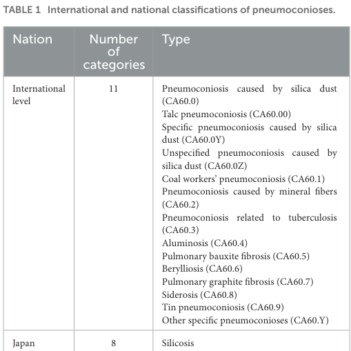
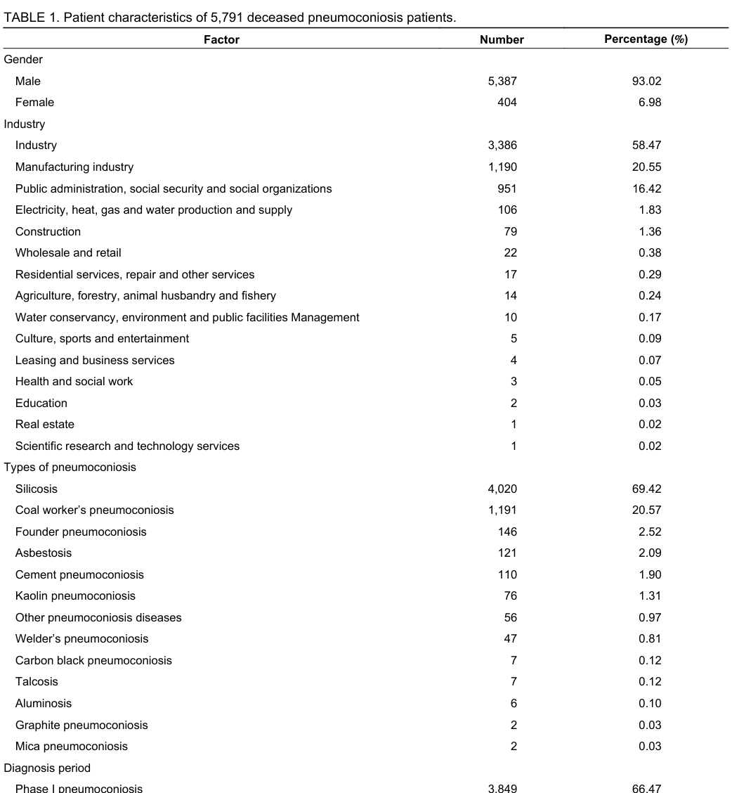
Reference Validation
Checked with linkml-reference-validator 0.3.0.
| Outcome | Count |
|---|---|
| References checked | 8 |
| Resolved | 7 |
| Unresolved (possible confabulation) | 1 |
| Unverifiable | 0 |
| References weighed for topical relevance | 7 |
| On topic | 1 |
| Off topic | 0 |
Unresolved references
These identifiers did not resolve to a record and may be fabricated. A lookup that failed for transport reasons is indistinguishable from one that failed because the record does not exist, so spot-check before acting on them:
DOI:10.46234/ccdcw2024.280(8 mentions) - Identifier did not resolve to a record