Cerebral Amyloid Angiopathy
Cerebral amyloid angiopathy (CAA) is an age-related cerebral small-vessel disease most commonly characterized by deposition of amyloid-beta (Abeta) in the walls of cortical and leptomeningeal small arteries, arterioles, and capillaries. Progressive vascular amyloid deposition with smooth muscle cell loss and vessel-wall fragility predisposes to spontaneous strictly lobar intracerebral hemorrhage (often recurrent), strictly lobar cerebral microbleeds, convexity subarachnoid hemorrhage, cortical superficial siderosis, transient focal neurological episodes ("amyloid spells"), and progressive cognitive decline. Non-hemorrhagic markers include white matter hyperintensities and enlarged centrum semiovale perivascular spaces. The common sporadic form increases in prevalence with age and is strongly associated with the APOE genotype (ε4 increases risk; ε2 is associated with vessel fragility/hemorrhage), while rare hereditary forms are caused by mutations in APP (e.g. Dutch-type HCHWA-D, APP p.E693Q) and in non-Abeta genes such as CST3 (cystatin C / Icelandic-type ACys). A rare iatrogenic form arises from prion-like Abeta "seeding" after exposure to cadaveric tissue (dura mater grafts, pituitary-derived growth hormone) decades earlier. Diagnosis in life relies on the Boston criteria v2.0 (clinical and MRI markers); definitive diagnosis requires histopathology. There is no targeted disease-modifying therapy; management is supportive and centered on hemorrhage-risk mitigation.
Ask OpenScientist
Ask a research question about Cerebral Amyloid Angiopathy. OpenScientist will conduct autonomous deep research using the Disorder Mechanisms Knowledge Base and PubMed literature (typically 10-30 minutes).
Do not include personal health information in your question. Questions and results are cached in your browser's local storage.
Subtypes
10Show evidence (1 reference)
Show evidence (3 references)
Show evidence (1 reference)
Show evidence (1 reference)
Show evidence (3 references)
Show evidence (3 references)
Show evidence (3 references)
Show evidence (4 references)
Show evidence (4 references)
Pathophysiology
14Show evidence (1 reference)
Show evidence (3 references)
Show evidence (1 reference)
Show evidence (2 references)
Show evidence (2 references)
Show evidence (1 reference)
Show evidence (2 references)
Show evidence (1 reference)
Show evidence (1 reference)
Show evidence (2 references)
Show evidence (1 reference)
Show evidence (2 references)
Show evidence (1 reference)
Pathograph
Phenotypes
14Blood 4
Show evidence (1 reference)
Show evidence (1 reference)
Show evidence (1 reference)
Show evidence (1 reference)
Cardiovascular 1
Show evidence (1 reference)
Nervous System 8
Show evidence (2 references)
Show evidence (1 reference)
Show evidence (1 reference)
Show evidence (2 references)
Show evidence (1 reference)
Show evidence (1 reference)
Show evidence (1 reference)
Show evidence (1 reference)
Other 1
Show evidence (2 references)
Genetic Associations
3Show evidence (2 references)
Show evidence (4 references)
Show evidence (2 references)
Medical Actions
14Show evidence (1 reference)
Show evidence (2 references)
Show evidence (2 references)
Show evidence (2 references)
Show evidence (1 reference)
Show evidence (2 references)
Show evidence (4 references)
Show evidence (2 references)
Show evidence (6 references)
Show evidence (4 references)
Show evidence (3 references)
Environmental Factors
2Show evidence (2 references)
Show evidence (1 reference)
Biochemical Markers
2Show evidence (2 references)
Show evidence (2 references)
Diagnosis
10Show evidence (1 reference)
Show evidence (3 references)
Show evidence (1 reference)
Show evidence (2 references)
Show evidence (1 reference)
Show evidence (3 references)
Show evidence (3 references)
Show evidence (3 references)
Show evidence (1 reference)
Progression
6Show evidence (2 references)
Show evidence (2 references)
Show evidence (2 references)
Show evidence (2 references)
Show evidence (1 reference)
Prevalence
2Show evidence (1 reference)
Show evidence (1 reference)
Epidemiology
1Show evidence (1 reference)
Related Datasets
4Show evidence (3 references)
Show evidence (1 reference)
Show evidence (2 references)
Show evidence (2 references)
Clinical Trials
8Show evidence (1 reference)
Show evidence (1 reference)
Show evidence (1 reference)
Show evidence (1 reference)
Show evidence (1 reference)
Show evidence (4 references)
Show evidence (4 references)
Show evidence (3 references)
Experimental Models
2Show evidence (2 references)
Show evidence (2 references)
Show evidence (1 reference)
Show evidence (2 references)
Animal Models
1Show evidence (1 reference)
Show evidence (1 reference)
Show evidence (3 references)
Source YAML
click to showname: Cerebral Amyloid Angiopathy
creation_date: "2026-06-03T12:00:00Z"
category: Complex
synonyms:
- CAA
- cerebral beta-amyloid angiopathy
- congophilic angiopathy
- hereditary cerebral hemorrhage with amyloidosis
description: >-
Cerebral amyloid angiopathy (CAA) is an age-related cerebral small-vessel disease most commonly characterized
by deposition of amyloid-beta (Abeta) in the walls of cortical and leptomeningeal small arteries, arterioles,
and capillaries. Progressive vascular amyloid deposition with smooth muscle cell loss and vessel-wall
fragility predisposes to spontaneous strictly lobar intracerebral hemorrhage (often recurrent), strictly
lobar cerebral microbleeds, convexity subarachnoid hemorrhage, cortical superficial siderosis, transient
focal neurological episodes ("amyloid spells"), and progressive cognitive decline. Non-hemorrhagic markers
include white matter hyperintensities and enlarged centrum semiovale perivascular spaces. The common
sporadic form increases in prevalence with age and is strongly associated with the APOE genotype (ε4
increases risk; ε2 is associated with vessel fragility/hemorrhage), while rare hereditary forms are
caused by mutations in APP (e.g. Dutch-type HCHWA-D, APP p.E693Q) and in non-Abeta genes such as CST3
(cystatin C / Icelandic-type ACys). A rare iatrogenic form arises from prion-like Abeta "seeding" after
exposure to cadaveric tissue (dura mater grafts, pituitary-derived growth hormone) decades earlier.
Diagnosis in life relies on the Boston criteria v2.0 (clinical and MRI markers); definitive diagnosis
requires histopathology. There is no targeted disease-modifying therapy; management is supportive and
centered on hemorrhage-risk mitigation.
disease_term:
preferred_term: Cerebral amyloid angiopathy
term:
id: MONDO:0005620
label: cerebral amyloid angiopathy
parents:
- cerebrovascular disorder
- amyloidosis
references:
- reference: PMID:40721902
title: >-
Diagnosis and management of cerebral amyloid angiopathy: a scientific
statement from the International CAA Association and the World Stroke
Organization.
- reference: PMID:37236210
title: "Progression of cerebral amyloid angiopathy: a pathophysiological framework."
- reference: PMID:37280119
title: "Clinical considerations in early-onset cerebral amyloid angiopathy."
- reference: url:https://pub.dzne.de/record/281348/files/DZNE-2025-01095.pdf
title: https://pub.dzne.de/record/281348/files/DZNE-2025-01095.pdf
- reference: url:https://discovery.ucl.ac.uk/id/eprint/10173198/1/Werring_Pathophysiological%20framework%20for%20CAA%20-%20FINAL.pdf
title: https://discovery.ucl.ac.uk/id/eprint/10173198/1/Werring_Pathophysiological%20framework%20for%20CAA%20-%20FINAL.pdf
- reference: PMID:31152566
title: Different microvascular alterations underlie microbleeds and microinfarcts.
- reference: PMID:36267331
title: Blood-brain barrier leakage and perivascular inflammation in cerebral amyloid angiopathy.
- reference: PMID:31810839
title: Vasomotion as a Driving Force for Paravascular Clearance in the Awake Mouse Brain.
- reference: PMID:39054254
title: 'The historical background of hereditary cystatin C amyloid angiopathy: Genealogical, pathological, and clinical manifestations.'
has_subtypes:
- name: Sporadic
display_name: Sporadic amyloid-beta CAA
description: >-
The commonest form of CAA, an age-related amyloid-beta cerebral small-vessel
disease that usually affects people in mid- to later life. APOE genotype is
the most significant common genetic risk factor.
inheritance:
- name: Multifactorial / sporadic
evidence:
- reference: PMID:37280119
reference_title: "Clinical considerations in early-onset cerebral amyloid angiopathy."
supports: SUPPORT
evidence_source: HUMAN_CLINICAL
snippet: "The commonest form, sporadic amyloid-β CAA, usually affects people in mid- to later life."
explanation: Defines sporadic amyloid-beta CAA as the commonest, later-life form.
- name: Dutch-type
display_name: Dutch-type hereditary CAA (HCHWA-D, APP p.E693Q)
description: >-
Autosomal dominant hereditary CAA caused by the APP E693Q (Dutch) amino acid
substitution. It is considered a "pure" form of CAA with minimal
Alzheimer-type plaques and tangles, with early-onset recurrent lobar
hemorrhage.
subtype_term:
preferred_term: Dutch-type hereditary CAA
term:
id: MONDO:0015033
label: ABeta amyloidosis, dutch type
inheritance:
- name: Autosomal dominant
inheritance_term:
preferred_term: Autosomal dominant inheritance
term:
id: HP:0000006
label: Autosomal dominant inheritance
genes:
- preferred_term: APP
term:
id: hgnc:620
label: APP
evidence:
- reference: PMID:36324420
reference_title: 'Main features of hereditary cerebral amyloid angiopathies: A systematic review.'
supports: SUPPORT
evidence_source: HUMAN_CLINICAL
quote_role: REVIEW_SYNTHESIS
snippet: >-
HCHWA-Dutch type is a rare autosomal dominant disorder caused by a mutation at codon 693 of the
APP gene (E693Q), resulting in the substitution of a glutamine for a glutamic acid at position 22
of the Aβ sequence
explanation: >-
Identifies the Dutch APP E693Q substitution and autosomal dominant inheritance directly.
- reference: PMID:37280119
reference_title: Clinical considerations in early-onset cerebral amyloid angiopathy.
supports: SUPPORT
evidence_source: HUMAN_CLINICAL
quote_role: REVIEW_SYNTHESIS
snippet: >-
D-CAA classically presents with recurrent ICH,90,91 although migraine with aura can also be a presenting
feature,50 as noted in some of the earliest case descriptions.51
explanation: >-
Supports the characteristic recurrent hemorrhage and migraine presentation.
- reference: PMID:37280119
reference_title: Clinical considerations in early-onset cerebral amyloid angiopathy.
supports: SUPPORT
evidence_source: HUMAN_CLINICAL
quote_role: REVIEW_SYNTHESIS
snippet: >-
Pathologically, there is severe CAA with a relative paucity of neuritic plaques and neurofibrillary
tangles rarely seen, although diffuse amyloid-β deposits may be found.94,95
explanation: >-
Qualifies the relative paucity of Alzheimer-type neuritic plaques and tangles.
- name: ACys
display_name: Icelandic-type hereditary CAA (HCHWA-I, CST3 / cystatin C)
description: >-
Hereditary non-amyloid-beta CAA caused by mutation in CST3 (cystatin C),
historically termed hereditary cerebral hemorrhage with amyloidosis,
Icelandic type (HCHWA-I), in which cystatin C amyloid deposits in cerebral
vessels.
subtype_term:
preferred_term: Icelandic-type hereditary cerebral amyloid angiopathy
term:
id: MONDO:0007098
label: ACys amyloidosis
inheritance:
- name: Autosomal dominant
inheritance_term:
preferred_term: Autosomal dominant inheritance
term:
id: HP:0000006
label: Autosomal dominant inheritance
review_notes: >-
ACys_Amyloidosis is the canonical standalone entry for CST3-related amyloidosis. This umbrella CAA
entry retains a concise comparative representation of its vascular cystatin C mechanisms, investigational
NAC treatment and trial, and HEK293T model so readers can distinguish protein-specific evidence from
amyloid-beta CAA. These shared findings describe the same disease and are not independent evidence
or a second disease entity. Systemic manifestations and comprehensive ACys-specific management belong
in the standalone entry.
genes:
- preferred_term: CST3
term:
id: hgnc:2475
label: CST3
evidence:
- reference: PMID:37280119
reference_title: "Clinical considerations in early-onset cerebral amyloid angiopathy."
supports: SUPPORT
evidence_source: HUMAN_CLINICAL
snippet: "non-amyloid-β CAA (associated with ITM2B, CST3, GSN, PRNP and TTR mutations)"
explanation: Supports CST3 as a monogenic cause of non-amyloid-beta hereditary CAA.
curated_in: ACys_Amyloidosis
- name: Iatrogenic
display_name: Iatrogenic CAA (acquired Abeta seeding)
description: >-
A rare, increasingly recognized acquired form attributed to prion-like
Abeta "seeding" after medical exposure to contaminated cadaveric tissue
(dura mater grafts/Lyodura, pituitary-derived human growth hormone,
neurosurgical instrumentation), typically presenting decades after exposure.
evidence:
- reference: PMID:37236210
reference_title: "Progression of cerebral amyloid angiopathy: a pathophysiological framework."
supports: SUPPORT
evidence_source: HUMAN_CLINICAL
snippet: "individuals with hereditary, sporadic, and iatrogenic forms of cerebral amyloid angiopathy"
explanation: Establishes iatrogenic CAA as a recognized form alongside hereditary and sporadic CAA.
- name: Italian-type
description: >-
APP E693K (amyloid-beta E22K) hereditary CAA, associated with recurrent lobar hemorrhage and cognitive
impairment; headache and seizures can occur. This form is distinct from the APP A713T variant also
sometimes called Italian.
display_name: Italian-type hereditary CAA
subtype_term:
preferred_term: ABeta amyloidosis, Italian type
term:
id: MONDO:0017947
label: ABeta amyloidosis, Italian type
inheritance:
- name: Autosomal dominant
inheritance_term:
preferred_term: Autosomal dominant inheritance
term:
id: HP:0000006
label: Autosomal dominant inheritance
genes:
- preferred_term: APP
term:
id: hgnc:620
label: APP
evidence:
- reference: PMID:36324420
reference_title: 'Main features of hereditary cerebral amyloid angiopathies: A systematic review.'
supports: SUPPORT
evidence_source: HUMAN_CLINICAL
quote_role: REVIEW_SYNTHESIS
snippet: >-
The Hereditary Cerebral Hemorrhage with Amyloidosis Italian (HCHWA-Italian) type is caused by an
APP mutation at codon 693 (E693K), resulting in substitution of a glutamic acid for a lysine at
residue 22 of Aβ.
explanation: >-
The review describes this hereditary CAA phenotype.
- reference: PMID:36324420
reference_title: 'Main features of hereditary cerebral amyloid angiopathies: A systematic review.'
supports: SUPPORT
evidence_source: HUMAN_CLINICAL
quote_role: REVIEW_SYNTHESIS
snippet: >-
It was identified in several members of three unrelated kindreds from Italy, who presented in the
sixth and seventh decade of life with progressive, predominantly amnestic, cognitive impairment
associated with recurrent lobar hemorrhages.
explanation: >-
The review describes this hereditary CAA phenotype.
- reference: PMID:36324420
reference_title: 'Main features of hereditary cerebral amyloid angiopathies: A systematic review.'
supports: SUPPORT
evidence_source: HUMAN_CLINICAL
quote_role: REVIEW_SYNTHESIS
snippet: >-
These familial CAA forms are caused by point mutation in the APP gene (Fig. 1), and generally are
inherited in an autosomal dominant pattern.
explanation: >-
The review establishes the shared APP point-mutation inheritance pattern; the adjacent evidence
supplies the variant-specific phenotype.
- name: Flemish-type
description: >-
APP A692G (amyloid-beta A21G) hereditary CAA, with dementia, lobar intracerebral hemorrhage or both,
often accompanied by prominent white-matter abnormalities. Vascular amyloid and Alzheimer-type parenchymal
pathology coexist.
display_name: Flemish-type hereditary CAA
subtype_term:
preferred_term: ABetaA21G amyloidosis
term:
id: MONDO:0017948
label: ABetaA21G amyloidosis
inheritance:
- name: Autosomal dominant
inheritance_term:
preferred_term: Autosomal dominant inheritance
term:
id: HP:0000006
label: Autosomal dominant inheritance
genes:
- preferred_term: APP
term:
id: hgnc:620
label: APP
evidence:
- reference: PMID:36324420
reference_title: 'Main features of hereditary cerebral amyloid angiopathies: A systematic review.'
supports: SUPPORT
evidence_source: HUMAN_CLINICAL
quote_role: REVIEW_SYNTHESIS
snippet: >-
The Flemish APP mutation (A692G) is located at codon 692 of the APP protein, and results in substitution
of an alanine by a glycine at residue 21 of the Aβ sequence.
explanation: >-
The review describes this hereditary CAA phenotype.
- reference: PMID:37280119
reference_title: Clinical considerations in early-onset cerebral amyloid angiopathy.
supports: SUPPORT
evidence_source: HUMAN_CLINICAL
quote_role: REVIEW_SYNTHESIS
snippet: >-
Flemish | c.2075C>G (p.Ala692Gly) | A21G | + | + | + | ICH, dementia or both.
explanation: >-
The genotype-specific table supports mixed hemorrhagic and cognitive presentation.
- reference: PMID:36324420
reference_title: 'Main features of hereditary cerebral amyloid angiopathies: A systematic review.'
supports: SUPPORT
evidence_source: HUMAN_CLINICAL
quote_role: REVIEW_SYNTHESIS
snippet: >-
These familial CAA forms are caused by point mutation in the APP gene (Fig. 1), and generally are
inherited in an autosomal dominant pattern.
explanation: >-
The review establishes the shared APP point-mutation inheritance pattern; the adjacent evidence
supplies the variant-specific phenotype.
- name: Iowa-type
description: >-
APP D694N (amyloid-beta D23N) hereditary CAA with variable dementia and hemorrhagic presentation.
Expressive language and personality changes, myoclonus and gait abnormalities are reported. Severe
vascular amyloid can coexist with plaques and neurofibrillary tangles.
display_name: Iowa-type hereditary CAA
subtype_term:
preferred_term: ABeta amyloidosis, Iowa type
term:
id: MONDO:0017946
label: ABeta amyloidosis, Iowa type
inheritance:
- name: Autosomal dominant
inheritance_term:
preferred_term: Autosomal dominant inheritance
term:
id: HP:0000006
label: Autosomal dominant inheritance
genes:
- preferred_term: APP
term:
id: hgnc:620
label: APP
evidence:
- reference: PMID:36324420
reference_title: 'Main features of hereditary cerebral amyloid angiopathies: A systematic review.'
supports: SUPPORT
evidence_source: HUMAN_CLINICAL
quote_role: REVIEW_SYNTHESIS
snippet: >-
The Iowa mutation at codon 694 within the Aβ region of APP results in substitution of an asparagine
for aspartic acid (D694N) at Aβ position 23
explanation: >-
The review describes this hereditary CAA phenotype.
- reference: PMID:37280119
reference_title: Clinical considerations in early-onset cerebral amyloid angiopathy.
supports: SUPPORT
evidence_source: HUMAN_CLINICAL
quote_role: REVIEW_SYNTHESIS
snippet: >-
Iowa | c.2080G>A (p.Asp694Asn) | D23N | + | + | + | ICH, dementia or both. Early 50s (range 38–67).68,77-82
| + | + | Seizures, expressive language dysfunction, personality change, myoclonus, gait abnormalities.
explanation: >-
The genotype-specific table supports the variable hemorrhagic, cognitive and neurological spectrum.
- reference: PMID:36324420
reference_title: 'Main features of hereditary cerebral amyloid angiopathies: A systematic review.'
supports: SUPPORT
evidence_source: HUMAN_CLINICAL
quote_role: REVIEW_SYNTHESIS
snippet: >-
These familial CAA forms are caused by point mutation in the APP gene (Fig. 1), and generally are
inherited in an autosomal dominant pattern.
explanation: >-
The review establishes the shared APP point-mutation inheritance pattern; the adjacent evidence
supplies the variant-specific phenotype.
- name: Piedmont-type
description: >-
APP L705V (amyloid-beta L34V) hereditary CAA, typically presenting with recurrent lobar hemorrhage.
Reported pathological samples show severe vascular amyloid without appreciable parenchymal amyloid
plaques or tau pathology; cognitive symptoms can follow repeated hemorrhage.
display_name: Piedmont-type hereditary CAA
subtype_term:
preferred_term: ABetaL34V amyloidosis
term:
id: MONDO:0017945
label: ABetaL34V amyloidosis
inheritance:
- name: Autosomal dominant
inheritance_term:
preferred_term: Autosomal dominant inheritance
term:
id: HP:0000006
label: Autosomal dominant inheritance
genes:
- preferred_term: APP
term:
id: hgnc:620
label: APP
evidence:
- reference: PMID:36324420
reference_title: 'Main features of hereditary cerebral amyloid angiopathies: A systematic review.'
supports: SUPPORT
evidence_source: HUMAN_CLINICAL
quote_role: REVIEW_SYNTHESIS
snippet: >-
The identified mutation (L705V) lies within the Aβ region of APP, but it is located further away
from the previously identified cluster of mutations in the 21 to 23 positions. This G to C transversion
in APP codon 705 leads to a valine for leucine substitution at Aβ residue 34.
explanation: >-
The review describes this hereditary CAA phenotype.
- reference: PMID:37280119
reference_title: Clinical considerations in early-onset cerebral amyloid angiopathy.
supports: SUPPORT
evidence_source: HUMAN_CLINICAL
quote_role: REVIEW_SYNTHESIS
snippet: >-
Piedmont | c.2113C>G (p.Leu705Val) | L34V | + | − | − | Recurrent ICH (range 45–72).84,85
explanation: >-
Names the genotype and hemorrhagic phenotype.
- reference: PMID:36324420
reference_title: 'Main features of hereditary cerebral amyloid angiopathies: A systematic review.'
supports: SUPPORT
evidence_source: HUMAN_CLINICAL
quote_role: REVIEW_SYNTHESIS
snippet: >-
Only one of the individuals described in the literature displayed evidence of cognitive impairment,
and only after having suffered at least four separate lobar hemorrhagic strokes
explanation: >-
The review limits reported cognitive impairment to a patient with repeated hemorrhages.
- reference: PMID:36324420
reference_title: 'Main features of hereditary cerebral amyloid angiopathies: A systematic review.'
supports: SUPPORT
evidence_source: HUMAN_CLINICAL
quote_role: REVIEW_SYNTHESIS
snippet: >-
These familial CAA forms are caused by point mutation in the APP gene (Fig. 1), and generally are
inherited in an autosomal dominant pattern.
explanation: >-
The review establishes the shared APP point-mutation inheritance pattern; the adjacent evidence
supplies the variant-specific phenotype.
- name: Arctic-type
description: >-
APP E693G (amyloid-beta E22G) hereditary cerebral amyloidosis with severe CAA, progressive predominantly
amnestic cognitive impairment and sometimes psychiatric manifestations. The cited series did not report
symptomatic hemorrhagic stroke; severe CAA pathology does not imply the Dutch-type hemorrhagic course.
display_name: Arctic-type hereditary CAA
subtype_term:
preferred_term: ABeta amyloidosis, Arctic type
term:
id: MONDO:0017949
label: ABeta amyloidosis, Arctic type
inheritance:
- name: Autosomal dominant
inheritance_term:
preferred_term: Autosomal dominant inheritance
term:
id: HP:0000006
label: Autosomal dominant inheritance
genes:
- preferred_term: APP
term:
id: hgnc:620
label: APP
evidence:
- reference: PMID:37280119
reference_title: Clinical considerations in early-onset cerebral amyloid angiopathy.
supports: SUPPORT
evidence_source: HUMAN_CLINICAL
quote_role: REVIEW_SYNTHESIS
snippet: >-
Arctic | c.2078A>G (p.Glu693Gly) | E22G | + | + | + | Memory-led cognitive impairment
explanation: >-
The table verifies the amino-acid substitution and cognitive phenotype; it corrects the inaccurate
amino-acid expansion in the older review.
- reference: PMID:37280119
reference_title: Clinical considerations in early-onset cerebral amyloid angiopathy.
supports: SUPPORT
evidence_source: HUMAN_CLINICAL
quote_role: REVIEW_SYNTHESIS
snippet: >-
The Arctic mutation causes progressive amnestic cognitive impairment.71,72 Pathologically, there
is severe CAA, together with distinctive amyloid-β plaques (lacking a dense core and described as
‘ring-like’ or ‘targetoid’) and neurofibrillary tangles.73,100,101
explanation: >-
Supports severe vascular pathology with accompanying parenchymal and tau pathology.
- reference: PMID:36324420
reference_title: 'Main features of hereditary cerebral amyloid angiopathies: A systematic review.'
supports: SUPPORT
evidence_source: HUMAN_CLINICAL
quote_role: REVIEW_SYNTHESIS
snippet: >-
Although clinical history was notable for no report of ischemic or hemorrhagic strokes, in the two
autopsy cases of Arctic mutations carriers were notable for severe vascular amyloid deposition in
leptomeningeal and cortical vessels, as well as numerous microinfarcts. Interestingly, no neuroimaging
or pathological evidence of amyloid-related hemorrhagic lesions were identified.
explanation: >-
Records the absence of observed hemorrhagic manifestations in the reported families, not universal
protection.
- reference: PMID:36324420
reference_title: 'Main features of hereditary cerebral amyloid angiopathies: A systematic review.'
supports: SUPPORT
evidence_source: HUMAN_CLINICAL
quote_role: REVIEW_SYNTHESIS
snippet: >-
These familial CAA forms are caused by point mutation in the APP gene (Fig. 1), and generally are
inherited in an autosomal dominant pattern.
explanation: >-
The review establishes the shared APP point-mutation inheritance pattern; the adjacent evidence
supplies the variant-specific phenotype.
- name: CAA-related inflammation
description: >-
Inflammatory complication of amyloid-beta CAA with perivascular inflammatory infiltrates or pathologically
demonstrated angio-destructive amyloid-beta-related angiitis (ABRA). Presentation may include subacute
cognitive change, encephalopathy, headache, seizures and asymmetric white-matter lesions. ABRA is
a histological descriptor within this inflammatory spectrum.
evidence:
- reference: url:https://pub.dzne.de/record/281348/files/DZNE-2025-01095.pdf
reference_title: https://pub.dzne.de/record/281348/files/DZNE-2025-01095.pdf
supports: SUPPORT
evidence_source: OTHER
quote_role: REVIEW_SYNTHESIS
snippet: >-
The term CAA-related inflammation (CAA-ri) is preferred for all cases, with the terms amyloid- beta
related angiitis (ABRA) or CAA-related vasculitis reserved (as an additional descriptor) only where
there is clear pathological evidence of an angio-destructive vasculitis.
explanation: >-
The guideline specifies the inflammatory umbrella term and reserves ABRA for destructive vasculitis.
- reference: PMID:40566003
reference_title: Clinical Management of Cerebral Amyloid Angiopathy.
supports: SUPPORT
evidence_source: HUMAN_CLINICAL
quote_role: REVIEW_SYNTHESIS
snippet: >-
CAA-ri presents with a variety of clinical manifestations, including cognitive decline as the most
prevalent, focal neurological deficits, encephalopathy, headache, epileptic seizures and psychiatric
symptoms [16].
explanation: >-
Summarizes the inflammatory clinical presentation.
pathophysiology:
- name: Impaired perivascular amyloid-beta clearance
description: >-
Impaired drainage of soluble amyloid-beta along perivascular pathways is a proposed contributor to
its vascular accumulation. Vascular dysfunction may further reduce clearance, creating a feedback
loop; the initiating factors in sporadic CAA remain incompletely understood.
biological_processes:
- preferred_term: amyloid-beta clearance
term:
id: GO:0097242
label: amyloid-beta clearance
modifier: DECREASED
downstream:
- target: Vascular amyloid-beta deposition
description: >-
Reduced perivascular removal can favor vascular amyloid-beta accumulation; the precise initiation
of sporadic deposits remains uncertain.
causal_link_type: INDIRECT_UNKNOWN_INTERMEDIATES
evidence:
- reference: PMID:40566003
reference_title: Clinical Management of Cerebral Amyloid Angiopathy.
supports: SUPPORT
evidence_source: HUMAN_CLINICAL
quote_role: REVIEW_SYNTHESIS
snippet: >-
Key-role in the evolution of CAA plays the impairment of the perivascular drainage system, leading
to an aging brain due to a progressively increasing Aβ entrapment and deposition in the walls
of small arteries [17,20].
explanation: >-
The review describes failed perivascular drainage as a proposed contributor to vascular amyloid
deposition.
- target: Enlarged centrum semiovale perivascular spaces
description: >-
CAA-related impaired perivascular fluid drainage is a proposed explanation for enlarged centrum
semiovale perivascular spaces; the spatial association does not prove causation.
causal_link_type: INDIRECT_UNKNOWN_INTERMEDIATES
evidence:
- reference: url:https://discovery.ucl.ac.uk/id/eprint/10173198/1/Werring_Pathophysiological%20framework%20for%20CAA%20-%20FINAL.pdf
reference_title: https://discovery.ucl.ac.uk/id/eprint/10173198/1/Werring_Pathophysiological%20framework%20for%20CAA%20-%20FINAL.pdf
supports: SUPPORT
evidence_source: HUMAN_CLINICAL
quote_role: REVIEW_SYNTHESIS
snippet: "The observed proximity of enlarged CSO-PVS to overlying CAA in individual arterioles63 raises the intriguing possibility that perivascular space enlargement may be a direct result of CAA- related impairment in perivascular fluid clearance."
explanation: >-
The framework explicitly proposes this link as a possibility.
evidence:
- reference: PMID:40566003
reference_title: Clinical Management of Cerebral Amyloid Angiopathy.
supports: SUPPORT
evidence_source: HUMAN_CLINICAL
quote_role: REVIEW_SYNTHESIS
snippet: >-
Key-role in the evolution of CAA plays the impairment of the perivascular drainage system, leading
to an aging brain due to a progressively increasing Aβ entrapment and deposition in the walls of
small arteries [17,20].
explanation: >-
The review describes failed perivascular drainage as a proposed contributor to vascular amyloid
deposition.
- name: Vascular amyloid-beta deposition
description: >-
Amyloid-beta accumulates in cortical and leptomeningeal vessel walls, principally arterioles and less
often capillaries and venules. This is deposition of an APP-derived peptide, not necessarily increased
peptide synthesis; substantial overproduction is not established for sporadic or Dutch-type disease.
genes:
- preferred_term: APP
term:
id: hgnc:620
label: APP
cell_types:
- preferred_term: smooth muscle cell of the brain vasculature
term:
id: CL:0002590
label: smooth muscle cell of the brain vasculature
downstream:
- target: Cerebral vascular smooth muscle cell degeneration
description: >-
Amyloid deposition is associated with loss of vascular smooth muscle cells; tissue studies and experimental
models support this step.
causal_link_type: INDIRECT_UNKNOWN_INTERMEDIATES
evidence:
- reference: PMID:36267331
reference_title: Blood-brain barrier leakage and perivascular inflammation in cerebral amyloid angiopathy.
supports: SUPPORT
evidence_source: HUMAN_CLINICAL
quote_role: PRIMARY_RESULT
snippet: >-
We observed overall lower SMA coverage with increasing vessel grade, starting at Grade 1
explanation: >-
Human autopsy vessels show reduced smooth muscle actin coverage even with early patchy amyloid.
This is a cross-sectional observation, not a longitudinal experiment.
- target: Blood-brain barrier leakage
description: >-
Barrier leakage is observed already in mildly amyloid-positive vessels, although the molecular steps
are unresolved.
causal_link_type: INDIRECT_UNKNOWN_INTERMEDIATES
evidence:
- reference: PMID:36267331
reference_title: Blood-brain barrier leakage and perivascular inflammation in cerebral amyloid angiopathy.
supports: SUPPORT
evidence_source: HUMAN_CLINICAL
quote_role: PRIMARY_RESULT
snippet: >-
Blood-brain barrier leakage and smooth muscle actin loss were observed in significantly more vessels
with mild amyloid-β deposition (Grade 1 vessels; P = 0.044 and P = 0.012, respectively) as compared
to vessels with no amyloid-β (Grade 0), and blood-brain barrier leakage was observed in 100% of
vessels with evidence of vessel remodelling (Grades 3 and 4).
explanation: >-
Shows barrier leakage across grades of amyloid-associated vessel pathology in seven autopsy cases;
timing is inferred cross-sectionally.
- target: CAA-related immune inflammation
description: >-
In a subset of patients, an immune response develops against cerebrovascular amyloid-beta; the trigger
is uncertain.
causal_link_type: INDIRECT_UNKNOWN_INTERMEDIATES
evidence:
- reference: PMID:40566003
reference_title: Clinical Management of Cerebral Amyloid Angiopathy.
supports: SUPPORT
evidence_source: HUMAN_CLINICAL
quote_role: REVIEW_SYNTHESIS
snippet: >-
The most prevalent hypothesis involves an inflammatory autoimmune response against cerebrovascular
Aβ-amyloid deposits [180,182].
explanation: >-
The review explicitly labels the response-to-amyloid mechanism as a hypothesis.
evidence:
- reference: PMID:40566003
reference_title: Clinical Management of Cerebral Amyloid Angiopathy.
supports: SUPPORT
evidence_source: HUMAN_CLINICAL
quote_role: REVIEW_SYNTHESIS
snippet: >-
In CAA, amyloid-β accumulation is detected primarily in the neocortical and leptomeningeal arterioles,
and to a lesser extent in capillaries and venules [17,18].
explanation: >-
Defines the vascular distribution of the amyloid-beta lesion.
- reference: PMID:36324420
reference_title: 'Main features of hereditary cerebral amyloid angiopathies: A systematic review.'
supports: SUPPORT
evidence_source: HUMAN_CLINICAL
quote_role: REVIEW_SYNTHESIS
snippet: >-
Sporadic CAA is caused by deposition of amyloid-β (Aβ), originating from proteolytic cleavage of
the Amyloid Precursor Protein (APP), within the walls of cerebral small caliber vessels.
explanation: >-
Establishes APP as the precursor of the deposited peptide.
- reference: url:https://discovery.ucl.ac.uk/id/eprint/10173198/1/Werring_Pathophysiological%20framework%20for%20CAA%20-%20FINAL.pdf
reference_title: https://discovery.ucl.ac.uk/id/eprint/10173198/1/Werring_Pathophysiological%20framework%20for%20CAA%20-%20FINAL.pdf
supports: SUPPORT
evidence_source: HUMAN_CLINICAL
quote_role: REVIEW_SYNTHESIS
snippet: >-
There is no evidence for substantial A overproduction in sporadic or Dutch-type hereditary CAA,23
suggesting that A deposition may instead reflect increased aggregation or impaired clearance.
explanation: >-
Prevents conflating deposition with overproduction in sporadic and Dutch-type CAA.
- name: Cerebral vascular smooth muscle cell degeneration
description: >-
Contractile smooth muscle cell coverage is reduced in affected cerebral arterioles. Amyloid-beta and
cystatin C angiopathies can converge on this loss of the mural cell layer.
cell_types:
- preferred_term: smooth muscle cell of the brain vasculature
term:
id: CL:0002590
label: smooth muscle cell of the brain vasculature
modifier: DECREASED
downstream:
- target: Impaired cerebrovascular reactivity
description: >-
Loss of contractile vascular smooth muscle is a proposed basis for impaired physiological reactivity.
causal_link_type: INDIRECT_UNKNOWN_INTERMEDIATES
evidence:
- reference: url:https://discovery.ucl.ac.uk/id/eprint/10173198/1/Werring_Pathophysiological%20framework%20for%20CAA%20-%20FINAL.pdf
reference_title: https://discovery.ucl.ac.uk/id/eprint/10173198/1/Werring_Pathophysiological%20framework%20for%20CAA%20-%20FINAL.pdf
supports: SUPPORT
evidence_source: MODEL_ORGANISM
quote_role: REVIEW_SYNTHESIS
snippet: >-
The reduction in vascular reactivity in these mice appeared to correlate more closely to loss
of vascular smooth muscle cells than CAA burden, suggesting that CAA-related loss of vascular
smooth muscle may be the direct mechanism for alterations in cerebrovascular physiology.
explanation: >-
The framework synthesizes mouse evidence linking loss of smooth muscle to reduced reactivity.
- target: Cerebral vessel-wall remodeling
description: >-
Smooth-muscle depletion accompanies later vessel-wall remodeling, but it is not sufficient by itself
to establish a bleeding mechanism.
causal_link_type: INDIRECT_UNKNOWN_INTERMEDIATES
evidence:
- reference: PMID:31152566
reference_title: Different microvascular alterations underlie microbleeds and microinfarcts.
supports: SUPPORT
evidence_source: HUMAN_CLINICAL
quote_role: PRIMARY_RESULT
snippet: >-
Collectively, these data suggest that extensive build-up of Aβ in the walls of arterioles alone
is not enough for the vessel to become fragile and rupture, but that it must undergo remodeling
as well.
explanation: >-
The authors distinguish amyloid accumulation from the additional remodeling associated with rupture.
evidence:
- reference: PMID:36267331
reference_title: Blood-brain barrier leakage and perivascular inflammation in cerebral amyloid angiopathy.
supports: SUPPORT
evidence_source: HUMAN_CLINICAL
quote_role: PRIMARY_RESULT
snippet: >-
We observed overall lower SMA coverage with increasing vessel grade, starting at Grade 1
explanation: >-
Human autopsy vessels show reduced smooth muscle actin coverage even with early patchy amyloid.
This is a cross-sectional observation, not a longitudinal experiment.
- name: Impaired cerebrovascular reactivity
description: >-
Reduced cerebrovascular responses to stimulation are found in sporadic and hereditary amyloid-beta
CAA. Mouse experiments link impaired reactivity to reduced paravascular tracer clearance, supporting
a possible self-reinforcing clearance defect.
downstream:
- target: Impaired perivascular amyloid-beta clearance
description: >-
Impaired evoked vascular reactivity corresponds to slower paravascular tracer clearance in APP/PS1
mice. Extension to amyloid-beta clearance and human CAA is a mechanistic inference; this study did
not directly measure amyloid-beta clearance.
causal_link_type: INDIRECT_UNKNOWN_INTERMEDIATES
evidence:
- reference: PMID:31810839
reference_title: Vasomotion as a Driving Force for Paravascular Clearance in the Awake Mouse Brain.
supports: SUPPORT
evidence_source: MODEL_ORGANISM
quote_role: PRIMARY_RESULT
snippet: >-
Evoked vascular reactivity was impaired in mice with CAA, which corresponded to slower clearance
rates.
explanation: >-
The mouse study directly measured impaired evoked reactivity and slower tracer clearance; it does
not establish a human treatment.
- &id016
reference: PMID:31810839
reference_title: Vasomotion as a Driving Force for Paravascular Clearance in the Awake Mouse Brain.
supports: SUPPORT
evidence_source: MODEL_ORGANISM
quote_role: PRIMARY_RESULT
snippet: >-
Whether our findings also apply to clearance of soluble Aβ remains to be assessed, since we did
not directly measure clearance of Aβ.
explanation: >-
The experiment measures surrogate tracers, not amyloid-beta clearance itself.
- target: Cortical microinfarction
description: >-
Impaired vascular function may cause local hypoperfusion and ischemic injury; direct occlusion was
not demonstrated in the autopsy study.
causal_link_type: INDIRECT_UNKNOWN_INTERMEDIATES
evidence:
- reference: PMID:31152566
reference_title: Different microvascular alterations underlie microbleeds and microinfarcts.
supports: SUPPORT
evidence_source: HUMAN_CLINICAL
quote_role: PRIMARY_RESULT
snippet: >-
These vessels likely predispose to hypoperfusion and/or occlusions. We did not observe individual
small thrombi or emboli in penetrating cortical arterioles that would have provided direct evidence
for occlusion.
explanation: >-
The primary study states the inferred mechanism and the absence of directly observed thrombi or
emboli.
- target: White matter microstructural injury
description: >-
CAA-associated vascular dysfunction is proposed to contribute to white-matter injury, with the detailed
injury mechanisms unresolved.
causal_link_type: INDIRECT_UNKNOWN_INTERMEDIATES
evidence:
- reference: url:https://discovery.ucl.ac.uk/id/eprint/10173198/1/Werring_Pathophysiological%20framework%20for%20CAA%20-%20FINAL.pdf
reference_title: https://discovery.ucl.ac.uk/id/eprint/10173198/1/Werring_Pathophysiological%20framework%20for%20CAA%20-%20FINAL.pdf
supports: SUPPORT
evidence_source: HUMAN_CLINICAL
quote_role: REVIEW_SYNTHESIS
snippet: >-
The time sequence of vascular dysfunction preceding evidence of non-haemorrhagic brain injury
is consistent with findings from a mediation analysis in sporadic CAA patients indicating that
the association between amyloid-PET signal and WMH volume is mediated by changes in BOLD fMRI
response.
explanation: >-
The framework uses temporal and mediation evidence, not proof of a universal causal sequence.
evidence:
- reference: url:https://discovery.ucl.ac.uk/id/eprint/10173198/1/Werring_Pathophysiological%20framework%20for%20CAA%20-%20FINAL.pdf
reference_title: https://discovery.ucl.ac.uk/id/eprint/10173198/1/Werring_Pathophysiological%20framework%20for%20CAA%20-%20FINAL.pdf
supports: SUPPORT
evidence_source: HUMAN_CLINICAL
quote_role: REVIEW_SYNTHESIS
snippet: >-
Impaired cerebrovascular reactivity, measured in humans by transcranial Doppler or blood- oxygen-level
dependent functional MRI (BOLD fMRI) response to visual stimulation, has been identified as a robust
feature of sporadic CAA.
explanation: >-
Summarizes human Doppler and BOLD-fMRI observations.
- reference: PMID:31810839
reference_title: Vasomotion as a Driving Force for Paravascular Clearance in the Awake Mouse Brain.
supports: SUPPORT
evidence_source: MODEL_ORGANISM
quote_role: PRIMARY_RESULT
snippet: >-
Evoked vascular reactivity was impaired in mice with CAA, which corresponded to slower clearance
rates.
explanation: >-
The mouse study directly measured impaired evoked reactivity and slower tracer clearance; it does
not establish a human treatment.
- name: Blood-brain barrier leakage
description: >-
Fibrin(ogen) in vessel walls and surrounding tissue indicates blood-brain barrier leakage. Human autopsy
studies detect leakage in mildly affected vessels as well as remodeled vessels. The observed association
does not establish exact temporal ordering in living patients.
downstream:
- target: Perivascular glial activation
description: >-
Leakage may trigger perivascular inflammation; the proposed ordering is based on cross-sectional
human tissue and remains uncertain.
causal_link_type: INDIRECT_UNKNOWN_INTERMEDIATES
evidence:
- reference: PMID:36267331
reference_title: Blood-brain barrier leakage and perivascular inflammation in cerebral amyloid angiopathy.
supports: SUPPORT
evidence_source: HUMAN_CLINICAL
quote_role: PRIMARY_RESULT
snippet: >-
These results suggest that early BBB leakage in CAA may in fact be a driver of perivascular inflammation.
explanation: >-
The authors present this direction as an inference from their observations.
evidence:
- reference: PMID:36267331
reference_title: Blood-brain barrier leakage and perivascular inflammation in cerebral amyloid angiopathy.
supports: SUPPORT
evidence_source: HUMAN_CLINICAL
quote_role: PRIMARY_RESULT
snippet: >-
Blood-brain barrier leakage and smooth muscle actin loss were observed in significantly more vessels
with mild amyloid-β deposition (Grade 1 vessels; P = 0.044 and P = 0.012, respectively) as compared
to vessels with no amyloid-β (Grade 0), and blood-brain barrier leakage was observed in 100% of
vessels with evidence of vessel remodelling (Grades 3 and 4).
explanation: >-
Shows barrier leakage across grades of amyloid-associated vessel pathology in seven autopsy cases;
timing is inferred cross-sectionally.
- reference: PMID:39039355
reference_title: Differences in the cerebral amyloid angiopathy proteome in Alzheimer's disease and mild cognitive impairment.
supports: SUPPORT
evidence_source: HUMAN_CLINICAL
quote_role: PRIMARY_RESULT
snippet: >-
Key BBB proteins that had robust expression in CAA(−) vessels were significantly decreased in CAA(+)
vessels (Supplementary Fig. 2), including OCLN (MCI: 2.7-fold decrease, p = 2.03 × 10⁻²; AD: not
detected), TJP1 (also known as zona occludens-1; ZO-1; MCI: 2.7-fold decrease, p = 4.45 × 10⁻³;
AD: 1.7-fold decrease, p = 1.76 × 10⁻³), and SLC2A1 (also known as GLUT1; MCI: 2.2-fold, p = 0.059;
AD: 1.7-fold, p = 0.030).
explanation: >-
Paired human vessel proteomics provides a molecular correlate of BBB dysfunction in the MCI/AD cohort,
without proving temporal ordering.
notes: >-
In addition to histological leakage markers, paired human vessel proteomics found reduced tight-junction-associated
proteins in CAA-positive vessels from MCI/AD donors. These postmortem associations do not establish
that protein loss precedes amyloid deposition.
- name: Perivascular glial activation
description: >-
Reactive astrocytes and activated microglia surround amyloid-affected arterioles, especially advanced
remodeled vessels. This local response is distinct from the overt clinical syndrome of CAA-related
inflammation.
cell_types:
- preferred_term: astrocyte
term:
id: CL:0000127
label: astrocyte
- preferred_term: microglial cell
term:
id: CL:0000129
label: microglial cell
downstream:
- target: Cerebral vessel-wall remodeling
description: >-
Local inflammation is a candidate contributor to remodeling; it has not been established as a necessary
driver in every vessel.
causal_link_type: INDIRECT_UNKNOWN_INTERMEDIATES
evidence:
- reference: PMID:36267331
reference_title: Blood-brain barrier leakage and perivascular inflammation in cerebral amyloid angiopathy.
supports: SUPPORT
evidence_source: HUMAN_CLINICAL
quote_role: PRIMARY_RESULT
snippet: >-
Inflammation may not in all cases be an essential driver of the vessel ‘cracking’ form of vascular
remodelling.
explanation: >-
Preserves the explicit limitation on causal necessity.
- reference: PMID:36267331
reference_title: Blood-brain barrier leakage and perivascular inflammation in cerebral amyloid angiopathy.
supports: SUPPORT
evidence_source: HUMAN_CLINICAL
quote_role: PRIMARY_RESULT
snippet: >-
The finding of perivascular inflammation around high-grade vessels suggests that this pathway
may be in part mediated by inflammation and that BBB leakage may be an important trigger for perivascular
inflammation and possible Aβ removal.
explanation: >-
The primary study proposes, rather than proves, inflammation-mediated remodeling.
evidence:
- reference: PMID:36267331
reference_title: Blood-brain barrier leakage and perivascular inflammation in cerebral amyloid angiopathy.
supports: SUPPORT
evidence_source: HUMAN_CLINICAL
quote_role: PRIMARY_RESULT
snippet: >-
Perivascular inflammation in the form of reactive astrocytes and activated microglia was observed
predominantly surrounding arterioles at later stages of vessel pathology (Grades 2-4) and consistently
around vessels with the same morphological features as ruptured vessel segments (Grade 4).
explanation: >-
Identifies the perivascular glial response in sampled human CAA vessels. This does not mean every
such case has clinical CAA-ri.
- name: Cerebral vessel-wall remodeling
description: >-
Affected arterioles can develop wall fragmentation, concentric splitting and fibrinoid-necrotic remodeling.
Human serial sections show that bleeding segments often have little residual amyloid-beta despite
deposits upstream or downstream; this is more specific than a simple relation between total amyloid
burden and bleeding.
downstream:
- target: Cerebral vascular hemorrhage
description: >-
Remodeling is proposed to render affected vessels prone to rupture. Cross-sectional pathology cannot
fully establish whether all remodeling precedes hemorrhage.
causal_link_type: INDIRECT_UNKNOWN_INTERMEDIATES
evidence:
- reference: PMID:31152566
reference_title: Different microvascular alterations underlie microbleeds and microinfarcts.
supports: SUPPORT
evidence_source: HUMAN_CLINICAL
quote_role: PRIMARY_RESULT
snippet: >-
Serial sectioning allowed us to make impactful observations at the single-vessel level. First,
our findings provide evidence for a scenario in which microbleeds happen in unhealthy enlarged
CAA-affected vessels due to Aβ-induced degeneration of SMCs, fibrin(ogen) build-up, extensive
vessel wall remodeling, and loss of Aβ locally.
explanation: >-
The primary study interprets the observed single-vessel changes as a mechanism of microbleeding.
- reference: PMID:31152566
reference_title: Different microvascular alterations underlie microbleeds and microinfarcts.
supports: SUPPORT
evidence_source: HUMAN_CLINICAL
quote_role: PRIMARY_RESULT
snippet: >-
An important limitation of post-mortem studies is their cross-sectional nature, which makes it
challenging to infer causes and consequences based on single neuropathological observations.
explanation: >-
States the limit on causal inference.
evidence:
- reference: PMID:31152566
reference_title: Different microvascular alterations underlie microbleeds and microinfarcts.
supports: SUPPORT
evidence_source: HUMAN_CLINICAL
quote_role: PRIMARY_RESULT
snippet: >-
qualitative analysis after single-vessel tracing over several serial sections revealed that vessels
involved in microbleeds were abnormally enlarged arterioles (sometimes showing microaneurysm-like
features), were associated with fibrinoid necrotic changes, and extensive remodeling of the vessel
wall extending upstream from the lesion site
explanation: >-
Serial human histopathology identifies enlarged, fibrinoid-necrotic remodeled arterioles at bleeding
sites.
- reference: PMID:39039355
reference_title: Differences in the cerebral amyloid angiopathy proteome in Alzheimer's disease and mild cognitive impairment.
supports: SUPPORT
evidence_source: HUMAN_CLINICAL
quote_role: PRIMARY_RESULT
snippet: >-
In CAA(+) vessels, proteins significantly increased in both AD and MCI were particularly associated
with collagen-containing extracellular matrix, while proteins associated with ribonucleoprotein
complex were significantly decreased in both AD and MCI.
explanation: >-
Human laser-capture proteomics independently supports a matrix-remodeling association, without establishing
a causal sequence to hemorrhage.
- name: Cortical microinfarction
description: >-
Small cortical ischemic lesions occur in association with amyloid-laden, smooth-muscle-depleted and
narrowed arterioles. Local hypoperfusion is a proposed mechanism; these lesions need not be microembolic.
downstream:
- target: Cerebral microinfarcts
description: >-
The cortical ischemic lesion constitutes the microinfarct phenotype.
causal_link_type: DIRECT
evidence:
- reference: PMID:31152566
reference_title: Different microvascular alterations underlie microbleeds and microinfarcts.
supports: SUPPORT
evidence_source: HUMAN_CLINICAL
quote_role: PRIMARY_RESULT
snippet: >-
qualitative analysis after single-vessel tracing over several serial sections revealed that vessels
involved in microinfarcts appeared as intact arterioles but were presumably ‘stiff’ (i.e. heavily
Aβ-occupied walls without SMCs) and with relatively narrow lumens
explanation: >-
Microinfarct-associated vessels were amyloid-laden and smooth-muscle-depleted with narrowed lumens,
distinct from remodeled bleeding vessels.
- target: Progressive cognitive decline
description: >-
Accumulated microscopic ischemic injury may contribute to cognitive decline; lesion-specific cognitive
effects are not fully resolved.
causal_link_type: INDIRECT_UNKNOWN_INTERMEDIATES
evidence:
- reference: url:https://discovery.ucl.ac.uk/id/eprint/10173198/1/Werring_Pathophysiological%20framework%20for%20CAA%20-%20FINAL.pdf
reference_title: https://discovery.ucl.ac.uk/id/eprint/10173198/1/Werring_Pathophysiological%20framework%20for%20CAA%20-%20FINAL.pdf
supports: SUPPORT
evidence_source: HUMAN_CLINICAL
quote_role: REVIEW_SYNTHESIS
snippet: >-
The mechanism for CAA-related cognitive decline has not been precisely identified, but appears
most closely associated with non-haemorrhagic forms of brain injury such as microinfarcts7 and
white matter ultrastructural changes.
explanation: >-
The framework identifies the strongest association with non-hemorrhagic injury while acknowledging
mechanistic uncertainty.
evidence:
- reference: PMID:31152566
reference_title: Different microvascular alterations underlie microbleeds and microinfarcts.
supports: SUPPORT
evidence_source: HUMAN_CLINICAL
quote_role: PRIMARY_RESULT
snippet: >-
qualitative analysis after single-vessel tracing over several serial sections revealed that vessels
involved in microinfarcts appeared as intact arterioles but were presumably ‘stiff’ (i.e. heavily
Aβ-occupied walls without SMCs) and with relatively narrow lumens
explanation: >-
Microinfarct-associated vessels were amyloid-laden and smooth-muscle-depleted with narrowed lumens,
distinct from remodeled bleeding vessels.
- name: White matter microstructural injury
description: >-
White-matter rarefaction and loss of axonal or myelin integrity accompany CAA. Diffusion abnormalities
correlate with tissue pathology and cognitive performance; the specific mechanisms can include impaired
perfusion and other small-vessel injury.
downstream:
- target: White matter hyperintensities
description: >-
White-matter injury contributes to non-hemorrhagic MRI abnormalities, which are not specific to
CAA.
causal_link_type: INDIRECT_UNKNOWN_INTERMEDIATES
evidence:
- reference: PMID:40566003
reference_title: Clinical Management of Cerebral Amyloid Angiopathy.
supports: SUPPORT
evidence_source: HUMAN_CLINICAL
quote_role: REVIEW_SYNTHESIS
snippet: >-
In stage three, these alterations lead to fragile vessels and non-hemorrhagic brain injury, manifested
as cerebral microinfarcts and white matter changes, with cognitive impairment being its prominent
clinical manifestation [29].
explanation: >-
The review links non-hemorrhagic tissue injury to white-matter changes.
- target: Progressive cognitive decline
description: >-
Distributed white-matter injury is associated with reduced cognitive performance in sporadic CAA.
causal_link_type: INDIRECT_UNKNOWN_INTERMEDIATES
evidence:
- reference: url:https://discovery.ucl.ac.uk/id/eprint/10173198/1/Werring_Pathophysiological%20framework%20for%20CAA%20-%20FINAL.pdf
reference_title: https://discovery.ucl.ac.uk/id/eprint/10173198/1/Werring_Pathophysiological%20framework%20for%20CAA%20-%20FINAL.pdf
supports: SUPPORT
evidence_source: HUMAN_CLINICAL
quote_role: REVIEW_SYNTHESIS
snippet: >-
Changes in PSMD and other DTI-based metrics correlate with cognitive performance (particularly
in areas such as executive function and processing speed) in sporadic CAA patients,
explanation: >-
Supports the injury-cognition association without identifying a unique sufficient mechanism.
evidence:
- reference: url:https://discovery.ucl.ac.uk/id/eprint/10173198/1/Werring_Pathophysiological%20framework%20for%20CAA%20-%20FINAL.pdf
reference_title: https://discovery.ucl.ac.uk/id/eprint/10173198/1/Werring_Pathophysiological%20framework%20for%20CAA%20-%20FINAL.pdf
supports: SUPPORT
evidence_source: HUMAN_CLINICAL
quote_role: REVIEW_SYNTHESIS
snippet: >-
Loss of FA in two white matter tracts (inferior longitudinal fasciculus and anterior thalamic radiation)
from 9 sporadic CAA brains was associated with tissue rarefaction and reduced axonal density, and
increased MD was associated with reduced myelin density.
explanation: >-
The framework summarizes ex vivo diffusion-histology associations with tissue rarefaction, axonal
loss and reduced myelin density.
- name: Cerebral vascular hemorrhage
description: >-
Extravasation of blood from diseased cerebral vessels produces hemorrhagic injury. The distribution
depends on the amyloid subtype: amyloid-beta CAA is predominantly lobar or leptomeningeal, whereas
cystatin C angiopathy can also cause severe deep hemorrhage.
downstream:
- target: Lobar intracerebral hemorrhage
description: >-
Parenchymal bleeding produces lobar hemorrhage.
causal_link_type: DIRECT
evidence:
- reference: url:https://pub.dzne.de/record/281348/files/DZNE-2025-01095.pdf
reference_title: https://pub.dzne.de/record/281348/files/DZNE-2025-01095.pdf
supports: SUPPORT
evidence_source: HUMAN_CLINICAL
quote_role: REVIEW_SYNTHESIS
snippet: >-
CAA is a major cause of spontaneous lobar intracerebral hemorrhage (ICH), and can also cause transient
focal neurological episodes, and convexity subarachnoid hemorrhage,
explanation: >-
The scientific statement identifies the lobar and convexity hemorrhagic manifestations.
- target: Cerebral microbleeds
description: >-
Small hemorrhagic lesions are linked to remodeled CAA vessels.
causal_link_type: DIRECT
evidence:
- reference: PMID:31152566
reference_title: Different microvascular alterations underlie microbleeds and microinfarcts.
supports: SUPPORT
evidence_source: HUMAN_CLINICAL
quote_role: PRIMARY_RESULT
snippet: >-
qualitative analysis after single-vessel tracing over several serial sections revealed that vessels
involved in microbleeds were abnormally enlarged arterioles (sometimes showing microaneurysm-like
features), were associated with fibrinoid necrotic changes, and extensive remodeling of the vessel
wall extending upstream from the lesion site
explanation: >-
Serial human histopathology identifies enlarged, fibrinoid-necrotic remodeled arterioles at bleeding
sites.
- target: Convexity subarachnoid hemorrhage
description: >-
Leptomeningeal bleeding can produce convexity subarachnoid hemorrhage.
causal_link_type: DIRECT
evidence:
- reference: url:https://pub.dzne.de/record/281348/files/DZNE-2025-01095.pdf
reference_title: https://pub.dzne.de/record/281348/files/DZNE-2025-01095.pdf
supports: SUPPORT
evidence_source: HUMAN_CLINICAL
quote_role: REVIEW_SYNTHESIS
snippet: >-
CAA is a major cause of spontaneous lobar intracerebral hemorrhage (ICH), and can also cause transient
focal neurological episodes, and convexity subarachnoid hemorrhage,
explanation: >-
The scientific statement identifies the lobar and convexity hemorrhagic manifestations.
- target: Cortical superficial siderosis
description: >-
Chronic blood products remain after cortical subarachnoid bleeding.
causal_link_type: INDIRECT_KNOWN_INTERMEDIATES
evidence:
- reference: PMID:40566003
reference_title: Clinical Management of Cerebral Amyloid Angiopathy.
supports: SUPPORT
evidence_source: HUMAN_CLINICAL
quote_role: REVIEW_SYNTHESIS
snippet: >-
cSS, which represent the radiological result of a chronic cSAH
explanation: >-
Connects siderosis to preceding cortical subarachnoid bleeding.
intermediate_mechanisms:
- Convexity subarachnoid bleeding and hemosiderin deposition.
- target: Transient focal neurological episodes
description: >-
Cortical hemorrhagic lesions are associated with transient focal episodes, potentially mediated
by cortical spreading depolarization.
causal_link_type: INDIRECT_KNOWN_INTERMEDIATES
evidence:
- reference: PMID:40566003
reference_title: Clinical Management of Cerebral Amyloid Angiopathy.
supports: SUPPORT
evidence_source: HUMAN_CLINICAL
quote_role: REVIEW_SYNTHESIS
snippet: >-
transient focal neurologic episodes attributed to convexity subarachnoid hemorrhage or cortical
superficial siderosis
explanation: >-
The clinical review attributes the episodes to the cortical hemorrhagic lesions.
intermediate_mechanisms:
- Convexity subarachnoid hemorrhage or cortical superficial siderosis.
- target: Deep intracerebral hemorrhage
description: >-
Cystatin C angiopathy can produce deep hemorrhages as well as lobar lesions.
causal_link_type: DIRECT
evidence:
- reference: PMID:39054254
reference_title: 'The historical background of hereditary cystatin C amyloid angiopathy: Genealogical, pathological, and clinical manifestations.'
supports: SUPPORT
evidence_source: HUMAN_CLINICAL
quote_role: REVIEW_SYNTHESIS
snippet: >-
The most severe hemorrhages are found in the basal ganglia, midbrain, and thalamus, often extending
to the ventricles
explanation: >-
Supports the ACys-scoped deep hemorrhage phenotype.
evidence:
- reference: url:https://pub.dzne.de/record/281348/files/DZNE-2025-01095.pdf
reference_title: https://pub.dzne.de/record/281348/files/DZNE-2025-01095.pdf
supports: SUPPORT
evidence_source: HUMAN_CLINICAL
quote_role: REVIEW_SYNTHESIS
snippet: >-
CAA is a major cause of spontaneous lobar intracerebral hemorrhage (ICH), and can also cause transient
focal neurological episodes, and convexity subarachnoid hemorrhage,
explanation: >-
The scientific statement identifies the lobar and convexity hemorrhagic manifestations.
- reference: PMID:39054254
reference_title: 'The historical background of hereditary cystatin C amyloid angiopathy: Genealogical, pathological, and clinical manifestations.'
supports: SUPPORT
evidence_source: HUMAN_CLINICAL
quote_role: REVIEW_SYNTHESIS
snippet: >-
The most severe hemorrhages are found in the basal ganglia, midbrain, and thalamus, often extending
to the ventricles
explanation: >-
The cystatin C review documents severe basal ganglia, midbrain and thalamic hemorrhages.
- name: CAA-related immune inflammation
description: >-
An inflammatory response around amyloid-beta-containing cerebral vessels produces the clinical CAA-ri
syndrome. Perivascular inflammation and angio-destructive ABRA occupy a spectrum; the triggers and
exact immune effectors remain incompletely established.
downstream:
- target: Encephalopathy
description: >-
Inflammatory CAA can manifest as subacute encephalopathy.
causal_link_type: INDIRECT_UNKNOWN_INTERMEDIATES
evidence:
- reference: PMID:40566003
reference_title: Clinical Management of Cerebral Amyloid Angiopathy.
supports: SUPPORT
evidence_source: HUMAN_CLINICAL
quote_role: REVIEW_SYNTHESIS
snippet: >-
CAA-ri presents with a variety of clinical manifestations, including cognitive decline as the
most prevalent, focal neurological deficits, encephalopathy, headache, epileptic seizures and
psychiatric symptoms [16].
explanation: >-
Supports the inflammatory clinical syndrome.
- target: Seizures
description: >-
CAA-related inflammatory brain injury can be accompanied by seizures.
causal_link_type: INDIRECT_UNKNOWN_INTERMEDIATES
evidence:
- reference: PMID:37179808
reference_title: 'Cerebral amyloid angiopathy related inflammation: An under recognized but treatable complication of cerebral amyloid angiopathy.'
supports: SUPPORT
evidence_source: HUMAN_CLINICAL
quote_role: PRIMARY_RESULT
snippet: >-
During her admission she was observed to have three generalized tonic clonic seizures.
explanation: >-
Seizures were directly observed in the CAA-ri case.
- target: Headache
description: >-
Headache is part of the inflammatory presentation.
causal_link_type: INDIRECT_UNKNOWN_INTERMEDIATES
evidence:
- reference: PMID:40566003
reference_title: Clinical Management of Cerebral Amyloid Angiopathy.
supports: SUPPORT
evidence_source: HUMAN_CLINICAL
quote_role: REVIEW_SYNTHESIS
snippet: >-
CAA-ri presents with a variety of clinical manifestations, including cognitive decline as the
most prevalent, focal neurological deficits, encephalopathy, headache, epileptic seizures and
psychiatric symptoms [16].
explanation: >-
Supports headache in the inflammatory syndrome.
- target: White matter hyperintensities
description: >-
Inflammatory edema produces asymmetric white-matter MRI lesions.
causal_link_type: INDIRECT_UNKNOWN_INTERMEDIATES
evidence:
- reference: url:https://pub.dzne.de/record/281348/files/DZNE-2025-01095.pdf
reference_title: https://pub.dzne.de/record/281348/files/DZNE-2025-01095.pdf
supports: SUPPORT
evidence_source: HUMAN_CLINICAL
quote_role: REVIEW_SYNTHESIS
snippet: >-
asymmetrical con- fluent fluid-attenuated inversion recovery (FLAIR) MRI hyperintensities due
to edema or inflammation
explanation: >-
The guideline attributes the characteristic FLAIR signal to edema or inflammation.
evidence:
- reference: PMID:40566003
reference_title: Clinical Management of Cerebral Amyloid Angiopathy.
supports: SUPPORT
evidence_source: HUMAN_CLINICAL
quote_role: REVIEW_SYNTHESIS
snippet: >-
CAA-ri is characterized by the predominant amyloid-Aβ deposition in the media and adventitia of
cortical and leptomingeal cerebral vessels, with an associated characteristic perivascular nondestructive
accumulation of inflammatory cells [180,182].
explanation: >-
Describes amyloid-associated perivascular inflammatory infiltrates.
subtypes:
- CAA-related inflammation
- name: Iatrogenic prion-like Abeta seeding
description: >-
Exogenous amyloid-beta assemblies introduced through certain historical cadaveric-tissue treatments
can seed cerebral vascular amyloid pathology after a prolonged latency. This is a templated-aggregation
mechanism, not evidence of increased amyloid-beta peptide synthesis. It is distinct from prion-protein
CJD.
evidence:
- &id003
reference: url:https://pub.dzne.de/record/281348/files/DZNE-2025-01095.pdf
reference_title: https://pub.dzne.de/record/281348/files/DZNE-2025-01095.pdf
supports: SUPPORT
evidence_source: HUMAN_CLINICAL
quote_role: REVIEW_SYNTHESIS
snippet: >-
Iatrogenic CAA is caused by seeding of the central nervous system with exogenous Aβ. Cases have
been linked to transplanted human dura mater, emboliza- tion of lyophilized dura mater, and human
cadaveric pituitary-derived growth hormone, typically with a latency period of several decades.
explanation: >-
The guideline directly connects exogenous seeding, relevant historical tissue exposures and long
latency.
downstream:
- target: Vascular amyloid-beta deposition
description: >-
Exogenous amyloid-beta seeding can initiate vascular amyloid accumulation.
causal_link_type: INDIRECT_UNKNOWN_INTERMEDIATES
evidence:
- reference: url:https://pub.dzne.de/record/281348/files/DZNE-2025-01095.pdf
reference_title: https://pub.dzne.de/record/281348/files/DZNE-2025-01095.pdf
supports: SUPPORT
evidence_source: HUMAN_CLINICAL
quote_role: REVIEW_SYNTHESIS
snippet: >-
Iatrogenic CAA is caused by seeding of the central nervous system with exogenous Aβ. Cases have
been linked to transplanted human dura mater, emboliza- tion of lyophilized dura mater, and human
cadaveric pituitary-derived growth hormone, typically with a latency period of several decades.
explanation: >-
The guideline directly connects exogenous seeding, relevant historical tissue exposures and long
latency.
subtypes:
- Iatrogenic
- name: Cystatin C amyloid aggregation
description: >-
The CST3 L68Q mature-protein variant destabilizes cystatin C and promotes dimer and oligomer formation.
These assemblies can aggregate as cystatin C amyloid, a distinct protein species from amyloid-beta.
genes:
- preferred_term: CST3
term:
id: hgnc:2475
label: CST3
biological_processes:
- preferred_term: Cystatin C amyloid fibril formation
term:
id: GO:1990000
label: amyloid fibril formation
modifier: INCREASED
downstream:
- target: Vascular cystatin C amyloid deposition
description: >-
Aggregation-prone cystatin C forms amyloid complexes that deposit in tissue.
causal_link_type: INDIRECT_UNKNOWN_INTERMEDIATES
evidence:
- reference: PMID:39054254
reference_title: 'The historical background of hereditary cystatin C amyloid angiopathy: Genealogical, pathological, and clinical manifestations.'
supports: SUPPORT
evidence_source: HUMAN_CLINICAL
quote_role: REVIEW_SYNTHESIS
snippet: >-
The mutated protein forms amyloid through stepwise process with formation of dimers, and oligomers
of different sizes ultimately aggregating into cystatin C amyloid complexes.
explanation: >-
Connects the variant-associated aggregation process to amyloid complex formation.
evidence:
- reference: PMID:39054254
reference_title: 'The historical background of hereditary cystatin C amyloid angiopathy: Genealogical, pathological, and clinical manifestations.'
supports: SUPPORT
evidence_source: HUMAN_CLINICAL
quote_role: REVIEW_SYNTHESIS
snippet: >-
The L68Q mutation makes the mutated cystatin C protein less structurally stable and more prone to
dimerization and aggregation compared to wild‐type cystatin C which is stable as a monomer
explanation: >-
The mature-protein L68Q substitution destabilizes cystatin C and increases its aggregation propensity.
- &id013
reference: PMID:33758187
reference_title: NAC blocks Cystatin C amyloid complex aggregation in a cell system and in skin of HCCAA patients.
supports: SUPPORT
evidence_source: IN_VITRO
quote_role: PRIMARY_RESULT
snippet: >-
we created stable transfectants of HEK293T cells overexpressing and secreting either WT- or L68Q-hCC.
We observed profound laddering on Western blots in lysates and the existence of only high-molecular-weight
species (HMW) in supernatants of cells expressing the mutant (Supplementary Fig. 1), but only if
the samples were run under nonreducing conditions.
explanation: >-
Defines the engineered human cell system and the aggregation assay.
subtypes:
- ACys
- name: Vascular cystatin C amyloid deposition
description: >-
Cystatin C amyloid deposits in cerebral vessel walls together with collagen IV-rich extracellular
matrix. The basement-membrane lesion is associated with smooth-muscle degeneration; the relative causal
roles of fibrosis and amyloid deposition remain under investigation.
downstream:
- target: Cerebral vascular smooth muscle cell degeneration
description: >-
Progressive cystatin C and collagen deposition is accompanied by degeneration and eventual loss
of medial smooth muscle cells.
causal_link_type: INDIRECT_UNKNOWN_INTERMEDIATES
evidence:
- reference: PMID:39054254
reference_title: 'The historical background of hereditary cystatin C amyloid angiopathy: Genealogical, pathological, and clinical manifestations.'
supports: SUPPORT
evidence_source: HUMAN_CLINICAL
quote_role: REVIEW_SYNTHESIS
snippet: >-
As the amyloid and collagen IV deposition progresses in the vessels, degeneration of SMA becomes
more evident, and in more affected arteries/arterioles, complete loss of SMA is seen
explanation: >-
Directly describes the progression of the cystatin C vascular lesion and smooth-muscle loss.
- target: Cerebral vascular hemorrhage
description: >-
Amyloid and fibrotic vessel-wall pathology can culminate in hemorrhage.
causal_link_type: INDIRECT_KNOWN_INTERMEDIATES
evidence:
- reference: PMID:39054254
reference_title: 'The historical background of hereditary cystatin C amyloid angiopathy: Genealogical, pathological, and clinical manifestations.'
supports: SUPPORT
evidence_source: HUMAN_CLINICAL
quote_role: REVIEW_SYNTHESIS
snippet: >-
HCCAA is therefore both amyloid and fibrotic disease, both take part in weakening the arterial
wall with SMC degeneration, which can lead to micro and macro hemorrhage/s and infarcts
explanation: >-
The review explicitly connects cystatin C-associated wall weakening to hemorrhage and infarction.
intermediate_mechanisms:
- Vessel-wall weakening with smooth muscle degeneration.
evidence:
- reference: PMID:39054254
reference_title: 'The historical background of hereditary cystatin C amyloid angiopathy: Genealogical, pathological, and clinical manifestations.'
supports: SUPPORT
evidence_source: HUMAN_CLINICAL
quote_role: REVIEW_SYNTHESIS
snippet: >-
Cystatin C amyloid complex deposition begins in the basement membrane around SMA cells in the media
of the vessel wall.
explanation: >-
The review localizes the initial cystatin C lesion to basement membranes around medial smooth muscle.
subtypes:
- ACys
phenotypes:
- name: Lobar intracerebral hemorrhage
category: Neurologic
description: >-
CAA is a major cause of spontaneous strictly lobar (cortical/subcortical)
intracerebral hemorrhage, which has high mortality, morbidity, and a high
recurrence rate.
phenotype_term:
preferred_term: Lobar intracerebral hemorrhage
term:
id: HP:0001342
label: Cerebral hemorrhage
evidence:
- reference: PMID:40721902
reference_title: "Diagnosis and management of cerebral amyloid angiopathy: a scientific statement from the International CAA Association and the World Stroke Organization."
supports: SUPPORT
evidence_source: HUMAN_CLINICAL
snippet: "CAA is a major cause of spontaneous lobar intracerebral hemorrhage \n(ICH), and can also cause transient focal neurological episodes, and convexity \nsubarachnoid hemorrhage, CAA-associated ICH has a high mortality, morbidity, and \nrecurrence rate."
explanation: >-
Directly supports lobar ICH as a major, high-recurrence manifestation of
CAA.
- name: Cerebral microbleeds
category: Neurologic
description: >-
Strictly lobar cortical or subcortical microbleeds are a hemorrhagic MRI marker used in the Boston
criteria. Their distribution and accompanying siderosis or prior hemorrhage influence diagnostic interpretation
and risk assessment.
phenotype_term:
preferred_term: Strictly lobar cerebral microbleeds
term:
id: HP:0001342
label: Cerebral hemorrhage
evidence:
- reference: PMID:40149580
reference_title: "Cerebral Amyloid Angiopathy: Clinical Presentation, Sequelae and Neuroimaging Features-An Update."
supports: SUPPORT
evidence_source: HUMAN_CLINICAL
snippet: "neuroradiologic findings of CAA include cortical and subcortical microbleeds \n(MB)"
explanation: >-
Supports cortical and subcortical microbleeds. The parenchymal-hemorrhage HPO term is retained with
a specific preferred term; it does not encode lesion size.
- name: Convexity subarachnoid hemorrhage
category: Neurologic
description: >-
Cortical (convexity) subarachnoid hemorrhage is a characteristic
hemorrhagic manifestation of CAA, often underlying transient focal
neurological episodes.
phenotype_term:
preferred_term: Cortical (convexity) subarachnoid hemorrhage
term:
id: HP:0002138
label: Subarachnoid hemorrhage
evidence:
- reference: PMID:40566003
reference_title: "Clinical Management of Cerebral Amyloid Angiopathy."
supports: SUPPORT
evidence_source: HUMAN_CLINICAL
snippet: "transient focal neurologic episodes attributed to convexity subarachnoid \nhemorrhage or cortical superficial siderosis, and progressive cognitive decline"
explanation: Supports convexity (cortical) subarachnoid hemorrhage as a CAA manifestation underlying transient focal neurologic episodes.
quote_role: REVIEW_SYNTHESIS
- name: Cortical superficial siderosis
category: Neurologic
description: >-
Cortical superficial siderosis (cSS) reflects chronic blood-breakdown
products over the cortical surface; disseminated cSS is among the strongest
predictors of future intracerebral hemorrhage in CAA.
phenotype_term:
preferred_term: Cortical superficial siderosis
term:
id: HP:6001256
label: Cortical superficial CNS hemosiderin deposition
notes: >-
Cortical superficial siderosis is a chronic hemosiderin-deposition finding, not active subarachnoid
hemorrhage. OAK HP:6001256 explicitly names cortical superficial siderosis as a synonym; it replaces
the previous HP:0002138 binding.
evidence:
- reference: PMID:40566003
reference_title: Clinical Management of Cerebral Amyloid Angiopathy.
supports: SUPPORT
evidence_source: HUMAN_CLINICAL
quote_role: REVIEW_SYNTHESIS
snippet: >-
cSS, which represent the radiological result of a chronic cSAH
explanation: >-
The review distinguishes chronic superficial siderosis from acute convexity subarachnoid hemorrhage.
- reference: url:https://pub.dzne.de/record/281348/files/DZNE-2025-01095.pdf
reference_title: https://pub.dzne.de/record/281348/files/DZNE-2025-01095.pdf
supports: SUPPORT
evidence_source: HUMAN_CLINICAL
quote_role: REVIEW_SYNTHESIS
snippet: >-
The pres- ence of cSS, particularly when it is disseminated, is the strongest risk factor for future
hemorrhagic stroke in patients with CAA.
explanation: >-
The guideline identifies disseminated siderosis as a major marker of subsequent hemorrhagic risk.
- name: Transient focal neurological episodes
category: Neurologic
description: >-
Recurrent, stereotyped, brief motor, sensory, visual or language symptoms, often associated with convexity
subarachnoid hemorrhage or cortical superficial siderosis. These episodes must be distinguished from
transient ischemic attacks; they are not themselves evidence of epilepsy.
phenotype_term:
preferred_term: Transient focal neurological episodes
temporality: TRANSIENT
evidence:
- reference: PMID:40566003
reference_title: Clinical Management of Cerebral Amyloid Angiopathy.
supports: SUPPORT
evidence_source: HUMAN_CLINICAL
quote_role: REVIEW_SYNTHESIS
snippet: >-
CAA-related TFNEs, previously referred also as “amyloid spells”, are recurrent, stereotyped, and
brief (usually lasting few minutes) episodes associated with disturbances in motor, sensory, visual,
or language functions [38,39].
explanation: >-
Defines the recurrent, brief motor, sensory, visual and language episodes.
- reference: url:https://pub.dzne.de/record/281348/files/DZNE-2025-01095.pdf
reference_title: https://pub.dzne.de/record/281348/files/DZNE-2025-01095.pdf
supports: SUPPORT
evidence_source: HUMAN_CLINICAL
quote_role: REVIEW_SYNTHESIS
snippet: >-
Although TFNEs are not epileptic in nature, their symptoms overlap with spreading cortical depolarization
phenomena observed in migraine and epilepsy.
explanation: >-
The guideline distinguishes TFNEs from epileptic events.
notes: >-
Needs term: CAA-related transient focal neurological episodes. The prior HP:0002326 binding asserts
cerebral ischemia, which this manifestation does not establish. OAK search 't~transient focal' returned
no match in the inspected HPO build.
- name: Progressive cognitive decline
category: Neurologic
description: >-
CAA contributes to vascular cognitive impairment and progressive cognitive
decline that may lead to dementia, and frequently coexists with Alzheimer's
disease pathology.
phenotype_term:
preferred_term: Cognitive impairment
term:
id: HP:0100543
label: Cognitive impairment
clinical_course: PROGRESSIVE
evidence:
- reference: PMID:36324420
reference_title: 'Main features of hereditary cerebral amyloid angiopathies: A systematic review.'
supports: SUPPORT
evidence_source: HUMAN_CLINICAL
quote_role: REVIEW_SYNTHESIS
snippet: >-
Specifically, deficits in perceptual speed and (less prominently) perceptual memory have been consistently
associated with CAA, independently of concomitant AD pathology.
explanation: >-
The review identifies a cognitive contribution of CAA independent of Alzheimer pathology; CAA is
not described as necessarily progressing to Alzheimer disease.
sequelae:
- target: Dementia
description: >-
Progressive cognitive impairment can reach dementia severity; this is not an inevitable outcome
for every person with CAA.
causal_link_type: DIRECT
evidence:
- &id001
reference: PMID:40566003
reference_title: Clinical Management of Cerebral Amyloid Angiopathy.
supports: SUPPORT
evidence_source: HUMAN_CLINICAL
quote_role: REVIEW_SYNTHESIS
snippet: >-
Clinically, CAA can present with spontaneous lobar intracerebral hemorrhage, transient focal neurologic
episodes attributed to convexity subarachnoid hemorrhage or cortical superficial siderosis, and
progressive cognitive decline leading to dementia.
explanation: >-
The review explicitly describes dementia as a possible outcome of progressive cognitive decline.
- name: Dementia
category: Neurologic
description: >-
Progressive cognitive decline in CAA can lead to dementia.
phenotype_term:
preferred_term: Dementia
term:
id: HP:0000726
label: Dementia
evidence:
- *id001
- name: Seizures
category: Neurologic
description: >-
Seizures occur in CAA-related inflammation and can also complicate hemorrhagic hereditary CAA. They
are distinct from non-epileptic transient focal neurological episodes.
phenotype_term:
preferred_term: Seizure
term:
id: HP:0001250
label: Seizure
evidence:
- reference: PMID:37179808
reference_title: 'Cerebral amyloid angiopathy related inflammation: An under recognized but treatable complication of cerebral amyloid angiopathy.'
supports: SUPPORT
evidence_source: HUMAN_CLINICAL
quote_role: PRIMARY_RESULT
snippet: >-
During her admission she was observed to have three generalized tonic clonic seizures.
explanation: >-
Direct clinical observation in the CAA-ri case, rather than a generalized introductory statement.
- reference: PMID:37280119
reference_title: Clinical considerations in early-onset cerebral amyloid angiopathy.
supports: SUPPORT
evidence_source: HUMAN_CLINICAL
quote_role: REVIEW_SYNTHESIS
snippet: >-
ICH can be followed by seizures (observed in around half of ICH survivors), cognitive decline and
dementia, although cases of cognitive impairment in the absence of ICH have also been described.22,90,92,93
explanation: >-
The Dutch-type review supports posthemorrhagic seizures as well as cognitive outcomes.
- name: White matter hyperintensities
category: Neurologic
description: >-
White matter hyperintensities, including a multispot pattern, are non-hemorrhagic MRI findings. The
multispot pattern is one white-matter feature in the Boston criteria v2.0; it is distinct from enlarged
perivascular spaces.
phenotype_term:
preferred_term: White matter hyperintensities on MRI
term:
id: HP:0030890
label: Hyperintensity of cerebral white matter on MRI
evidence:
- &id002
reference: PMID:40566003
reference_title: Clinical Management of Cerebral Amyloid Angiopathy.
supports: SUPPORT
evidence_source: HUMAN_CLINICAL
quote_role: REVIEW_SYNTHESIS
snippet: >-
Visible enlarged perivascular spaces in the centrum semiovale detected in T2-weighted sequences
and white matter hyperintensities in a multispot pattern detected in FLAIR sequences represent also
common neuroimaging findings and were incorporated in the recently revised Boston criteria for the
diagnosis of CAA, enhancing their sensitivity [10,23,51,52].
explanation: >-
The review identifies both non-hemorrhagic markers and their incorporation in the revised criteria.
- name: Enlarged centrum semiovale perivascular spaces
description: >-
Dilated perivascular spaces in the centrum semiovale are a non-hemorrhagic MRI marker. Severe involvement
is a white-matter feature in the Boston criteria v2.0.
category: Neurologic
phenotype_term:
preferred_term: Enlarged centrum semiovale perivascular spaces
term:
id: HP:0012520
label: Dilation of Virchow-Robin spaces
evidence:
- *id002
- name: Cerebral microinfarcts
description: >-
Small cortical infarcts are a non-hemorrhagic feature of CAA. Standard MRI underestimates their total
pathological burden.
category: Neurologic
phenotype_term:
preferred_term: Cerebral cortical microinfarcts
term:
id: HP:0025722
label: Cerebral infarct
evidence:
- reference: PMID:31152566
reference_title: Different microvascular alterations underlie microbleeds and microinfarcts.
supports: SUPPORT
evidence_source: HUMAN_CLINICAL
quote_role: PRIMARY_RESULT
snippet: >-
In fact, microinfarcts appear to greatly outnumber microbleeds in CAA cases.
explanation: >-
Human pathology finds substantial microscopic ischemic injury; this comparison is not a population
prevalence estimate.
- name: Deep intracerebral hemorrhage
description: >-
Cystatin C amyloid angiopathy can involve the basal ganglia, thalamus and midbrain with ventricular
extension. Strictly lobar hemorrhage criteria developed for sporadic amyloid-beta CAA should not be
generalized to this subtype.
category: Neurologic
subtype: ACys
phenotype_term:
preferred_term: Deep intracerebral hemorrhage
term:
id: HP:0001342
label: Cerebral hemorrhage
evidence:
- reference: PMID:39054254
reference_title: 'The historical background of hereditary cystatin C amyloid angiopathy: Genealogical, pathological, and clinical manifestations.'
supports: SUPPORT
evidence_source: HUMAN_CLINICAL
quote_role: REVIEW_SYNTHESIS
snippet: >-
The most severe hemorrhages are found in the basal ganglia, midbrain, and thalamus, often extending
to the ventricles
explanation: >-
Supports the ACys-specific deep hemorrhagic distribution.
- name: Encephalopathy
description: >-
CAA-related inflammation may present with subacute altered mental state or rapidly progressive cognitive
dysfunction.
category: Neurologic
subtype: CAA-related inflammation
phenotype_term:
preferred_term: Encephalopathy
term:
id: HP:0001298
label: Encephalopathy
evidence:
- reference: PMID:40566003
reference_title: Clinical Management of Cerebral Amyloid Angiopathy.
supports: SUPPORT
evidence_source: HUMAN_CLINICAL
quote_role: REVIEW_SYNTHESIS
snippet: >-
CAA-ri presents with a variety of clinical manifestations, including cognitive decline as the most
prevalent, focal neurological deficits, encephalopathy, headache, epileptic seizures and psychiatric
symptoms [16].
explanation: >-
Includes encephalopathy in the inflammatory presentation.
- name: Headache
description: >-
Headache can accompany CAA-related inflammation.
category: Neurologic
subtype: CAA-related inflammation
phenotype_term:
preferred_term: Headache
term:
id: HP:0002315
label: Headache
evidence:
- reference: PMID:40566003
reference_title: Clinical Management of Cerebral Amyloid Angiopathy.
supports: SUPPORT
evidence_source: HUMAN_CLINICAL
quote_role: REVIEW_SYNTHESIS
snippet: >-
CAA-ri presents with a variety of clinical manifestations, including cognitive decline as the most
prevalent, focal neurological deficits, encephalopathy, headache, epileptic seizures and psychiatric
symptoms [16].
explanation: >-
Includes headache in the inflammatory syndrome.
genetic:
- name: APOE
association: Genetic Risk Factor
relationship_type: RISK_FACTOR
subtype: Sporadic
gene_term:
preferred_term: APOE
term:
id: hgnc:613
label: APOE
notes: >-
APOE epsilon4 is associated with greater vascular amyloid burden and CAA severity. Epsilon2 is associated
with vessel fragility and hemorrhage despite different effects on amyloid aggregation; it should not
be described as uniformly protective against clinical CAA. APOE is a susceptibility modifier, not
a Mendelian diagnostic test.
evidence:
- reference: PMID:40566003
reference_title: Clinical Management of Cerebral Amyloid Angiopathy.
supports: SUPPORT
evidence_source: HUMAN_CLINICAL
quote_role: REVIEW_SYNTHESIS
snippet: >-
Despite that APOE2 has been associated with the inhibition of Aβ oligomers and protofibrils deposition,
APOE2 is related to increased vascular fragility and bleeding
explanation: >-
Separates effects on amyloid deposition from the vascular fragility and bleeding associated with
epsilon2.
- reference: PMID:40566003
reference_title: Clinical Management of Cerebral Amyloid Angiopathy.
supports: SUPPORT
evidence_source: HUMAN_CLINICAL
quote_role: REVIEW_SYNTHESIS
snippet: >-
Similarly, APOE4 has also been associated with increased risk of sporadic CAA and CAA-related cerebral
micro- or macrohemorrhages, as well as with increased risk of a more severe CAA pathology [93,95].
explanation: >-
Supports epsilon4 as a modifier of CAA risk and severity.
- name: APP
association: Genetic Mutation
relationship_type: CAUSATIVE
gene_term:
preferred_term: APP
term:
id: hgnc:620
label: APP
inheritance:
- name: Autosomal dominant
notes: >-
Pathogenic APP missense variants within the amyloid-beta coding region include Dutch E693Q, Italian
E693K, Flemish A692G, Iowa D694N, Piedmont L705V and Arctic E693G, with different hemorrhagic and
cognitive phenotypes. APP duplications and rare triplications can produce early-onset Alzheimer disease
with prominent CAA. Copy-number analysis must be included in the diagnostic method; not all sequencing
panels detect duplications.
evidence:
- reference: PMID:37280119
reference_title: Clinical considerations in early-onset cerebral amyloid angiopathy.
supports: SUPPORT
evidence_source: HUMAN_CLINICAL
quote_role: REVIEW_SYNTHESIS
snippet: >-
There are six mutations with confirmed pathogenicity (Fig. 3) and several of uncertain pathogenicity,
described in detail below.
explanation: >-
Defines the established variant group, with identities supported individually in the subtype section.
- reference: PMID:37280119
reference_title: Clinical considerations in early-onset cerebral amyloid angiopathy.
supports: SUPPORT
evidence_source: HUMAN_CLINICAL
quote_role: REVIEW_SYNTHESIS
snippet: >-
APP duplications are associated with early onset Alzheimer’s disease and prominent CAA,5,112-118
with ICH described in approximately one-third of cases.119
explanation: >-
Identifies a copy-number mechanism and its mixed clinical context.
- reference: PMID:37280119
reference_title: Clinical considerations in early-onset cerebral amyloid angiopathy.
supports: SUPPORT
evidence_source: HUMAN_CLINICAL
quote_role: REVIEW_SYNTHESIS
snippet: >-
More recently, two related cases of APP triplication have been described,126 both of whom presented
with early onset dementia; in one of these cases, seizures and recurrent ICH developed during the
disease course.
explanation: >-
Supports the rare triplication observation without assuming a broad prevalence.
- reference: PMID:37280119
reference_title: Clinical considerations in early-onset cerebral amyloid angiopathy.
supports: SUPPORT
evidence_source: HUMAN_CLINICAL
quote_role: REVIEW_SYNTHESIS
snippet: >-
In cases where gene panel testing is requested, it is important to ensure that the method allows
duplications (and other copy number variants) to be identified, as this is not universally the case.
explanation: >-
Directly supports explicit copy-number coverage in genetic testing.
- name: CST3
association: Genetic Mutation
relationship_type: CAUSATIVE
subtype: ACys
gene_term:
preferred_term: CST3
term:
id: hgnc:2475
label: CST3
inheritance:
- name: Autosomal dominant
notes: >-
CST3 (cystatin C) mutation causes hereditary non-amyloid-beta CAA
(Icelandic-type, HCHWA-I / ACys).
evidence:
- reference: PMID:37280119
reference_title: "Clinical considerations in early-onset cerebral amyloid angiopathy."
supports: SUPPORT
evidence_source: HUMAN_CLINICAL
snippet: "non-amyloid-β CAA (associated with ITM2B, CST3, GSN, PRNP and TTR mutations)"
explanation: >-
Supports CST3 as a cause of hereditary non-amyloid-beta CAA. The sentence
lists five genes together, so it establishes CST3's membership of that
group rather than anything specific to CST3; the item below carries the
gene-specific claim.
- reference: PMID:39054254
reference_title: "The historical background of hereditary cystatin C amyloid angiopathy: Genealogical, pathological, and clinical manifestations."
supports: SUPPORT
evidence_source: HUMAN_CLINICAL
snippet: "HCCAA is dominantly inherited, caused by L68Q mutation in the cystatin C gene, leading to aggregation of the cystatin C protein."
explanation: >-
Gene-specific and mode-specific: names the CST3 L68Q founder substitution as
the dominantly inherited cause of hereditary cystatin C amyloid angiopathy.
environmental:
- name: Iatrogenic Abeta exposure
description: >-
Medical exposure to Abeta-contaminated cadaveric tissue — cadaveric dura
mater grafts (e.g. Lyodura), pituitary-derived human growth hormone, and
neurosurgical instrumentation — can transmit Abeta pathology and cause
iatrogenic CAA after a latency of decades.
evidence:
- reference: PMID:37214406
reference_title: "Case report of iatrogenic cerebral amyloid angiopathy after exposure to Lyodura: an Australian perspective."
supports: SUPPORT
evidence_source: HUMAN_CLINICAL
snippet: "Archived surgical notes confirmed exposure to Lyodura in 1985 and 1986."
explanation: >-
Case report documenting Lyodura (cadaveric dura mater) exposure preceding
biopsy-confirmed iatrogenic CAA.
- reference: DOI:10.1038/s41591-023-02729-2
reference_title: Iatrogenic Alzheimer’s disease in recipients of cadaveric pituitary-derived growth hormone
supports: SUPPORT
evidence_source: HUMAN_CLINICAL
quote_role: BACKGROUND
snippet: >-
We previously reported human transmission of Aβ pathology and CAA in relatively young adults who
had died of iatrogenic Creutzfeldt–Jakob disease (iCJD) after childhood treatment with cadaver-derived
pituitary growth hormone (c-hGH) contaminated with both CJD prions and Aβ seeds.
explanation: >-
The primary clinical report summarizes earlier transmission evidence from c-hGH recipients with
iatrogenic CJD. Its new AD-spectrum cases are not all treated as confirmed clinical CAA.
influences_mechanisms:
- target: Iatrogenic prion-like Abeta seeding
environmental_effect: TRIGGERS
causal_link_type: DIRECT
description: >-
Historical contaminated cadaveric-tissue exposure introduces exogenous amyloid-beta seeds.
evidence:
- *id003
exposure_term:
preferred_term: Iatrogenic exposure to amyloid-beta
term:
id: ECTO:9000501
label: exposure to beta-amyloid
notes: >-
ECTO:9000501 binds the beta-amyloid exposure itself; the cadaveric-tissue route and historical medical
context are specified in the description and evidence.
- name: Hypertension
description: >-
Inadequate blood-pressure control is associated with recurrent lobar intracerebral hemorrhage. Blood
pressure is a modifiable hemorrhagic-risk factor; this does not establish hypertension as the cause
of vascular amyloid deposition.
evidence:
- &id004
reference: url:https://pub.dzne.de/record/281348/files/DZNE-2025-01095.pdf
reference_title: https://pub.dzne.de/record/281348/files/DZNE-2025-01095.pdf
supports: SUPPORT
evidence_source: HUMAN_CLINICAL
quote_role: REVIEW_SYNTHESIS
snippet: >-
Long-term BP control is inadequate in ICH patients, including those with CAA, 62 and is asso- ciated
with higher risk for ICH recurrence.
explanation: >-
The guideline relates inadequate pressure control to recurrence, without asserting that hypertension
initiates amyloid deposition.
influences_mechanisms:
- target: Cerebral vascular hemorrhage
environmental_effect: EXACERBATES
causal_link_type: INDIRECT_UNKNOWN_INTERMEDIATES
description: >-
Poor blood-pressure control increases recurrent hemorrhagic risk in established CAA.
evidence:
- *id004
treatments:
- name: Vascular risk factor control (blood pressure management)
description: >-
For intracerebral hemorrhage attributed to CAA, the 2025 scientific statement recommends regular blood-pressure
monitoring and a long-term target at or below 130/80 mmHg to reduce recurrence. Home monitoring can
support adherence and detection of variability. This is secondary hemorrhage prevention, not an amyloid-clearing
therapy.
treatment_term:
preferred_term: Antihypertensive Therapy
term:
id: NCIT:C172184
label: Antihypertensive Therapy
evidence:
- reference: url:https://pub.dzne.de/record/281348/files/DZNE-2025-01095.pdf
reference_title: https://pub.dzne.de/record/281348/files/DZNE-2025-01095.pdf
supports: SUPPORT
evidence_source: OTHER
quote_role: REVIEW_SYNTHESIS
snippet: >-
In patients with ICH attributed to CAA, blood pressure (BP) should be regularly monitored to maintain
a long-term target of ⩽130/80 mm Hg to reduce risk of ICH recurrence. Home BP monitoring may be
helpful to empower patients, improve medication adherence, and allow more frequent and accurate
BP measurements to avoid BP variability.
explanation: >-
Directly supports the target, monitoring and recurrence-prevention indication.
- name: Immunosuppressive therapy for CAA-related inflammation
description: >-
Early corticosteroid immunosuppression is recommended for diagnosed CAA-related inflammation, followed
by a gradual oral taper. Clinical and MRI response should be monitored, with blood monitoring and
prevention of steroid complications as appropriate. Observational evidence supports this approach;
efficacy is not established by a randomized CAA-ri trial.
treatment_term:
preferred_term: immunosuppressive therapy
term:
id: NCIT:C15261
label: Immunosuppressive Therapy
therapeutic_agent:
- preferred_term: corticosteroid
term:
id: CHEBI:50858
label: corticosteroid
evidence:
- reference: url:https://pub.dzne.de/record/281348/files/DZNE-2025-01095.pdf
reference_title: https://pub.dzne.de/record/281348/files/DZNE-2025-01095.pdf
supports: SUPPORT
evidence_source: OTHER
quote_role: REVIEW_SYNTHESIS
snippet: >-
Early immunosuppression (as soon as possible after diagnosis) with corticosteroids is recommended
(e.g. intravenous or oral methylprednisolone 1 g/ day for 5 days, followed by an oral steroid taper,
typically over 3–6 months.
explanation: >-
The guideline recommends initial corticosteroids followed by a taper.
- reference: url:https://pub.dzne.de/record/281348/files/DZNE-2025-01095.pdf
reference_title: https://pub.dzne.de/record/281348/files/DZNE-2025-01095.pdf
supports: SUPPORT
evidence_source: OTHER
quote_role: REVIEW_SYNTHESIS
snippet: >-
Response to treatment of CAA-ri should be monitored with clinical assessments (neurological and
cognitive examination), repeat MRI, and blood test monitoring as needed.
explanation: >-
Specifies clinical, MRI and laboratory follow-up.
- reference: PMID:40566003
reference_title: Clinical Management of Cerebral Amyloid Angiopathy.
supports: SUPPORT
evidence_source: HUMAN_CLINICAL
quote_role: REVIEW_SYNTHESIS
snippet: >-
Unfortunately, there are no data from randomized-controlled clinical trials investigating therapeutic
options for CAA-ri. Evidence from observational studies suggest that immunosuppression may be beneficial,
especially when initiated early [197].
explanation: >-
States the observational basis and randomized-trial evidence gap.
target_mechanisms:
- target: CAA-related immune inflammation
treatment_effect: INHIBITS
description: >-
Corticosteroids suppress the inflammatory syndrome; this does not establish removal of the underlying
vascular amyloid.
evidence:
- reference: url:https://pub.dzne.de/record/281348/files/DZNE-2025-01095.pdf
reference_title: https://pub.dzne.de/record/281348/files/DZNE-2025-01095.pdf
supports: SUPPORT
evidence_source: OTHER
quote_role: REVIEW_SYNTHESIS
snippet: >-
Early immunosuppression (as soon as possible after diagnosis) with corticosteroids is recommended
(e.g. intravenous or oral methylprednisolone 1 g/ day for 5 days, followed by an oral steroid
taper,
typically over 3–6 months.
explanation: >-
The guideline recommends initial corticosteroids followed by a taper.
- name: Supportive care
description: >-
Acute CAA-related lobar hemorrhage follows established intracerebral-hemorrhage management, including
blood-pressure management, anticoagulant reversal when indicated, supportive care and rehabilitation.
CAA alone is not a contraindication to indicated hematoma evacuation.
treatment_term:
preferred_term: Supportive Care
term:
id: NCIT:C15747
label: Supportive Care
evidence:
- reference: url:https://pub.dzne.de/record/281348/files/DZNE-2025-01095.pdf
reference_title: https://pub.dzne.de/record/281348/files/DZNE-2025-01095.pdf
supports: SUPPORT
evidence_source: OTHER
quote_role: REVIEW_SYNTHESIS
snippet: >-
Standard acute ICH management guidelines apply to CAA-related lobar ICH.
explanation: >-
Applies standard acute hemorrhage care to CAA.
- reference: url:https://pub.dzne.de/record/281348/files/DZNE-2025-01095.pdf
reference_title: https://pub.dzne.de/record/281348/files/DZNE-2025-01095.pdf
supports: SUPPORT
evidence_source: OTHER
quote_role: REVIEW_SYNTHESIS
snippet: >-
In patients who are eligible for hematoma evacuation, CAA should not be considered a contraindication.
explanation: >-
CAA does not by itself exclude hematoma evacuation.
- name: Genetic counseling
description: >-
Offer clinical genetics involvement for a monogenic diagnosis, including counseling for relatives
who wish to consider predictive or diagnostic testing and reproductive options. Pre-test counseling
for exome or genome sequencing should address uncertain and secondary findings.
treatment_term:
preferred_term: Genetic Counseling
term:
id: NCIT:C15240
label: Genetic Counseling
evidence:
- reference: PMID:37280119
reference_title: Clinical considerations in early-onset cerebral amyloid angiopathy.
supports: SUPPORT
evidence_source: HUMAN_CLINICAL
quote_role: REVIEW_SYNTHESIS
snippet: >-
The diagnosis of a monogenic form of CAA has implications for the patient but also for their families,
who may benefit from early involvement of a clinical genetics team. Related individuals should,
if they wish, be given access to genetic counselling to explore their choices in a variety of areas
including predictive (pre-symptomatic) or diagnostic genetic testing, and reproductive options such
as pre-implantation genetic diagnosis.
explanation: >-
Directly supports counseling for patients and relatives, predictive testing and reproductive choices.
- reference: PMID:37280119
reference_title: Clinical considerations in early-onset cerebral amyloid angiopathy.
supports: SUPPORT
evidence_source: HUMAN_CLINICAL
quote_role: REVIEW_SYNTHESIS
snippet: >-
For both whole exome and whole genome approaches, careful pre-test discussions regarding variants
of uncertain significance and secondary findings (mutations unrelated to the condition being investigated,
which could have implications for future health) should be included as part of the informed consent
process.272
explanation: >-
Supports informed consent about uncertain variants and secondary findings.
- name: Individualized antithrombotic management
description: >-
Balance the established ischemic indication, prior ICH or convexity SAH, imaging markers, bleeding
risk and patient preferences. Antiplatelets are not recommended solely for primary prevention in probable
CAA. Antithrombotics can be reasonable for a compelling indication in selected lower-risk patients;
this is not a blanket prohibition.
treatment_term:
preferred_term: Individualized antithrombotic management
evidence:
- reference: url:https://pub.dzne.de/record/281348/files/DZNE-2025-01095.pdf
reference_title: https://pub.dzne.de/record/281348/files/DZNE-2025-01095.pdf
supports: SUPPORT
evidence_source: OTHER
quote_role: REVIEW_SYNTHESIS
snippet: >-
In patients with probable CAA, we do not recommend antiplatelet treatment in the absence of prior
symptomatic ischemic stroke, cardiovascular disease, or peripheral vascular disease (i.e. for primary
prevention).
explanation: >-
Specifies the primary-prevention restriction.
- reference: url:https://pub.dzne.de/record/281348/files/DZNE-2025-01095.pdf
reference_title: https://pub.dzne.de/record/281348/files/DZNE-2025-01095.pdf
supports: SUPPORT
evidence_source: OTHER
quote_role: REVIEW_SYNTHESIS
snippet: >-
In patients with probable CAA who have not experienced a prior symptomatic ICH or cSAH and are not
known to have disseminated or multifocal cSS on MRI, antiplatelet or anticoagulation is reasonable
where there is an established indication for secondary prevention of major ischemic vascular events
or ischemic stroke prevention
explanation: >-
Preserves the qualified secondary-prevention recommendation for patients without the listed high-risk
hemorrhagic features.
- name: Anticoagulation decisions after CAA-related hemorrhage
description: >-
For CAA-related ICH or convexity SAH with high-risk atrial fibrillation, the benefit of resuming anticoagulation
remains uncertain and avoidance may be considered. If anticoagulation is chosen, the guideline favors
a direct oral anticoagulant over a vitamin K antagonist. Mechanical valves and other compelling indications
require separate individualized multidisciplinary assessment.
treatment_term:
preferred_term: Anticoagulation decisions after CAA-related hemorrhage
evidence:
- reference: url:https://pub.dzne.de/record/281348/files/DZNE-2025-01095.pdf
reference_title: https://pub.dzne.de/record/281348/files/DZNE-2025-01095.pdf
supports: SUPPORT
evidence_source: OTHER
quote_role: REVIEW_SYNTHESIS
snippet: >-
it is unclear whether anticoagulation should be resumed or avoided. Based on the current evidence,
anticoagulation may be avoided in this situation. If anticoagulation is considered, a direct oral
anticoagulant (DOAC) is preferable to a vitamin K antagonist (VKA).
explanation: >-
Records uncertainty after hemorrhage and the conditional DOAC preference.
- reference: url:https://pub.dzne.de/record/281348/files/DZNE-2025-01095.pdf
reference_title: https://pub.dzne.de/record/281348/files/DZNE-2025-01095.pdf
supports: SUPPORT
evidence_source: OTHER
quote_role: REVIEW_SYNTHESIS
snippet: >-
In patients with probable CAA and a mechanical heart valve and additional cardioembolic risk factors
(such as greater age or prior valve-related stroke), VKAs should be considered because the high
risk of cardiogenic emboli likely outweighs the risk of ICH.
explanation: >-
Preserves the important mechanical-valve exception.
- name: Left atrial appendage closure in selected atrial fibrillation
description: >-
May be considered for selected patients with CAA-related ICH and high-risk atrial fibrillation when
balancing long-term anticoagulation risks. Its net benefit over medical management in this setting
remains uncertain.
evidence:
- reference: url:https://pub.dzne.de/record/281348/files/DZNE-2025-01095.pdf
reference_title: https://pub.dzne.de/record/281348/files/DZNE-2025-01095.pdf
supports: SUPPORT
evidence_source: OTHER
quote_role: REVIEW_SYNTHESIS
snippet: >-
clinicians might consider left atrial appendage closure, although it is unclear whether left atrial
appendage closure provides greater net benefit than medical management in this setting.
explanation: >-
Provides the weak conditional recommendation and its uncertainty.
treatment_term:
preferred_term: Left Atrial Appendage Closure
term:
id: NCIT:C157905
label: Left Atrial Appendage Closure
- name: Symptomatic treatment of distressing recurrent TFNEs
description: >-
A short course of an antiseizure drug such as topiramate, lamotrigine or levetiracetam may be considered
for frequent distressing episodes. The rationale is symptom control; TFNEs are not necessarily seizures
and randomized trials of this use are lacking.
treatment_term:
preferred_term: Pharmacotherapy
term:
id: NCIT:C15986
label: Pharmacotherapy
therapeutic_agent:
- preferred_term: topiramate
term:
id: CHEBI:63631
label: topiramate
- preferred_term: lamotrigine
term:
id: CHEBI:6367
label: lamotrigine
- preferred_term: levetiracetam
term:
id: CHEBI:6437
label: levetiracetam
evidence:
- reference: url:https://pub.dzne.de/record/281348/files/DZNE-2025-01095.pdf
reference_title: https://pub.dzne.de/record/281348/files/DZNE-2025-01095.pdf
supports: SUPPORT
evidence_source: OTHER
quote_role: REVIEW_SYNTHESIS
snippet: >-
While CAA-related TFNEs are usually self- limited, in patients with multiple attacks causing distress,
it is reasonable to consider a short course (e.g. 3–6 months) of an antiseizure drug, particularly
those also effective against migraine (e.g. topiramate, lamotrigine, and levetiracetam).
explanation: >-
Scopes treatment to recurrent distressing episodes.
- reference: url:https://pub.dzne.de/record/281348/files/DZNE-2025-01095.pdf
reference_title: https://pub.dzne.de/record/281348/files/DZNE-2025-01095.pdf
supports: SUPPORT
evidence_source: OTHER
quote_role: REVIEW_SYNTHESIS
snippet: >-
However, no randomized trials have evaluated antiseizure therapy for TFNEs, and treatment should
be individualized based on symptom burden.
explanation: >-
Limits certainty of treatment benefit.
target_mechanisms:
- target: Transient focal neurological episodes
treatment_effect: MODULATES
description: >-
Symptomatic reduction of recurrent distressing episodes, with no randomized efficacy evidence.
evidence:
- reference: url:https://pub.dzne.de/record/281348/files/DZNE-2025-01095.pdf
reference_title: https://pub.dzne.de/record/281348/files/DZNE-2025-01095.pdf
supports: SUPPORT
evidence_source: OTHER
quote_role: REVIEW_SYNTHESIS
snippet: >-
While CAA-related TFNEs are usually self- limited, in patients with multiple attacks causing distress,
it is reasonable to consider a short course (e.g. 3–6 months) of an antiseizure drug, particularly
those also effective against migraine (e.g. topiramate, lamotrigine, and levetiracetam).
explanation: >-
Scopes treatment to recurrent distressing episodes.
notes: >-
NCIT:C64172 Anticonvulsant Therapy is defined for seizure or convulsion control. Pharmacotherapy with
explicit agents is retained here because the indication is a non-epileptic transient focal neurological
syndrome. The listed agents are alternatives, not a combination regimen.
- name: Second-line immunosuppression for refractory CAA-related inflammation
description: >-
With a secure diagnosis, consider cyclophosphamide, azathioprine or mycophenolate for inadequate steroid
response, poor tolerability or relapse. Benefit and toxicity require individualized assessment. Efficacy
of rituximab, plasma exchange and intravenous immunoglobulin remains uncertain.
treatment_term:
preferred_term: Immunosuppressive Therapy
term:
id: NCIT:C15261
label: Immunosuppressive Therapy
therapeutic_agent:
- preferred_term: cyclophosphamide
term:
id: CHEBI:4027
label: cyclophosphamide
- preferred_term: azathioprine
term:
id: CHEBI:2948
label: azathioprine
- preferred_term: Mycophenolate
term:
id: NCIT:C29272
label: Mycophenolate
evidence:
- reference: url:https://pub.dzne.de/record/281348/files/DZNE-2025-01095.pdf
reference_title: https://pub.dzne.de/record/281348/files/DZNE-2025-01095.pdf
supports: SUPPORT
evidence_source: OTHER
quote_role: REVIEW_SYNTHESIS
snippet: >-
If there is a limited response, poor tolerability, or relapse with corticosteroids, and the diagnosis
is secure, then alternative second-line longer- term immunosuppression should be considered; reasonable
options include cyclophosphamide, azathioprine, or mycophenolate.
explanation: >-
Defines the indication and candidate second-line agents.
- reference: url:https://pub.dzne.de/record/281348/files/DZNE-2025-01095.pdf
reference_title: https://pub.dzne.de/record/281348/files/DZNE-2025-01095.pdf
supports: SUPPORT
evidence_source: OTHER
quote_role: REVIEW_SYNTHESIS
snippet: >-
The efficacy of monoclonal antibodies (e.g. rituximab), plasma exchange and intravenous immunoglobulin
in CAA-ri are unknown; further research is required.
explanation: >-
Avoids implying established benefit for other immune treatments.
notes: >-
The agents are alternative second-line choices; the list does not assert simultaneous combination
treatment.
- name: Medication and lifestyle risk review
description: >-
The guideline advises avoiding regular long-term NSAID use, avoiding excessive alcohol, and smoking
cessation. Decisions on statins and SSRIs remain individualized because CAA-specific benefit-risk
evidence is uncertain; significant depression and established cardiovascular indications remain relevant.
treatment_term:
preferred_term: Medication and lifestyle risk review
evidence:
- reference: url:https://pub.dzne.de/record/281348/files/DZNE-2025-01095.pdf
reference_title: https://pub.dzne.de/record/281348/files/DZNE-2025-01095.pdf
supports: SUPPORT
evidence_source: OTHER
quote_role: REVIEW_SYNTHESIS
snippet: >-
Regular long-term use of non-steroidal anti- inflammatory drugs (NSAIDs) in patients with CAA should
be avoided because of increased ICH risk.
explanation: >-
Supports the specific NSAID precaution.
- reference: url:https://pub.dzne.de/record/281348/files/DZNE-2025-01095.pdf
reference_title: https://pub.dzne.de/record/281348/files/DZNE-2025-01095.pdf
supports: SUPPORT
evidence_source: OTHER
quote_role: REVIEW_SYNTHESIS
snippet: >-
In patients with CAA, healthy lifestyle modifications including avoidance of excessive alcohol consumption
and smoking cessation are advisable to reduce stroke risk
explanation: >-
Supports lifestyle counseling.
- reference: url:https://pub.dzne.de/record/281348/files/DZNE-2025-01095.pdf
reference_title: https://pub.dzne.de/record/281348/files/DZNE-2025-01095.pdf
supports: SUPPORT
evidence_source: OTHER
quote_role: REVIEW_SYNTHESIS
snippet: >-
There are insufficient data to assess the benefits versus risks of lipid-lowering agents, (including
statins) relative to prevention of cardiovascular events and hemorrhage occurrence in patients with
CAA.
explanation: >-
Records uncertainty for lipid-lowering treatment rather than a categorical prohibition.
- reference: url:https://pub.dzne.de/record/281348/files/DZNE-2025-01095.pdf
reference_title: https://pub.dzne.de/record/281348/files/DZNE-2025-01095.pdf
supports: SUPPORT
evidence_source: OTHER
quote_role: REVIEW_SYNTHESIS
snippet: >-
The risks of continuation or initiation of selective serotonin reuptake inhibitors (SSRIs) on hemorrhage
occurrence in patients with CAA are uncertain. It may be reasonable to use SSRIs in CAA patients
with significant depressive symptoms.
explanation: >-
Balances uncertain hemorrhagic risk with treating significant depression.
- name: Symptomatic cognitive treatment with coexisting Alzheimer disease
description: >-
Acetylcholinesterase inhibitors or memantine may be considered for concomitant Alzheimer disease,
including familial forms. They are not established disease-modifying CAA treatments and are not routinely
offered for isolated CAA on this evidence.
treatment_term:
preferred_term: Pharmacotherapy
term:
id: NCIT:C15986
label: Pharmacotherapy
therapeutic_agent:
- preferred_term: donepezil
term:
id: CHEBI:53289
label: donepezil
- preferred_term: rivastigmine
term:
id: CHEBI:8874
label: rivastigmine
- preferred_term: galantamine
term:
id: CHEBI:42944
label: galanthamine
- preferred_term: memantine
term:
id: CHEBI:64312
label: memantine
evidence:
- reference: PMID:37280119
reference_title: Clinical considerations in early-onset cerebral amyloid angiopathy.
supports: SUPPORT
evidence_source: HUMAN_CLINICAL
quote_role: REVIEW_SYNTHESIS
snippet: >-
Pharmacological treatments such as acetylcholinesterase inhibitors (donepezil, rivastigmine, galantamine)
and the NMDA receptor antagonist memantine289 are widely licensed for symptomatic management in
Alzheimer’s disease and should be offered to individuals with familial Alzheimer’s disease.
explanation: >-
The indication is Alzheimer disease, not CAA itself.
- reference: PMID:37280119
reference_title: Clinical considerations in early-onset cerebral amyloid angiopathy.
supports: SUPPORT
evidence_source: HUMAN_CLINICAL
quote_role: REVIEW_SYNTHESIS
snippet: >-
Given their relatively benign side effect profile, treatment with these agents can be considered
in early-onset cases suspected to have both diagnoses.
explanation: >-
Scopes consideration in early-onset CAA to suspected concomitant Alzheimer disease.
notes: >-
These agents are alternatives used according to the concomitant Alzheimer disease indication; the
list is not a four-drug regimen.
- name: Investigational N-acetylcysteine for cystatin C amyloid angiopathy
description: >-
Oral N-acetylcysteine (NAC) has been investigated in ACys/HCCAA, not established as a disease-modifying
treatment. An open-label phase IIa study enrolled 17 CST3 L68Q carriers and administered 2400 mg daily
for nine months. Plasma high-molecular-weight cystatin C and several skin fibrosis/cell-activation
biomarkers decreased, but the reduction in skin cystatin C staining was not statistically significant
(P=0.27). Five participants had cerebral bleeds during treatment. The small uncontrolled study, prior
NAC use by nine participants and subjective biomarker scoring limit inference; prevention of hemorrhage,
dementia or death and benefit in amyloid-beta CAA remain unproven.
action_category: THERAPEUTIC
treatment_term:
preferred_term: Investigational N-acetylcysteine therapy
term:
id: NCIT:C15986
label: Pharmacotherapy
therapeutic_agent:
- preferred_term: N-acetyl-L-cysteine
term:
id: CHEBI:28939
label: N-acetyl-L-cysteine
target_mechanisms:
- target: Cystatin C amyloid aggregation
treatment_effect: INHIBITS
description: >-
NAC disrupts L68Q cystatin C aggregates in cell-derived preparations; the corresponding cerebral
disease-modifying effect is investigational.
evidence:
- &id014
reference: PMID:33758187
reference_title: NAC blocks Cystatin C amyloid complex aggregation in a cell system and in skin of HCCAA patients.
supports: SUPPORT
evidence_source: IN_VITRO
quote_role: PRIMARY_RESULT
snippet: >-
These results provide evidence, from an independent biochemical method, that NAC is capable of
breaking HMW complexes of L68Q-hCC into smaller or less-dense species.
explanation: >-
Sucrose-gradient analysis supports disruption of cystatin C aggregates in vitro.
- reference: PMID:33758187
reference_title: NAC blocks Cystatin C amyloid complex aggregation in a cell system and in skin of HCCAA patients.
supports: SUPPORT
evidence_source: HUMAN_CLINICAL
quote_role: PRIMARY_RESULT
snippet: >-
Whether or not NAC treatment affects fully mature amyloid deposits found in brain and other tissues
remains to be investigated.
explanation: >-
Separates skin/cell biomarkers from mature cerebral amyloid and clinical benefit.
notes: >-
The six carriers in the 2021 pilot (PMID:33758187) were included in the later 17-person trial, so
these are not independent clinical cohorts. Findings concern the cystatin C subtype and must not be
extrapolated to sporadic amyloid-beta CAA.
evidence:
- &id005
reference: PMID:40163249
reference_title: 'N-Acetylcysteine for Hereditary Cystatin C Amyloid Angiopathy: A Nonrandomized Clinical Trial.'
supports: SUPPORT
evidence_source: HUMAN_CLINICAL
quote_role: PRIMARY_RESULT
snippet: >-
This phase 2a open-label clinical trial was conducted from March 2019 to December 2021 at a single
study center at Landspitali University Hospital in Reykjavik, Iceland, and included 17 confirmed
carriers of the L68Q-CST3 sequence variation.
explanation: >-
Establishes the phase, uncontrolled design and actual enrollment.
- reference: PMID:40163249
reference_title: 'N-Acetylcysteine for Hereditary Cystatin C Amyloid Angiopathy: A Nonrandomized Clinical Trial.'
supports: SUPPORT
evidence_source: HUMAN_CLINICAL
quote_role: PRIMARY_RESULT
snippet: >-
High-dose NAC treatment was administered at 2400 mg daily for 9 months.
explanation: >-
Records the studied regimen, not a clinical prescribing recommendation.
- &id006
reference: PMID:40163249
reference_title: 'N-Acetylcysteine for Hereditary Cystatin C Amyloid Angiopathy: A Nonrandomized Clinical Trial.'
supports: SUPPORT
evidence_source: HUMAN_CLINICAL
quote_role: PRIMARY_RESULT
snippet: >-
The quantification of the staining intensity and distribution (percentage per ROI) was lower in
biopsy from V4 in 12 of 15 patients—however, the difference between V1 and V4 median (IQR) values
was not significant (V1: 0.89% [0.51%-4.75%]; V4: 0.65% [0.19%-4.10%]; P = .27) (Figure 2A).
explanation: >-
The within-person reduction of skin cystatin C staining was not statistically significant.
- reference: PMID:40163249
reference_title: 'N-Acetylcysteine for Hereditary Cystatin C Amyloid Angiopathy: A Nonrandomized Clinical Trial.'
supports: SUPPORT
evidence_source: HUMAN_CLINICAL
quote_role: PRIMARY_RESULT
snippet: >-
long-term treatment with NAC notably reduced the presence of HMW species of hCC in L68Q-hCC carriers
at 3 (V2), 6 (V3), and 9 (V4) months compared to baseline.
explanation: >-
Documents reduced plasma high-molecular-weight cystatin C after treatment; this biomarker comparison
does not establish stroke prevention.
- &id007
reference: PMID:40163249
reference_title: 'N-Acetylcysteine for Hereditary Cystatin C Amyloid Angiopathy: A Nonrandomized Clinical Trial.'
supports: SUPPORT
evidence_source: HUMAN_CLINICAL
quote_role: PRIMARY_RESULT
snippet: >-
Five cerebral bleeds occurred during the treatment period without permanent neurological sequela;
no death occurred.
explanation: >-
Hemorrhages still occurred; absence of a control arm prevents claims of stroke prevention.
- reference: PMID:40163249
reference_title: 'N-Acetylcysteine for Hereditary Cystatin C Amyloid Angiopathy: A Nonrandomized Clinical Trial.'
supports: SUPPORT
evidence_source: HUMAN_CLINICAL
quote_role: PRIMARY_RESULT
snippet: >-
While the analysis of the data was masked to the best of our ability, the potential for biased scoring
is high, particularly when using a subjective scale, such as the skin pathology scoring, and is
acknowledged as a potential limitation.
explanation: >-
The authors acknowledge possible subjective scoring bias.
- name: Investigational ponezumab immunotherapy
description: >-
The anti-amyloid-beta 1-40 antibody ponezumab was tested in a phase II randomized placebo-controlled
trial in 36 adults with probable CAA. It did not meet the prespecified cerebrovascular-reactivity
efficacy criteria at day 2 or day 90. The day-90 fMRI trend was opposite to the hypothesized benefit,
but did not establish clinical worsening. One asymptomatic ARIA-E event occurred in the treated group.
This study does not establish prevention of hemorrhage or cognitive decline, and ponezumab is not
an established CAA treatment.
action_category: THERAPEUTIC
treatment_term:
preferred_term: Investigational ponezumab immunotherapy
term:
id: NCIT:C15986
label: Pharmacotherapy
therapeutic_agent:
- preferred_term: Ponezumab
term:
id: NCIT:C97941
label: Ponezumab
evidence:
- &id008
reference: PMID:31020004
reference_title: Immunotherapy with ponezumab for probable cerebral amyloid angiopathy.
supports: SUPPORT
evidence_source: HUMAN_CLINICAL
quote_role: PRIMARY_RESULT
snippet: >-
Thirty-six participants aged 55-80 years with probable CAA received intravenous placebo (n = 12)
or ponezumab (n = 24).
explanation: >-
Defines the actual randomized clinical population and comparator.
- &id009
reference: PMID:31020004
reference_title: Immunotherapy with ponezumab for probable cerebral amyloid angiopathy.
supports: SUPPORT
evidence_source: HUMAN_CLINICAL
quote_role: PRIMARY_RESULT
snippet: >-
Thus, the predefined efficacy criteria for increased vascular reactivity were not met either at
Day 2 or Day 90.
explanation: >-
The trial did not demonstrate its prespecified vascular-reactivity benefit.
- &id010
reference: PMID:31020004
reference_title: Immunotherapy with ponezumab for probable cerebral amyloid angiopathy.
supports: SUPPORT
evidence_source: HUMAN_CLINICAL
quote_role: PRIMARY_RESULT
snippet: >-
There was one asymptomatic occurrence of amyloid-related imaging abnormality-edema in the ponezumab
group. The total number of new cerebral microbleeds from baseline to day 90 did not differ between
groups.
explanation: >-
Records the observed ARIA-E event and absence of a detected difference in new microbleeds in this
small study.
- reference: PMID:31020004
reference_title: Immunotherapy with ponezumab for probable cerebral amyloid angiopathy.
supports: SUPPORT
evidence_source: HUMAN_CLINICAL
quote_role: PRIMARY_RESULT
snippet: >-
The large gamut of clinical, radiologic, and laboratory data obtained longitudinally did not show
any concerning changes in the ponezumab group, so there is no indication that the reduced slope
observed on fMRI represents a clinically meaningful worsening.
explanation: >-
A surrogate trend opposite to the hypothesis is not proof of clinically meaningful harm.
- name: Investigational tramiprosate therapy
description: >-
Tramiprosate was evaluated in a 12-week phase II study of 24 adults with possible or probable CAA
and prior lobar hemorrhage. Participants were randomized among three oral doses. The study assessed
safety, tolerability and pharmacokinetics; nausea and vomiting were more frequent at higher doses,
and nine participants developed symptomatic or asymptomatic hemorrhages. It did not establish prevention
of hemorrhage or cognitive decline and does not support routine CAA treatment.
action_category: THERAPEUTIC
treatment_term:
preferred_term: Investigational tramiprosate therapy
term:
id: NCIT:C15986
label: Pharmacotherapy
therapeutic_agent:
- preferred_term: Tramiprosate
term:
id: CHEBI:1457
label: 3-aminopropanesulfonic acid
evidence:
- &id011
reference: PMID:17132972
reference_title: A phase 2 study of tramiprosate for cerebral amyloid angiopathy.
supports: SUPPORT
evidence_source: HUMAN_CLINICAL
quote_role: PRIMARY_RESULT
snippet: >-
Twenty-four subjects age > or =55 years with possible or probable CAA were randomized to receive
12 weeks of tramiprosate at 1 of 3 oral doses (50, 100, or 150 mg twice daily).
explanation: >-
Defines the population, randomized dose groups and exposure duration.
- &id012
reference: PMID:17132972
reference_title: A phase 2 study of tramiprosate for cerebral amyloid angiopathy.
supports: SUPPORT
evidence_source: HUMAN_CLINICAL
quote_role: PRIMARY_RESULT
snippet: >-
Nausea and vomiting were the most common adverse events and were more frequent at high doses. Nine
subjects had new symptomatic or asymptomatic hemorrhages during treatment; all occurred in subjects
with advanced baseline disease, with no apparent effect of drug dosing assignment.
explanation: >-
Captures dose-related gastrointestinal intolerance and observed hemorrhages without attributing
causality or prevention.
- reference: PMID:17132972
reference_title: A phase 2 study of tramiprosate for cerebral amyloid angiopathy.
supports: SUPPORT
evidence_source: HUMAN_CLINICAL
quote_role: PRIMARY_RESULT
snippet: >-
These data suggest that tramiprosate can be given safely to subjects with suspected CAA and support
future efficacy trials.
explanation: >-
The authors call for future efficacy trials; this safety and pharmacokinetic dose study does not
establish clinical benefit.
clinical_trials:
- name: NCT05709314
description: >-
Diagnostic retinal-tracer study of AMDX-2011P in CAA, assessing safety, tolerability, pharmacokinetics
and biologic activity. This is diagnostic development, not disease-modifying treatment.
status: RECRUITING
evidence:
- reference: clinicaltrials:NCT05709314
reference_title: A Phase 2, Open-Label Study of AMDX-2011P as a Retinal Tracer in Participants With Cerebral Amyloid Angiopathy (CAA)
supports: SUPPORT
evidence_source: OTHER
quote_role: PRIMARY_RESULT
snippet: >-
The purpose of this study is to assess safety, tolerability, plasma pharmacokinetics and biologic
activity of a single intravenous dose of AMDX-2011P in participants with cerebral amyloid angiopathy
(CAA).
explanation: >-
The registry describes the study purpose; it does not report a demonstrated treatment benefit.
phase: PHASE_II
notes: >-
ClinicalTrials.gov API checked 2026-10-05; last posted update 2026-02-13. Study type: INTERVENTIONAL.
- name: NCT06393712
description: >-
Phase 2 randomized placebo-controlled study of intrathecal ALN-APP (mivelsiran) in sporadic and Dutch-type
CAA, measuring disease progression, safety, tolerability and pharmacodynamics. Investigational; the
registry summary does not establish clinical efficacy.
status: ACTIVE_NOT_RECRUITING
evidence:
- reference: clinicaltrials:NCT06393712
reference_title: A Phase 2, Randomized, Double-blind, Placebo-controlled Study to Evaluate the Efficacy, Safety, Tolerability, and Pharmacodynamics of Intrathecally Administered ALN-APP in Patients With Cerebral Amyloid Angiopathy (CAA)
supports: SUPPORT
evidence_source: OTHER
quote_role: PRIMARY_RESULT
snippet: >-
The purpose of the study is to evaluate the effect of ALN-APP on measures of CAA disease progression
and to characterize the safety, tolerability, and pharmacodynamics (PD) of ALN-APP in adult patients
with sporadic CAA (sCAA) and Dutch-type CAA (D-CAA).
explanation: >-
The registry describes the study purpose; it does not report a demonstrated treatment benefit.
phase: PHASE_II
notes: >-
ClinicalTrials.gov API checked 2026-10-05; last posted update 2026-09-18. Study type: INTERVENTIONAL.
- name: NCT03969732
description: >-
Diagnostic biomarker study combining MRI and amyloid PET with plasma and genetic markers to study
CAA diagnosis, biology, progression and prognosis. Its registered phase 3 designation refers to diagnostic
development, not a phase 3 therapeutic trial.
status: UNKNOWN
evidence:
- reference: clinicaltrials:NCT03969732
reference_title: Multimodal Biomarkers for Diagnosis and Prognosis in Cerebral Amyloid Angiopathy
supports: SUPPORT
evidence_source: OTHER
quote_role: PRIMARY_RESULT
snippet: >-
Enhance the diagnostic potentials of the radiological biomarkers by combining MRI and amyloid PET
in CAA patients.
explanation: >-
The registry describes the study purpose; it does not report a demonstrated treatment benefit.
phase: PHASE_III
notes: >-
ClinicalTrials.gov API checked 2026-10-05; last posted update 2021-03-12. Study type: INTERVENTIONAL.
Registry status is UNKNOWN; the historical RECRUITING status is not treated as current.
- name: NCT06128824
description: >-
Prospective observational HIFI-CAA cohort using monthly MRI to investigate cortical superficial siderosis,
acute ischemic lesions and their temporal evolution. No therapeutic intervention or trial phase is
assigned.
status: ACTIVE_NOT_RECRUITING
evidence:
- reference: clinicaltrials:NCT06128824
reference_title: High Frequency Imaging in Patients With Cerebral Amyloid Angiopathy
supports: SUPPORT
evidence_source: OTHER
quote_role: PRIMARY_RESULT
snippet: >-
The HIFI-CAA cohort study aims to provide novel insights into cSS, acute ischemic lesions and other
relevant brain alterations in CAA through high-frequency (monthly) serial magnetic resonance imaging.
explanation: >-
The registry describes the study purpose; it does not report a demonstrated treatment benefit.
notes: >-
ClinicalTrials.gov API checked 2026-10-05; last posted update 2025-09-12. Study type: OBSERVATIONAL.
- name: NCT03542656
description: >-
Completed diagnostic imaging study combining amyloid PET with susceptibility-weighted and perfusion
MRI and assessing clinical diagnostic criteria. The registered phase 3 designation is diagnostic;
completion does not establish therapeutic benefit.
status: COMPLETED
evidence:
- reference: clinicaltrials:NCT03542656
reference_title: Application of Amyloid PET in Cerebral Amyloid Angiopathy
supports: SUPPORT
evidence_source: OTHER
quote_role: PRIMARY_RESULT
snippet: >-
In this project, we will try to enhance the diagnostic potentials of amyloid PET in CAA by combination
of dynamic amyloid PET with MRI SWI and MR perfusion images.
explanation: >-
The registry describes the study purpose; it does not report a demonstrated treatment benefit.
phase: PHASE_III
notes: >-
ClinicalTrials.gov API checked 2026-10-05; last posted update 2019-05-30. Study type: INTERVENTIONAL.
- name: EUCTR2017-004776-56-IS
description: >-
AT1-HCCAA: phase IIa single-center open-label NAC biomarker study in 17 confirmed CST3 L68Q carriers,
with nine months of treatment. Published results report tolerability and changes in plasma aggregates
and skin fibrosis markers; skin cystatin C reduction was not statistically significant and five cerebral
bleeds occurred. There was no randomized or untreated comparator to establish clinical benefit.
phase: PHASE_II
status: UNKNOWN
notes: >-
The study has reported final results. Status is left UNKNOWN because the official EUCTR protocol page
labels it Prematurely Ended, whereas the final results page says the trial was not ended prematurely
and gives 3 September 2020 as the global end date (checked 2026-10-05). The ICTRP snapshot was last
refreshed 30 April 2019 and says Not Recruiting. The 2021 sponsor synopsis also contains inconsistent
completion counts (16 completed, yet two withdrawals among 17); numerical outcomes here follow the
detailed 2025 publication, which reports 15 completing all four visits. Sources: https://www.clinicaltrialsregister.eu/ctr-search/trial/2017-004776-56/IS
and https://www.clinicaltrialsregister.eu/ctr-search/trial/2017-004776-56/results.
evidence:
- reference: ICTRP:EUCTR2017-004776-56-IS
reference_title: Phase IIa biomarker study for evaluating the effect, tolerability and safety of study drug, N-acetýlcýsteine in patients with hereditary stroke due to L68Q mutations in the cystatin C gene (HCCAA).
supports: SUPPORT
evidence_source: OTHER
quote_role: PRIMARY_RESULT
snippet: >-
| Main ID | EUCTR2017-004776-56-IS |
explanation: >-
Verifies the non-NCT registration identity.
- *id005
- *id006
- *id007
- name: NCT01821118
description: >-
Completed phase II randomized double-blind placebo-controlled ponezumab study: 24 treated participants
and 12 placebo participants, aged 55-80 years with probable CAA. The protocol used 10 mg/kg intravenously
on day 1 and 7.5 mg/kg on days 30 and 60. The visual-cortex BOLD fMRI reactivity endpoint failed its
prespecified efficacy criteria at days 2 and 90. The small, short study did not establish clinical
benefit.
phase: PHASE_II
status: COMPLETED
notes: >-
ClinicalTrials.gov API status COMPLETED, primary record last updated 2017-05-10; checked 2026-10-05.
Published results are PMID:31020004. The fMRI endpoint is a surrogate and should not be described
as a demonstrated reduction in clinical events.
evidence:
- reference: clinicaltrials:NCT01821118
reference_title: A Phase 2, Randomized, Double Blind Placebo Controlled Trial To Evaluate The Safety, Tolerability, Pharmacokinetics And Efficacy Of Pf-04360365 (Ponezumab) In Adult Subjects With Probable Cerebral Amyloid Angiopathy
supports: SUPPORT
evidence_source: OTHER
quote_role: PRIMARY_RESULT
snippet: >-
This study will examine the study drug (PF-04360365) vs. placebo (saline) at 10 mg/kg - Day 1 and
the maintenance dose of the study drug (PF-04360365) vs. placebo (saline) at 7.5mg/kg on Days 30
and 60.
explanation: >-
The registry establishes the study drug, comparator and protocol dosing.
- *id008
- *id009
- *id010
- name: Tramiprosate phase II CAA study (2006)
description: >-
Published five-center phase II double-blind dose-comparison study in 24 adults aged at least 55 years
with possible or probable CAA and lobar intracerebral hemorrhage. Participants received 50, 100 or
150 mg twice daily for 12 weeks. Primary goals were safety, tolerability and pharmacokinetics. Nine
participants had new symptomatic or asymptomatic hemorrhages, all with advanced baseline disease.
Clinical efficacy was not established.
phase: PHASE_II
status: COMPLETED
notes: >-
The published CAA study is identified by PMID:17132972; a registry identifier was not established
from the publication and registry search. It is distinct from later Alzheimer disease trials of tramiprosate
or its prodrug.
evidence:
- *id011
- *id012
- reference: PMID:17132972
reference_title: A phase 2 study of tramiprosate for cerebral amyloid angiopathy.
supports: SUPPORT
evidence_source: HUMAN_CLINICAL
quote_role: PRIMARY_RESULT
snippet: >-
These data suggest that tramiprosate can be given safely to subjects with suspected CAA and support
future efficacy trials.
explanation: >-
The authors call for future efficacy trials; this safety and pharmacokinetic dose study does not
establish clinical benefit.
notes: >-
The entry includes common sporadic amyloid-beta CAA and hereditary vascular amyloidoses with a CAA-predominant
phenotype. Other neurodegenerative amyloidoses can include vascular amyloid but require their own disease
context. Boston criteria address sporadic clinical-radiological diagnosis and do not exclude early-onset
genetic or iatrogenic disease when their age or imaging requirements are unmet. The DZNE PDF is the
published 2025 International CAA Association/World Stroke Organization statement (PMID:40721902). The
UCL eprint is the accepted manuscript of Progression of cerebral amyloid angiopathy: a pathophysiological
framework (PMID:37236210). PSEN1/PSEN2 familial Alzheimer disease, ITM2B familial dementias, gelsolin,
inherited prion and transthyretin amyloidoses are relevant differential or overlapping diagnoses; their
systemic and parenchymal manifestations are not generalized to sporadic CAA. The mapped APP subtypes
are retained even when a cognitive rather than hemorrhagic phenotype predominates, and that distinction
is stated explicitly.
datasets:
- accession: geo:GSE278723
title: 'Differential Roles of Astrocytic CSF1 in Alzheimer''s Disease and Cerebral Amyloid Angiopathy: Insights from Transcriptomic Analysis'
description: >-
Bulk RNA sequencing of 248 postmortem hippocampal-entorhinal tissue samples from 50 donors. The study
compares normal controls (13), AD without the combined pathology (15), CAA (6), and AD with CAA (16),
with repeated subfields per donor. Exploratory analysis identifies different CSF1 expression patterns
in CA4; follow-up cell experiments are separate from the human bulk dataset.
organism:
preferred_term: human
term:
id: NCBITaxon:9606
label: Homo sapiens
data_type: BULK_RNA_SEQ
sample_count: 248
publication: PMID:39571157
notes: >-
Sample count denotes tissue libraries, not 248 independent people. Full-text PMID:39571157 distinguishes
the initial 34-donor comparison from 16 additional AD+CAA donors and identifies accession GSE278723.
CSF1 findings are exploratory and do not establish a validated diagnostic biomarker or causal human
therapeutic target. Accession verified with just verify-datasets.
evidence:
- reference: PMID:39571157
reference_title: "Differential Roles of Astrocytic CSF1 in Alzheimer's Disease and Cerebral Amyloid Angiopathy: Insights from Transcriptomic Analysis."
supports: SUPPORT
evidence_source: HUMAN_CLINICAL
quote_role: PRIMARY_RESULT
snippet: >-
Through transcriptomic analysis of 248 brain tissue samples from the hippocampal-entorhinal system
of 50 individuals, we identified a significant increase in CSF1 expression in the CA4 subfield of
AD patients, contrasting with a marked decrease in CAA.
explanation: >-
Establishes tissue-library and donor counts; these are observational expression findings.
- reference: PMID:39571157
reference_title: "Differential Roles of Astrocytic CSF1 in Alzheimer's Disease and Cerebral Amyloid Angiopathy: Insights from Transcriptomic Analysis."
supports: SUPPORT
evidence_source: HUMAN_CLINICAL
quote_role: PRIMARY_RESULT
snippet: >-
Thirty-four individuals were divided into three groups: Normal Control (NC; n=13), AD (n=15), and
CAA (n=6),
explanation: >-
Specifies the initial donor groups rather than counting tissue regions as separate people.
- reference: PMID:39571157
reference_title: "Differential Roles of Astrocytic CSF1 in Alzheimer's Disease and Cerebral Amyloid Angiopathy: Insights from Transcriptomic Analysis."
supports: SUPPORT
evidence_source: HUMAN_CLINICAL
quote_role: PRIMARY_RESULT
snippet: >-
We also performed RNA-Seq analysis on CA1-CA4 and EC tissues from 16 donors with AD+CAA pathology
explanation: >-
Accounts for the additional combined-pathology donor group.
- accession: geo:GSE289809
title: Exploration of common pathogenic genes between cerebral amyloid angiopathy and insomnia based on bioinformatics and experimental validation
description: >-
Peripheral-blood bulk RNA sequencing from 11 clinically diagnosed CAA patients and 11 neurologically
healthy controls. The associated study compares this CAA dataset with a separate insomnia microarray
cohort, GSE208668, to nominate shared expression signatures; GSE289809 itself is not a cohort of patients
jointly diagnosed with CAA and insomnia.
organism:
preferred_term: human
term:
id: NCBITaxon:9606
label: Homo sapiens
data_type: BULK_RNA_SEQ
sample_count: 22
publication: PMID:40685483
notes: >-
PMID:40685483 full-text methods verify peripheral venous blood and the 11+11 design, with additional
small validation groups outside the discovery cohort. CBX5 and POLR1B are exploratory expression candidates,
not established inherited causes or validated clinical diagnostic tests. Accession verified with just
verify-datasets.
evidence:
- reference: PMID:40685483
reference_title: Exploration of common pathogenic genes between cerebral amyloid angiopathy and insomnia based on bioinformatics and experimental validation.
supports: SUPPORT
evidence_source: HUMAN_CLINICAL
quote_role: PRIMARY_RESULT
snippet: >-
Blood samples were collected from 11 CAA patients and 11 healthy controls, followed by RNA sequencing
(RNA-seq). Additionally, the microarray dataset GSE208668 for the insomnia cohort was downloaded
from the Gene Expression Omnibus (GEO) database.
explanation: >-
Separates the CAA blood dataset from the independently sourced insomnia cohort.
- accession: geo:GSE150394
title: A1-Reactive Astrocytes and a loss of TREM2 are associated to an early state of pathology in a mouse model of Cerebral Amyloid Angiopathy
description: >-
Cerebellar bulk RNA sequencing of three Tg-FDD mice and three wild-type controls at nine months. The
familial Danish dementia model expresses mutant human BRI2/ITM2B and deposits ADan amyloid in vessels.
It investigates early astroglial, immune and lipid-processing changes associated with vascular amyloid.
organism:
preferred_term: mouse
term:
id: NCBITaxon:10090
label: Mus musculus
data_type: BULK_RNA_SEQ
sample_count: 6
publication: PMID:32711525
notes: >-
PMID:32711525 full-text methods identify cerebellum, three mice per group, nine-month-old Tg-FDD comparisons
and accession GSE150394. This is a non-amyloid-beta model from a related cerebral amyloidosis; it
must not be presented as direct evidence for identical glial responses in sporadic amyloid-beta CAA
or CST3 angiopathy. Accession verified with just verify-datasets.
evidence:
- reference: PMID:32711525
reference_title: A1 reactive astrocytes and a loss of TREM2 are associated with an early stage of pathology in a mouse model of cerebral amyloid angiopathy.
supports: SUPPORT
evidence_source: MODEL_ORGANISM
quote_role: PRIMARY_RESULT
snippet: >-
Three mice per group were used for RNA sequencing experiments. The cerebellum was homogenized in
Trizol, and total RNA was isolated by chloroform extraction as previously described
explanation: >-
Verifies the cerebellar material and biological replicate counts.
- reference: PMID:32711525
reference_title: A1 reactive astrocytes and a loss of TREM2 are associated with an early stage of pathology in a mouse model of cerebral amyloid angiopathy.
supports: SUPPORT
evidence_source: MODEL_ORGANISM
quote_role: PRIMARY_RESULT
snippet: >-
The Tg-FDD mouse model expresses an FDD-associated human mutant BRI2 transgene that leads to the
vascular accumulation of ADan amyloid
explanation: >-
Establishes ADan rather than amyloid-beta as the deposited peptide.
- accession: massive:MSV000094400
title: Differences in the Cerebral Amyloid Angiopathy Proteome in Alzheimers Disease and Mild Cognitive Impairment
description: >-
Label-free quantitative proteomics of laser-captured cerebral vessels from inferior temporal cortex
of 20 autopsy donors: ten controls, four with mild cognitive impairment and six with sporadic Alzheimer
disease. CAA-positive and neighboring CAA-negative vessels were collected separately from MCI and
AD cases; control cases contributed CAA-negative vessels. The design examines local vascular amyloid
in an AD/MCI context rather than a separate pure-CAA patient cohort.
notes: >-
The MassIVE landing page and full-text PMID:39039355 identify accession MSV000094400. The paper describes
paired vessel fractions and excludes leptomeningeal and white-matter vessels. Twenty denotes donors,
not the number of vessel-fraction measurements; sample_count is omitted to avoid conflating these
units. Small cohorts and selection for sufficient inferior temporal CAA constrain generalization.
Protein associations, including SEMA3G, are exploratory rather than validated clinical tests.
organism:
preferred_term: human
term:
id: NCBITaxon:9606
label: Homo sapiens
data_type: PROTEOMICS
publication: PMID:39039355
evidence:
- reference: PMID:39039355
reference_title: Differences in the cerebral amyloid angiopathy proteome in Alzheimer's disease and mild cognitive impairment.
supports: SUPPORT
evidence_source: HUMAN_CLINICAL
quote_role: PRIMARY_RESULT
snippet: >-
CAA(+) and CAA(-) vessels were microdissected from paraffin-embedded autopsy temporal cortex of
age-matched Control (n = 10), mild cognitive impairment (MCI; n = 4), and sporadic AD (n = 6) cases,
followed by label-free quantitative mass spectrometry.
explanation: >-
Records the anatomical material, comparator design and donor-group sizes.
- reference: PMID:39039355
reference_title: Differences in the cerebral amyloid angiopathy proteome in Alzheimer's disease and mild cognitive impairment.
supports: SUPPORT
evidence_source: HUMAN_CLINICAL
quote_role: PRIMARY_RESULT
snippet: >-
Raw data is available on the MassIVE server (https://massive.ucsd.edu/) under accession MSV000094400.
explanation: >-
Connects this publication to the accession.
diagnosis:
- name: Brain MRI with hemorrhage-sensitive sequences
description: >-
Clinical investigation of suspected CAA uses brain MRI including T2*-weighted gradient echo or susceptibility-weighted
imaging, together with sequences that assess white-matter and ischemic lesions.
evidence:
- reference: url:https://pub.dzne.de/record/281348/files/DZNE-2025-01095.pdf
reference_title: https://pub.dzne.de/record/281348/files/DZNE-2025-01095.pdf
supports: SUPPORT
evidence_source: OTHER
quote_role: REVIEW_SYNTHESIS
snippet: >-
Patients suspected of CAA should have brain MRI with T2*-weighted sequences sensitive to hemorrhagic
lesions
explanation: >-
Specifies the hemorrhage-sensitive MRI requirement.
- name: Boston criteria v2.0 for probable sporadic CAA
description: >-
In a symptomatic person aged at least 50 years, probable CAA requires either at least two strictly
lobar hemorrhagic lesions in any combination, or one such lesion plus a qualifying white-matter feature:
severe centrum semiovale perivascular spaces or multispot white-matter hyperintensities. Deep hemorrhagic
lesions and alternative explanations must be absent. Cerebellar hemorrhages count as neither lobar
nor deep. These are clinical-radiological criteria, not a genetic or histopathological definition.
evidence:
- reference: PMID:40566003
reference_title: Clinical Management of Cerebral Amyloid Angiopathy.
supports: SUPPORT
evidence_source: HUMAN_CLINICAL
quote_role: REVIEW_SYNTHESIS
snippet: >-
Age ≥ 50 years • Presentation with spontaneous ICH, TFNEs, or CI/Dementia • ≥2 of the following
strictly lobar hemorrhagic lesions on T2*-weighted MRI, in any combination: ICH, CMB, cSS/cSAH foci
OR • 1 lobar hemorrhagic lesion + 1 white matter feature (Severe CSO-PVS or WMH-MS)
explanation: >-
Reproduces the age, presentation and alternative imaging combinations for probable CAA.
- reference: PMID:40566003
reference_title: Clinical Management of Cerebral Amyloid Angiopathy.
supports: SUPPORT
evidence_source: HUMAN_CLINICAL
quote_role: REVIEW_SYNTHESIS
snippet: >-
Absence of any deep hemorrhagic lesion
explanation: >-
Requires absence of deep hemorrhagic lesions; cerebellar classification is supplied separately by
the original criteria.
- reference: PMID:35841910
reference_title: 'The Boston criteria version 2.0 for cerebral amyloid angiopathy: a multicentre, retrospective, MRI-neuropathology diagnostic accuracy study.'
supports: SUPPORT
evidence_source: HUMAN_CLINICAL
quote_role: PRIMARY_RESULT
snippet: >-
Haemorrhagic lesion in cerebellum not counted as either lobar or deep haemorrhagic lesion
explanation: >-
Table 2 of the original Boston v2.0 study states the cerebellar counting rule directly.
- name: Boston criteria v2.0 for possible sporadic CAA
description: >-
The possible category requires the age and clinical presentation criteria with one strictly lobar
hemorrhagic lesion or one qualifying white-matter feature, no deep hemorrhagic lesions, and no other
cause. It conveys lower diagnostic certainty than probable CAA.
evidence:
- reference: PMID:40566003
reference_title: Clinical Management of Cerebral Amyloid Angiopathy.
supports: SUPPORT
evidence_source: HUMAN_CLINICAL
quote_role: REVIEW_SYNTHESIS
snippet: >-
1 strictly lobar hemorrhagic lesion on T2*-weighted MRI: ICH, CMB, cSS/cSAH focus OR • 1 white matter
feature (Severe CSO-PVS or WMH-MS) • Absence of any deep hemorrhagic lesions (ICH, CMB) on T2*-weighted
MRI
explanation: >-
Specifies the lower imaging threshold for possible CAA, not probable CAA.
- name: Population-dependent diagnostic performance
description: >-
Boston v2.0 sensitivity is limited in community-based older populations. In the Rush community-based
study of 134 individuals, including 49 pathology-proven definite cases, sensitivity was 38.8% and
specificity 83.5%. These estimates must not be generalized to symptomatic hospital cohorts.
evidence:
- reference: PMID:38165367
reference_title: Sensitivity and Specificity of the Boston Criteria Version 2.0 for the Diagnosis of Cerebral Amyloid Angiopathy in a Community-Based Sample.
supports: SUPPORT
evidence_source: HUMAN_CLINICAL
quote_role: PRIMARY_RESULT
snippet: >-
In total, 134 individuals were included in the study (mean age = 82.4 ± 6.0 years; 69.4% F), and
49 of them were considered pathology-proven definite cases with CAA
explanation: >-
Defines the community-sample denominator and pathology-positive group.
- reference: PMID:38165367
reference_title: Sensitivity and Specificity of the Boston Criteria Version 2.0 for the Diagnosis of Cerebral Amyloid Angiopathy in a Community-Based Sample.
supports: SUPPORT
evidence_source: HUMAN_CLINICAL
quote_role: PRIMARY_RESULT
snippet: >-
The recently released Boston criteria v2.0 offered higher sensitivity (38.8%) and slightly lower
specificity (83.5%).
explanation: >-
Reports performance in this sample, not a universal diagnostic accuracy.
- name: CT-based simplified Edinburgh criteria
description: >-
For lobar intracerebral hemorrhage when MRI is not feasible, the simplified Edinburgh criteria provide
a CT-based alternative. They use hemorrhage morphology and subarachnoid extension; they do not replace
MRI assessment in all CAA presentations.
evidence:
- reference: url:https://pub.dzne.de/record/281348/files/DZNE-2025-01095.pdf
reference_title: https://pub.dzne.de/record/281348/files/DZNE-2025-01095.pdf
supports: SUPPORT
evidence_source: OTHER
quote_role: REVIEW_SYNTHESIS
snippet: >-
For patients with a lobar ICH and CT only, where MRI is not feasible, the simplified Edinburgh Criteria
are a reasonable alternative for diagnosis
explanation: >-
Scopes the CT alternative to lobar ICH when MRI is unavailable.
- name: Amyloid biomarkers as selected adjuncts
description: >-
CSF amyloid-beta measurements and amyloid PET are generally unnecessary for routine CAA diagnosis.
They may help selected cases in which Boston criteria cannot be applied or competing causes remain.
Amyloid PET cannot distinguish vascular from parenchymal deposition, and neither PET nor CSF anti-amyloid
antibodies establishes CAA-related inflammation as a routine validated test.
evidence:
- reference: url:https://pub.dzne.de/record/281348/files/DZNE-2025-01095.pdf
reference_title: https://pub.dzne.de/record/281348/files/DZNE-2025-01095.pdf
supports: SUPPORT
evidence_source: OTHER
quote_role: REVIEW_SYNTHESIS
snippet: >-
Amyloid biomarker testing, by CSF or PET, is generally not needed to diagnose CAA. However, amyloid
biomarker testing could be considered in cases where the Boston criteria cannot be applied (e.g.
in patients with mixed lobar and deep ICH or patients less than 50 years old with neurosurgical
history) or when competing causes are possible.
explanation: >-
The guideline limits biomarker use to selected diagnostic problems.
- reference: PMID:40566003
reference_title: Clinical Management of Cerebral Amyloid Angiopathy.
supports: SUPPORT
evidence_source: HUMAN_CLINICAL
quote_role: REVIEW_SYNTHESIS
snippet: >-
Unfortunately, it is known that vascular amyloid deposition, a hallmark of CAA, cannot be discriminated
from the parenchymal amyloid deposition, characteristic of AD, on amyloid-PET [13].
explanation: >-
Explains why a positive amyloid PET is not specific for CAA.
- reference: url:https://pub.dzne.de/record/281348/files/DZNE-2025-01095.pdf
reference_title: https://pub.dzne.de/record/281348/files/DZNE-2025-01095.pdf
supports: SUPPORT
evidence_source: OTHER
quote_role: REVIEW_SYNTHESIS
snippet: >-
The detection of CSF anti-Aβ antibodies in CAA-ri is an important research topic, but testing is
not validated for routine clinical diagnostic use.
explanation: >-
Distinguishes a research antibody marker from a validated clinical test.
- name: Early-onset and familial CAA investigation
description: >-
A detailed family history and consideration of genetic testing are indicated for suggestive clinical
and imaging findings before age 50 or multiple affected first-degree relatives. Prior exposure to
cadaveric tissue is also relevant to early-onset cases. APOE genotyping is not routinely indicated
to diagnose CAA or predict hemorrhage risk, except in the specific Edinburgh-criteria or anti-amyloid-immunotherapy
contexts described in the guideline. APP sequencing and copy-number coverage are important; concomitant
cognitive symptoms or family dementia history broaden consideration to PSEN1 and PSEN2 and other amyloidoses
according to phenotype.
evidence:
- reference: url:https://pub.dzne.de/record/281348/files/DZNE-2025-01095.pdf
reference_title: https://pub.dzne.de/record/281348/files/DZNE-2025-01095.pdf
supports: SUPPORT
evidence_source: OTHER
quote_role: REVIEW_SYNTHESIS
snippet: >-
For patients with potential CAA-related clinical and imaging findings who are <50 years of age or
have multiple affected first-degree family members, a detailed family history should be taken with
consideration of genetic testing for autosomal dominant mutations that cause CAA.
explanation: >-
Defines the clinical context for family history and monogenic testing.
- reference: url:https://pub.dzne.de/record/281348/files/DZNE-2025-01095.pdf
reference_title: https://pub.dzne.de/record/281348/files/DZNE-2025-01095.pdf
supports: SUPPORT
evidence_source: OTHER
quote_role: REVIEW_SYNTHESIS
snippet: >-
Testing for APOE genotype is not indicated to diagnose CAA or predict CAA-related ICH risk, except
when applying the Edinburgh criteria or evaluating patients with AD for anti-Aβ immunotherapies
such as lecanemab or donanemab.
explanation: >-
Limits susceptibility-genotype testing; it is not a Mendelian diagnostic assay.
- reference: PMID:37280119
reference_title: Clinical considerations in early-onset cerebral amyloid angiopathy.
supports: SUPPORT
evidence_source: HUMAN_CLINICAL
quote_role: REVIEW_SYNTHESIS
snippet: >-
In people with haemorrhagic markers of CAA but no cognitive symptoms, and no family history of dementia,
focused initial testing of APP (to include both missense and copy number variants) is likely to
have the highest yield. In people with cognitive symptoms as well as haemorrhagic CAA, or cases
where there is a family history, testing of APP (missense mutations and copy number variants), PSEN1
and PSEN2 is needed.
explanation: >-
Specifies the phenotype-dependent testing strategy and inclusion of APP copy-number changes.
- name: CAA-related inflammation investigation
description: >-
Suspect CAA-related inflammation with subacute encephalopathy, cognitive change, headache, seizures
or focal findings and asymmetric corticosubcortical FLAIR lesions, lobar hemorrhagic markers or leptomeningeal
enhancement. MRI and CSF investigation should exclude competing infectious, autoimmune and neoplastic
causes. Consider brain biopsy for unresolved diagnostic doubt or failure of first-line immunotherapy.
Reserve ABRA for pathologically demonstrated angio-destructive vasculitis.
evidence:
- reference: url:https://pub.dzne.de/record/281348/files/DZNE-2025-01095.pdf
reference_title: https://pub.dzne.de/record/281348/files/DZNE-2025-01095.pdf
supports: SUPPORT
evidence_source: OTHER
quote_role: REVIEW_SYNTHESIS
snippet: >-
Patients suspected of CAA-ri should have MRI including FLAIR, T2*-weighted imaging (e.g. susceptibility
sensitive SWI sequences), diffusion- weighted imaging, and contrast-enhanced T1- weighted or FLAIR
MR imaging.
explanation: >-
Specifies multimodal MRI for the inflammatory presentation.
- reference: url:https://pub.dzne.de/record/281348/files/DZNE-2025-01095.pdf
reference_title: https://pub.dzne.de/record/281348/files/DZNE-2025-01095.pdf
supports: SUPPORT
evidence_source: OTHER
quote_role: REVIEW_SYNTHESIS
snippet: >-
In suspected CAA-ri, CSF testing should be performed to seek evidence of alternative diagnoses including
other autoimmune or infectious diseases.
explanation: >-
Exclusion of alternative causes is part of diagnosis.
- reference: url:https://pub.dzne.de/record/281348/files/DZNE-2025-01095.pdf
reference_title: https://pub.dzne.de/record/281348/files/DZNE-2025-01095.pdf
supports: SUPPORT
evidence_source: OTHER
quote_role: REVIEW_SYNTHESIS
snippet: >-
If there is diagnostic doubt regarding CAA-ri after detailed non-invasive tests, or a lack of response
to first-line immunotherapy, then a brain biopsy should be considered after evaluating the risks
and benefits.
explanation: >-
Scopes the indication for tissue confirmation.
- reference: url:https://pub.dzne.de/record/281348/files/DZNE-2025-01095.pdf
reference_title: https://pub.dzne.de/record/281348/files/DZNE-2025-01095.pdf
supports: SUPPORT
evidence_source: OTHER
quote_role: REVIEW_SYNTHESIS
snippet: >-
The term CAA-related inflammation (CAA-ri) is preferred for all cases, with the terms amyloid- beta
related angiitis (ABRA) or CAA-related vasculitis reserved (as an additional descriptor) only where
there is clear pathological evidence of an angio-destructive vasculitis.
explanation: >-
Distinguishes the umbrella inflammatory syndrome from histologically confirmed ABRA.
- name: Neuropathological confirmation
description: >-
Histopathology demonstrates vascular amyloid and associated vasculopathy. Definite CAA under Boston
criteria requires full postmortem examination; cortical biopsy or tissue obtained during hematoma
evacuation can support probable CAA. If evacuation is undertaken, tissue should be submitted for analysis.
evidence:
- reference: PMID:40566003
reference_title: Clinical Management of Cerebral Amyloid Angiopathy.
supports: SUPPORT
evidence_source: HUMAN_CLINICAL
quote_role: REVIEW_SYNTHESIS
snippet: >-
Full post-mortem examination demonstrating the following: • Presentation with spontaneous ICH, TFNEs,
cSAH, or CI/Dementia • Severe CAA with vasculopathy • Absence of other diagnostic lesion
explanation: >-
Defines the definite pathological category.
- reference: PMID:40566003
reference_title: Clinical Management of Cerebral Amyloid Angiopathy.
supports: SUPPORT
evidence_source: HUMAN_CLINICAL
quote_role: REVIEW_SYNTHESIS
snippet: >-
Clinical data and pathologic tissue (evacuated hematoma or cortical biopsy) demonstrating the following:
• Presentation with spontaneous ICH, TFNEs, cSAH, or CI/Dementia • Some degree of CAA in specimen
• Absence of other diagnostic lesion
explanation: >-
Distinguishes supporting biopsy/evacuation pathology from the definite autopsy category.
- reference: url:https://pub.dzne.de/record/281348/files/DZNE-2025-01095.pdf
reference_title: https://pub.dzne.de/record/281348/files/DZNE-2025-01095.pdf
supports: SUPPORT
evidence_source: OTHER
quote_role: REVIEW_SYNTHESIS
snippet: >-
When hematoma evacuation is performed, obtaining tissue for histopathological analysis
explanation: >-
Recommends obtaining tissue during an indicated procedure.
- name: Anti-amyloid immunotherapy safety assessment
description: >-
Anti-amyloid monoclonal antibodies should not be used to treat CAA outside research trials. In concomitant
early Alzheimer disease, assess CAA-related hemorrhagic markers and ARIA risk against treatment-specific
eligibility recommendations; this entry does not represent them as established CAA therapy.
evidence:
- reference: url:https://pub.dzne.de/record/281348/files/DZNE-2025-01095.pdf
reference_title: https://pub.dzne.de/record/281348/files/DZNE-2025-01095.pdf
supports: SUPPORT
evidence_source: OTHER
quote_role: REVIEW_SYNTHESIS
snippet: >-
CAA appears to be associated with increased risk for adverse effects of anti-Aβ immunotherapy. As
such, these treatments should not be used for the purpose of treating CAA outside the context of
a research trial.
explanation: >-
The guideline explicitly restricts CAA-directed use to research settings.
experimental_models:
- name: HEK293T cells expressing wild-type or L68Q cystatin C
description: >-
Stable engineered HEK293T lines overexpress and secrete wild-type or L68Q cystatin C. Nonreducing
immunoblots and sucrose gradients detect mutant high-molecular-weight species; NAC, glutathione and
NAC derivatives reduce aggregate readouts in cell-derived preparations or cultures. This system tests
aggregation and compound effects, not human cerebral hemorrhage.
experimental_model_type: CELL_LINE
cell_source: Engineered immortalized human HEK293T cells
publication: PMID:33758187
modeled_mechanisms:
- target: Cystatin C amyloid aggregation
relationship: PARTIALLY_RECAPITULATES
description: >-
Models secretion and oligomerization of overexpressed mutant cystatin C and permits pharmacological
perturbation.
fidelity: MODERATE
model_scale: MOLECULAR
limitations: >-
Nonvascular cells with supraphysiological expression lack cerebral vessel architecture and do not
establish formation or removal of mature brain amyloid or prevention of stroke.
evidence:
- *id013
- *id014
evidence:
- *id013
- *id014
- name: Astrocyte Csf1-knockdown and endothelial co-culture
description: >-
Mouse C8-D1A astrocytes with siRNA-mediated Csf1 knockdown were co-cultured with bEnd.3 brain microvascular
endothelial cells. Endothelial ZO-1, occludin, VE-cadherin and CD31 expression decreased. This perturbation
tests a candidate astrocyte-support mechanism for vascular dysfunction suggested by the human transcriptomic
study; it does not reproduce cerebral amyloid deposition or demonstrate human genetic causation.
experimental_model_type: CO_CULTURE
cell_source: Mouse C8-D1A astrocyte and bEnd.3 endothelial cell lines
publication: PMID:39571157
modeled_mechanisms:
- target: Blood-brain barrier leakage
relationship: PARTIALLY_RECAPITULATES
description: >-
Recapitulates reduced expression of endothelial junction-associated proteins, an upstream molecular
component of barrier dysfunction.
fidelity: LOW
model_scale: CELLULAR
limitations: >-
The reported readouts are expression markers, not direct barrier permeability, vessel-wall amyloid
or cerebral hemorrhage. The model does not establish that CSF1 deficiency initiates CAA in humans.
Human AD and CAA cohorts showed different CSF1-expression patterns.
evidence:
- &id015
reference: PMID:39571157
reference_title: "Differential Roles of Astrocytic CSF1 in Alzheimer's Disease and Cerebral Amyloid Angiopathy: Insights from Transcriptomic Analysis."
supports: SUPPORT
evidence_source: IN_VITRO
quote_role: PRIMARY_RESULT
snippet: >-
Transcription and protein levels of endothelial tight junction markers, including ZO-1, Occludin,
VE-Cadherin, and the endothelial cell marker CD31, were significantly downregulated in bEnd.3
cells co-cultured with C8-D1A-siCSF1 cells (Fig. 5G).
explanation: >-
Reports reduced junction-associated marker expression after astrocyte Csf1 knockdown; it is not
a direct permeability or hemorrhage measurement.
evidence:
- reference: PMID:39571157
reference_title: "Differential Roles of Astrocytic CSF1 in Alzheimer's Disease and Cerebral Amyloid Angiopathy: Insights from Transcriptomic Analysis."
supports: SUPPORT
evidence_source: IN_VITRO
quote_role: PRIMARY_RESULT
snippet: >-
Using C8-D1A and bEnd.3 (mouse brain microvascular endothelial cells), we established a co-culture
to assess how the significant reduction of CSF1 in astrocytes may affect endothelial cell function
(Fig. 5F).
explanation: >-
Defines the mouse astrocyte/endothelial co-culture and astrocytic Csf1 perturbation.
- *id015
animal_models:
- name: APPswe/PSEN1dE9 mouse model of vascular amyloid and clearance
species: Mouse
genotype: APPswe/PSEN1dE9
publication: PMID:31810839
description: >-
Transgenic mice expressing mutant human APP and PSEN1 develop parenchymal and vascular amyloid. Awake
two-photon imaging at 8–10 months linked impaired visually evoked vascular reactivity to slower paravascular
tracer clearance; spontaneous resting vasomotion and resting clearance were relatively preserved at
that age.
modeled_mechanisms:
- target: Impaired cerebrovascular reactivity
relationship: PARTIALLY_RECAPITULATES
description: >-
Evoked vascular responses are impaired during visual stimulation, while the neuronal response to
the same stimulus is preserved.
fidelity: MODERATE
limitations: >-
A transgenic amyloid-overproduction model with parenchymal plaques differs from typical sporadic
human CAA. Age and stimulation conditions affect the findings.
evidence:
- &id017
reference: PMID:31810839
reference_title: Vasomotion as a Driving Force for Paravascular Clearance in the Awake Mouse Brain.
supports: SUPPORT
evidence_source: MODEL_ORGANISM
quote_role: PRIMARY_RESULT
snippet: >-
In contrast, evoked vascular reactivity recorded over 5 minutes was significantly impaired in
8–10 months old Tg mice compared to WT, as suggested by the averaged hemodynamic response functions
to the checkerboard stimulation (Figure 5A), and the quantification of the maximum amplitude of
the power at 0.05 Hz after Fourier transform (0.043 ± 0.034 1/Hz (n=22 vessels in 5 Tg mice) vs.
0.13 ± 0.069 1/Hz (n=18 vessels in 9 WT mice), p<0.0001) (Figure 5B).
explanation: >-
Shows impaired evoked responses in APP/PS1 mice, with age and stimulation conditions specified.
- target: Impaired perivascular amyloid-beta clearance
relationship: MEASURES
description: >-
Fluorescent dextran decay near arterioles provides a surrogate of paravascular solute drainage.
fidelity: LOW
limitations: >-
Amyloid-beta clearance was not directly measured. Optical resolution did not resolve intramural
drainage; focal laser irradiation and tracer delivery can alter local physiology.
evidence:
- *id016
evidence:
- reference: PMID:31810839
reference_title: Vasomotion as a Driving Force for Paravascular Clearance in the Awake Mouse Brain.
supports: SUPPORT
evidence_source: MODEL_ORGANISM
quote_role: PRIMARY_RESULT
snippet: >-
The APP/PS1 mouse model expresses the human mutant amyloid precursor protein (APP) gene containing
the Swedish mutation K594N/M595L and the human presenilin 1 gene lacking exon 9, both under the
control of the PrP promotor
explanation: >-
Defines the genetic model rather than assuming it is sporadic CAA.
- *id017
- *id016
prevalence:
- population: Autopsied individuals aged 60–69 years
measure_type: POINT_PREVALENCE
prevalence_class: ABOVE_1_IN_1000
rate_low: 5000.0
rate_high: 9000.0
rate_denominator: POPULATION
notes: >-
Autopsy-detected CAA; the denominator is the age-stratified autopsied population. These series are
selected populations and do not estimate symptomatic CAA in living community residents.
evidence:
- &id018
reference: PMID:40149580
reference_title: 'Cerebral Amyloid Angiopathy: Clinical Presentation, Sequelae and Neuroimaging Features-An Update.'
supports: SUPPORT
evidence_source: HUMAN_CLINICAL
quote_role: REVIEW_SYNTHESIS
snippet: >-
Autopsy studies have shown a CAA prevalence of 5–9% in individuals aged 60–69 years, which increases
progressively to 43–58% in those over 90 years of age [31,32].
explanation: >-
Reports age-stratified prevalence of neuropathological CAA in autopsy series, not prevalence of
symptomatic clinical CAA.
- population: Autopsied individuals older than 90 years
measure_type: POINT_PREVALENCE
prevalence_class: ABOVE_1_IN_1000
rate_low: 43000.0
rate_high: 58000.0
rate_denominator: POPULATION
notes: >-
Autopsy-detected CAA; the denominator is the age-stratified autopsied population. These series are
selected populations and do not estimate symptomatic CAA in living community residents.
evidence:
- *id018
epidemiology:
- name: CAA-attributed fraction of spontaneous intracerebral hemorrhage
description: >-
Approximately 12% of spontaneous ICH cases are attributed to sporadic CAA in the cited review. This
is a fraction among ICH cases, not general-population CAA prevalence.
minimum_value: 12
maximum_value: 12
unit: percent of spontaneous ICH cases
evidence:
- reference: PMID:40566003
reference_title: Clinical Management of Cerebral Amyloid Angiopathy.
supports: SUPPORT
evidence_source: HUMAN_CLINICAL
quote_role: REVIEW_SYNTHESIS
snippet: >-
Sporadic CAA represents one of the most common forms of cerebral small vessel disease and is a major
cause of non-traumatic lobar intracerebral hemorrhage (ICH), accounting for 12% of cases with spontaneous
ICH cases [2,4].
explanation: >-
The denominator is spontaneous ICH cases; the approximate causal fraction is distinct from age-stratified
autopsy prevalence.
progression:
- phase: Initial vascular amyloid deposition
notes: >-
Stage 1 of the proposed amyloid-beta CAA framework. The overall sequence is proposed to evolve over
two to three decades, drawing heavily on Dutch-type biomarker data. Stages may overlap and timings
are not fixed for individual patients or validated across all CAA forms; the framework does not apply
to CST3-related ACys.
evidence:
- &id019
reference: PMID:37236210
reference_title: 'Progression of cerebral amyloid angiopathy: a pathophysiological framework.'
supports: SUPPORT
evidence_source: OTHER
quote_role: REVIEW_SYNTHESIS
snippet: >-
Key stages that appear to evolve sequentially over two to three decades are (stage one) initial
vascular amyloid deposition, (stage two) alteration of cerebrovascular physiology, (stage three)
non-haemorrhagic brain injury, and (stage four) appearance of haemorrhagic brain lesions.
explanation: >-
The authors propose four sequential stages over two to three decades; this is a framework rather
than a validated fixed trajectory for every patient.
- &id020
reference: url:https://discovery.ucl.ac.uk/id/eprint/10173198/1/Werring_Pathophysiological%20framework%20for%20CAA%20-%20FINAL.pdf
reference_title: https://discovery.ucl.ac.uk/id/eprint/10173198/1/Werring_Pathophysiological%20framework%20for%20CAA%20-%20FINAL.pdf
supports: SUPPORT
evidence_source: OTHER
quote_role: REVIEW_SYNTHESIS
snippet: >-
A second notable limitation is that findings from D-CAA may not be fully generalizable to the more
common condition of sporadic CAA. D-CAA appears to have more aggressive progression of its haemorrhagic
stage,13 potentially foreshortening the period of non-haemorrhagic brain injury, as well as a relative
paucity of AD pathology.14
explanation: >-
The authors explicitly limit extrapolation of the Dutch-type timeline to sporadic CAA.
- phase: Alteration of cerebrovascular physiology
notes: >-
Stage 2 of the proposed amyloid-beta CAA framework. The overall sequence is proposed to evolve over
two to three decades, drawing heavily on Dutch-type biomarker data. Stages may overlap and timings
are not fixed for individual patients or validated across all CAA forms; the framework does not apply
to CST3-related ACys.
evidence:
- *id019
- *id020
- phase: Non-hemorrhagic brain injury
notes: >-
Stage 3 of the proposed amyloid-beta CAA framework. The overall sequence is proposed to evolve over
two to three decades, drawing heavily on Dutch-type biomarker data. Stages may overlap and timings
are not fixed for individual patients or validated across all CAA forms; the framework does not apply
to CST3-related ACys.
evidence:
- *id019
- *id020
- phase: Appearance of hemorrhagic brain lesions
notes: >-
Stage 4 of the proposed amyloid-beta CAA framework. The overall sequence is proposed to evolve over
two to three decades, drawing heavily on Dutch-type biomarker data. Stages may overlap and timings
are not fixed for individual patients or validated across all CAA forms; the framework does not apply
to CST3-related ACys.
evidence:
- *id019
- *id020
- phase: Dutch-type symptomatic hemorrhagic disease
subtype: Dutch-type
age_range: Mean first lobar ICH at approximately 54 years
notes: >-
The cited Dutch-type cohorts have annual recurrence greater than 20% after the first ICH. This is
a cohort summary, not a prediction for an individual carrier.
evidence:
- reference: url:https://discovery.ucl.ac.uk/id/eprint/10173198/1/Werring_Pathophysiological%20framework%20for%20CAA%20-%20FINAL.pdf
reference_title: https://discovery.ucl.ac.uk/id/eprint/10173198/1/Werring_Pathophysiological%20framework%20for%20CAA%20-%20FINAL.pdf
supports: SUPPORT
evidence_source: HUMAN_CLINICAL
quote_role: REVIEW_SYNTHESIS
snippet: >-
First lobar ICH in D-CAA mutation carriers occurs at a mean age of approximately 54 with annual
recurrence of greater than 20%.13
explanation: >-
Gives the Dutch-type mean onset and annual recurrence, scoped to mutation carriers.
- phase: Recurrent CAA-related lobar hemorrhage
notes: >-
The review summarizes annual lobar-ICH recurrence at approximately 7.4% in CAA-related lobar ICH cohorts.
This post-hemorrhage recurrence estimate is distinct from incidence of a first hemorrhage in an asymptomatic
carrier.
evidence:
- reference: url:https://discovery.ucl.ac.uk/id/eprint/10173198/1/Werring_Pathophysiological%20framework%20for%20CAA%20-%20FINAL.pdf
reference_title: https://discovery.ucl.ac.uk/id/eprint/10173198/1/Werring_Pathophysiological%20framework%20for%20CAA%20-%20FINAL.pdf
supports: SUPPORT
evidence_source: HUMAN_CLINICAL
quote_role: REVIEW_SYNTHESIS
snippet: >-
CAA-related lobar ICH carries high morbidity and mortality3 and recurs at an annual incidence of
approximately 7.4%,4 higher than almost any other identified stroke subtype.
explanation: >-
Reports a recurrence estimate among patients already affected by CAA-related lobar ICH.
biochemical:
- name: CSF amyloid-beta 40
presence: Decreased
subtype: Dutch-type
context: CSF in presymptomatic and symptomatic Dutch-type CAA
notes: >-
Reduced CSF concentrations are an early group-level biomarker in Dutch-type carriers, reported from
the mid-20s. The precise onset is undetermined and these observations do not establish a stand-alone
clinical diagnostic cutoff.
evidence:
- reference: url:https://discovery.ucl.ac.uk/id/eprint/10173198/1/Werring_Pathophysiological%20framework%20for%20CAA%20-%20FINAL.pdf
reference_title: https://discovery.ucl.ac.uk/id/eprint/10173198/1/Werring_Pathophysiological%20framework%20for%20CAA%20-%20FINAL.pdf
supports: SUPPORT
evidence_source: HUMAN_CLINICAL
quote_role: REVIEW_SYNTHESIS
snippet: >-
Abnormally low concentrations of A42 and A40 have been consistently reported in sporadic CAA26
and in both the presymptomatic and symptomatic stages of D-CAA.20,23,27
explanation: >-
The framework reports reductions of both CSF amyloid-beta species in presymptomatic and symptomatic
Dutch-type CAA.
- reference: url:https://discovery.ucl.ac.uk/id/eprint/10173198/1/Werring_Pathophysiological%20framework%20for%20CAA%20-%20FINAL.pdf
reference_title: https://discovery.ucl.ac.uk/id/eprint/10173198/1/Werring_Pathophysiological%20framework%20for%20CAA%20-%20FINAL.pdf
supports: SUPPORT
evidence_source: HUMAN_CLINICAL
quote_role: REVIEW_SYNTHESIS
snippet: >-
Decreases in both A species are detectable in D-CAA mutation carriers in their mid-20s, close to
30 years prior to the average age of symptomatic ICH in this population, and then further decrease
as the carriers move closer to the age of ICH (Fig. 1). Indeed no study to date has reported an
age at which CSF A concentrations are normal in mutation carriers, precluding a determination of
the precise age at which CSF A reductions begin.
explanation: >-
The observed mid-20s reductions precede mean first ICH, but no normal baseline age has been established.
- name: CSF amyloid-beta 42
presence: Decreased
subtype: Dutch-type
context: CSF in presymptomatic and symptomatic Dutch-type CAA
notes: >-
Reduced CSF concentrations are an early group-level biomarker in Dutch-type carriers, reported from
the mid-20s. The precise onset is undetermined and these observations do not establish a stand-alone
clinical diagnostic cutoff.
evidence:
- reference: url:https://discovery.ucl.ac.uk/id/eprint/10173198/1/Werring_Pathophysiological%20framework%20for%20CAA%20-%20FINAL.pdf
reference_title: https://discovery.ucl.ac.uk/id/eprint/10173198/1/Werring_Pathophysiological%20framework%20for%20CAA%20-%20FINAL.pdf
supports: SUPPORT
evidence_source: HUMAN_CLINICAL
quote_role: REVIEW_SYNTHESIS
snippet: >-
Abnormally low concentrations of A42 and A40 have been consistently reported in sporadic CAA26
and in both the presymptomatic and symptomatic stages of D-CAA.20,23,27
explanation: >-
The framework reports reductions of both CSF amyloid-beta species in presymptomatic and symptomatic
Dutch-type CAA.
- reference: url:https://discovery.ucl.ac.uk/id/eprint/10173198/1/Werring_Pathophysiological%20framework%20for%20CAA%20-%20FINAL.pdf
reference_title: https://discovery.ucl.ac.uk/id/eprint/10173198/1/Werring_Pathophysiological%20framework%20for%20CAA%20-%20FINAL.pdf
supports: SUPPORT
evidence_source: HUMAN_CLINICAL
quote_role: REVIEW_SYNTHESIS
snippet: >-
Decreases in both A species are detectable in D-CAA mutation carriers in their mid-20s, close to
30 years prior to the average age of symptomatic ICH in this population, and then further decrease
as the carriers move closer to the age of ICH (Fig. 1). Indeed no study to date has reported an
age at which CSF A concentrations are normal in mutation carriers, precluding a determination of
the precise age at which CSF A reductions begin.
explanation: >-
The observed mid-20s reductions precede mean first ICH, but no normal baseline age has been established.
References & Deep Research
References
9Deep Research
1Deep research results are used as seeds for research; they do not undergo the same validation as the main records and may contain errors. How we use deep research.
1. Disease Information
1.1 Definition and current understanding
CAA is described as “a common neuropathologic finding characterized by the deposition of β-amyloid in the walls of cortical and leptomeningeal blood vessels,” and is a major cause of recurrent lobar ICH and a contributor to cognitive impairment/dementia. (zotin2024sensitivityandspecificity pages 1-2)
A mechanistic definition used in clinical guidance/reviews emphasizes that Aβ accumulates in leptomeningeal and cortical arterioles/capillaries, leading to vascular cell loss, impaired vascular physiology, white matter injury, and later hemorrhagic lesions (cerebral microbleeds [CMB], convexity SAH [cSAH], cSS, lobar ICH). (cordonnier2025diagnosisandmanagement pages 8-11)
1.2 Key identifiers (best available in this run)
- Open Targets disease concept: cerebral amyloid angiopathy EFO_0006790 (OpenTargets Search: cerebral amyloid angiopathy)
- MONDO terms retrieved indirectly via OpenTargets associations (not a complete identifier set):
- MONDO_0011583: cerebral amyloid angiopathy, APP-related (OpenTargets Search: cerebral amyloid angiopathy)
- Other related amyloidosis MONDO terms appeared in association outputs (e.g., ABetaA21G amyloidosis), indicating linkage to specific APP variants/amyloid entities. (OpenTargets Search: cerebral amyloid angiopathy)
Not available from retrieved evidence in this run: ICD-10/ICD-11 codes, MeSH ID, Orphanet ID, OMIM disease entry numbers for “CAA” as a concept (note that specific hereditary CAA entities are often OMIM-classified by gene/variant and were not pulled as ontology records here).
1.3 Synonyms and alternative names (commonly used)
Commonly used synonymous phrasing in the retrieved literature includes: * “cerebral β-amyloid angiopathy” / “amyloid-β CAA” (banerjee2023clinicalconsiderationsin pages 1-1) * “congophilic angiopathy” (conventional clinicopathologic synonym; not explicitly enumerated in the retrieved excerpts, but consistent with standard neuropathology terminology)
1.4 Evidence sources
The information summarized here is largely from aggregated disease-level sources (consensus statement/reviews and large observational datasets) rather than EHR case series, except where explicitly noted (iatrogenic CAA case reports and transmissibility discussions). (cordonnier2025diagnosisandmanagement pages 8-11, muller2023casereportof pages 1-2, zhao2023intracerebralhemorrhageamong pages 1-5)
2. Etiology
2.1 Disease causal factors
Core causal mechanism: Aβ deposition in small/medium cortical and leptomeningeal vessel walls, progressively disrupting vessel structure/function and causing downstream hemorrhagic and ischemic injury. (cordonnier2025diagnosisandmanagement pages 8-11, weidauer2025cerebralamyloidangiopathy pages 1-2)
Genetic and acquired etiologies are especially relevant for early-onset disease.
2.2 Risk factors
2.2.1 Genetic risk factors
Common susceptibility * APOE is consistently highlighted as a key genetic factor in CAA; both ε2 and ε4 alleles are associated with CAA, and APOE4 is emphasized as strongly linked to CAA pathogenesis through modulation of Aβ aggregation/clearance and neurovascular dysfunction. (hu2025decipheringtherole pages 9-11, banerjee2023clinicalconsiderationsin pages 2-3)
Monogenic causes of early-onset CAA (2023 emphasis) Banerjee et al. (Brain, 2023) explicitly summarize early-onset causes, including: * Amyloid-β CAA genes: APP missense mutations and copy-number variants; PSEN1 and PSEN2 mutations. (banerjee2023clinicalconsiderationsin pages 1-1) * Non–amyloid-β CAA genes: ITM2B, CST3, GSN, PRNP, TTR mutations. (banerjee2023clinicalconsiderationsin pages 1-1)
Dutch-type hereditary CAA (D-CAA) Koemans et al. (Lancet Neurology, 2023) describe D-CAA as caused by an APP E693Q substitution and as a “pure form of CAA” with minimal Alzheimer-type plaques/tangles. (koemans2023progressionofcerebral pages 6-9)
2.2.2 Environmental/iatrogenic risk factors
Iatrogenic Aβ seeding (2023–2024 focus) CAA can be acquired via iatrogenic Aβ “seeding” after medical exposures. The 2023 Lancet Neurology framework reports iatrogenic CAA cases linked to “growth hormone preparations, cadaveric dura, and neurosurgical instrumentation,” with mean latency 34 years (range 25–46) among 23 published cases. (koemans2023progressionofcerebral pages 6-9)
A 2024 Nature Medicine study on cadaveric pituitary-derived growth hormone (c-hGH) recipients supports iatrogenic Aβ transmission and links Aβ deposition patterns to CAA, noting that Aβ appears as “parenchymal and leptomeningeal vascular aggregation, corresponding to CAA.” (banerjee2024iatrogenicalzheimer’sdisease pages 2-3)
Direct quote (abstract, Nature Medicine 2024): “Alzheimer’s disease (AD) is characterized pathologically by amyloid-beta (Aβ) deposition in brain parenchyma and blood vessels (as cerebral amyloid angiopathy (CAA)) …” (banerjee2024iatrogenicalzheimer’sdisease pages 2-3)
Latency (Nature Medicine 2024): “latency from c-hGH exposure was three to four decades” with symptom onset between ages 38 and 55 in described cases. (banerjee2024iatrogenicalzheimer’sdisease pages 2-3)
Hypertension Hypertension is identified as a major non-genetic trigger that may promote vessel-wall weakening and hemorrhage. (weidauer2025cerebralamyloidangiopathy pages 1-2, weidauer2025cerebralamyloidangiopathy pages 2-4)
2.3 Protective factors
Protective genetic/environmental factors were not robustly extractable from the retrieved evidence. A review focusing on APOE states (at a high level) that ε2 can confer a “protective effect relative to the common ε3 allele,” but this claim is presented in a 2025 review and is not accompanied by extractable quantitative protective estimates in the evidence gathered here. (weidauer2025cerebralamyloidangiopathy pages 1-2, hu2025decipheringtherole pages 9-11)
2.4 Gene–environment interaction
The retrieved evidence supports plausible interaction between genetic background (e.g., APOE genotype) and acquired Aβ seeding exposures (iatrogenic forms) but does not provide formal interaction effect estimates. (koemans2023progressionofcerebral pages 6-9, banerjee2024iatrogenicalzheimer’sdisease pages 2-3)
3. Phenotypes (Clinical spectrum)
3.1 Core manifestations
Common clinical manifestations summarized across guidance/reviews include: * Spontaneous lobar ICH (often recurrent) (cordonnier2025diagnosisandmanagement pages 8-11, theodorou2025clinicalmanagementof pages 1-3) * Convexity subarachnoid hemorrhage (cSAH) and cortical superficial siderosis (cSS) (cordonnier2025diagnosisandmanagement pages 8-11, weidauer2025cerebralamyloidangiopathy pages 4-6) * Transient focal neurologic episodes (TFNE; “amyloid spells”) attributed to cSAH/cSS (weidauer2025cerebralamyloidangiopathy pages 4-6) * Cognitive impairment / vascular cognitive impairment / dementia (cordonnier2025diagnosisandmanagement pages 8-11, theodorou2025clinicalmanagementof pages 1-3) * CAA-related inflammation (CAA-ri) with subacute neuropsychiatric/cognitive symptoms, seizures, and asymmetric white matter lesions (theodorou2025clinicalmanagementof pages 3-5)
3.2 Age of onset and progression
CAA is predominantly mid- to late-life in sporadic forms, while early-onset forms may be monogenic or iatrogenic and require targeted investigation. (banerjee2023clinicalconsiderationsin pages 1-1)
Koemans et al. propose a multi-decade timeline (“two-to-three decade timeline”) with staged transition from deposition → vascular dysfunction → non-hemorrhagic injury → hemorrhagic lesions. (koemans2023progressionofcerebral pages 1-6)
3.3 Suggested HPO terms (non-exhaustive)
- Lobar intracerebral hemorrhage: Intracerebral hemorrhage (HP:0001342)
- Cerebral microbleeds: Cerebral microbleeds (HP:0033818)
- Subarachnoid hemorrhage: Subarachnoid hemorrhage (HP:0002133)
- Transient focal neurologic episodes: Transient focal neurological symptoms (candidate; map via transient ischemic attack-like terms such as HP:0002326 Seizure is distinct—TFNE may require custom mapping in KB)
- Cognitive impairment: Cognitive impairment (HP:0100543); Dementia: Dementia (HP:0000726)
- Seizures (in CAA-ri): Seizure (HP:0001250) (theodorou2025clinicalmanagementof pages 3-5)
- White matter lesions: Abnormality of cerebral white matter (HP:0002505) (theodorou2025clinicalmanagementof pages 3-5)
Note: HPO mappings are suggested for knowledge-base normalization; the evidence excerpts provide the clinical entities but not HPO IDs.
3.4 Frequency data (available)
- In a 2025 update review compiling prior autopsy literature, CAA prevalence is reported as ~5–9% at ages 60–69 and 43–58% over age 90; among those >80 years, 20–40% in cognitively normal and 50–60% with cognitive impairment. (weidauer2025cerebralamyloidangiopathy pages 2-4)
- A 2025 clinical management review states CAA accounts for approximately ~12% of spontaneous ICH and co-exists with Alzheimer’s pathology in ~85% of cases. (theodorou2025clinicalmanagementof pages 1-3)
4. Genetic / Molecular Information
4.1 Causal genes (monogenic CAA)
From early-onset CAA review: * APP, PSEN1, PSEN2 (amyloid-β CAA) (banerjee2023clinicalconsiderationsin pages 1-1) * ITM2B, CST3, GSN, PRNP, TTR (non–amyloid-β CAA syndromes) (banerjee2023clinicalconsiderationsin pages 1-1)
4.2 Pathogenic variants (examples explicitly named)
- APP E693Q (Dutch-type hereditary CAA). (koemans2023progressionofcerebral pages 6-9)
4.3 Modifier genes / additional loci
Additional genetic factors listed (without effect sizes in retrieved excerpts) include TGF-β1, neprilysin, α1-antichymotrypsin, LRP, ACE. (weidauer2025cerebralamyloidangiopathy pages 1-2, weidauer2025cerebralamyloidangiopathy pages 2-4)
4.4 Epigenetics / chromosomal abnormalities
Not available from the retrieved evidence.
5. Environmental Information
5.1 Non-genetic contributing factors
Iatrogenic exposures (see §2.2.2) are the most salient non-genetic contributors highlighted in 2023–2024 literature. (koemans2023progressionofcerebral pages 6-9, banerjee2024iatrogenicalzheimer’sdisease pages 2-3)
Hypertension is highlighted as a trigger for hemorrhagic events in CAA. (weidauer2025cerebralamyloidangiopathy pages 1-2, weidauer2025cerebralamyloidangiopathy pages 2-4)
5.2 Lifestyle factors / infectious agents
Not specifically addressed in retrieved evidence.
6. Mechanism / Pathophysiology
6.1 Causal chain (current synthesis)
Aβ accumulates in cortical and leptomeningeal vessel walls → vascular smooth muscle cell loss / wall thickening / impaired vascular physiology → non-hemorrhagic injury (white matter hyperintensities, microinfarcts) → vessel fragility and hemorrhagic lesions (microbleeds, cSAH, cSS, lobar ICH) and cognitive decline. (cordonnier2025diagnosisandmanagement pages 8-11, theodorou2025clinicalmanagementof pages 3-5)
6.2 Staged progression framework (2023 Lancet Neurology)
Koemans et al. propose four stages over a “two-to-three decade timeline”: 1) cerebrovascular amyloid deposition 2) altered cerebrovascular physiology 3) non-haemorrhagic brain injury 4) haemorrhagic brain lesions (koemans2023progressionofcerebral pages 1-6)
6.3 Iatrogenic “seeding” mechanism
The 2023 framework and 2024 Nature Medicine work support that exogenous Aβ assemblies can seed vascular Aβ pathology after long incubation periods, consistent with prion-like templated misfolding. (koemans2023progressionofcerebral pages 6-9, banerjee2024iatrogenicalzheimer’sdisease pages 2-3)
6.4 Immune involvement (CAA-ri)
CAA-related inflammation is described as a treatable subtype; probable/possible CAA-ri diagnosis integrates clinical symptoms (headache, behavioral change, focal deficits, seizures) with asymmetric white matter hyperintensities and hemorrhagic markers, while definitive diagnosis requires biopsy. (theodorou2025clinicalmanagementof pages 3-5)
6.5 Suggested ontology terms
GO biological process (examples) * Amyloid-beta clearance: GO:0097242 (amyloid-beta clearance) (suggested) * Inflammatory response: GO:0006954 (suggested) * Response to oxidative stress: GO:0006979 (suggested) * Regulation of vascular permeability: GO:0043114 (suggested)
Cell Ontology (CL) cell types (examples) * Vascular smooth muscle cell: CL:0000192 (suggested; implicated by smooth muscle loss/wall pathology) (theodorou2025clinicalmanagementof pages 3-5) * Endothelial cell: CL:0000115 (suggested) (theodorou2025clinicalmanagementof pages 3-5) * Astrocyte: CL:0000127 and microglia CL:0000129 (suggested; APOE-related immune/glial functional changes discussed in APOE review) (hu2025decipheringtherole pages 9-11)
7. Anatomical Structures Affected
7.1 Organ/tissue localization
CAA primarily affects brain vessels—especially cortical and leptomeningeal arterioles/capillaries. (theodorou2025clinicalmanagementof pages 3-5, zotin2024sensitivityandspecificity pages 1-2)
UBERON suggestions * Brain: UBERON:0000955 * Cerebral cortex: UBERON:0000956 * Leptomeninx: UBERON:0001630 * Cerebral blood vessel: UBERON:0007610 (or more specific arterial terms)
7.2 Localization patterns
Hemorrhagic lesions are classically strictly lobar/cortical rather than deep (basal ganglia, thalamus), a key discriminant in Boston criteria frameworks. (weidauer2025cerebralamyloidangiopathy pages 4-6, theodorou2025clinicalmanagementof pages 3-5)
8. Temporal Development (Natural history)
Long natural history: CAA pathology may begin decades before symptomatic hemorrhage, consistent with a two-to-three decade progression framework. (koemans2023progressionofcerebral pages 1-6)
Early biomarker deviations in hereditary CAA: In Dutch-type hereditary CAA, CSF Aβ40/Aβ42 are detectably low in mid-20s (~30 years before average symptomatic ICH), with amyloid PET positivity later. (koemans2023progressionofcerebral pages 6-9)
9. Inheritance and Population
9.1 Epidemiology (quantitative)
Autopsy-based prevalence estimates compiled in a 2025 update review: * 5–9% (ages 60–69) * 43–58% (>90) * >80 years: 20–40% cognitively normal; 50–60% cognitively impaired * CAA present histopathologically in ~90% of Alzheimer’s disease cases (weidauer2025cerebralamyloidangiopathy pages 2-4)
9.2 Inheritance patterns
- Sporadic CAA is most common (age-related). (theodorou2025clinicalmanagementof pages 1-3)
- Monogenic early-onset forms include autosomal dominant hereditary CAA (e.g., Dutch-type APP variant). (koemans2023progressionofcerebral pages 1-6, banerjee2023clinicalconsiderationsin pages 1-1)
10. Diagnostics
10.1 Clinical-radiologic diagnosis and Boston criteria v2.0
CAA is usually diagnosed in life using clinical and MRI markers; definitive diagnosis requires histopathology. (weidauer2025cerebralamyloidangiopathy pages 2-4, theodorou2025clinicalmanagementof pages 1-3)
Boston criteria v2.0 non-hemorrhagic markers (as operationalized in an autopsy-validated community sample): * Severe CSO-PVS: >20 visible PVS in centrum semiovale (one slice, one hemisphere) * WMH-MS: ≥10 small round/ovoid subcortical T2-FLAIR hyperintense lesions across the whole brain (zotin2024sensitivityandspecificity pages 1-2, zotin2024sensitivityandspecificity media 692b4dd3)
Diagnostic performance (autopsy-validated, 2024 Neurology) In a community-based sample with autopsy confirmation (n=134; definite CAA n=49), Boston criteria v2.0 showed: * Sensitivity 38.8% and specificity 83.5% (probable CAA) * Earlier versions (v1.0/v1.5): sensitivity 26.5%, specificity ~90% (zotin2024sensitivityandspecificity pages 1-2, zotin2024sensitivityandspecificity media 692b4dd3)
(Visual evidence: Table reporting sensitivity/specificity and marker definitions.) (zotin2024sensitivityandspecificity media 692b4dd3)
10.2 Imaging biomarkers (core)
- Hemorrhagic: strictly lobar ICH, strictly lobar microbleeds, cSAH, cSS. (weidauer2025cerebralamyloidangiopathy pages 4-6, theodorou2025clinicalmanagementof pages 3-5)
- Non-hemorrhagic: centrum semiovale enlarged perivascular spaces; multispot WMH pattern. (cordonnier2025diagnosisandmanagement pages 8-11, zotin2024sensitivityandspecificity pages 1-2)
10.3 Fluid/PET biomarkers
Biomarker information in retrieved evidence is strongest for hereditary CAA (CSF Aβ40/Aβ42 reductions; PET timing) and for iatrogenic AD/CAA pathology (AT(N) biomarker patterns), while routine diagnostic use is not always necessary in guidelines. (koemans2023progressionofcerebral pages 6-9, cordonnier2025diagnosisandmanagement pages 8-11, banerjee2024iatrogenicalzheimer’sdisease pages 2-3)
10.4 Differential diagnosis
Not systematically extractable from retrieved excerpts; typical clinical practice differentials include hypertensive arteriopathy for deep hemorrhages and macrovascular causes for lobar hemorrhage, but this run did not retrieve differential tables.
11. Outcome / Prognosis
11.1 Hemorrhage recurrence and risk markers
- CAA-related lobar ICH recurrence is estimated at ~7.4% per year in a pathophysiologic framework review. (koemans2023progressionofcerebral pages 1-6)
- In Dutch-type hereditary CAA, the first lobar ICH occurs at mean age ~54 with annual recurrence >20%. (koemans2023progressionofcerebral pages 1-6)
11.2 Prognostic imaging markers
The guideline-style statement emphasizes that prior hemorrhagic lesion burden—particularly disseminated cSS and multiple prior ICHs—identifies high future ICH risk; microbleeds-only phenotypes imply lower risk. (cordonnier2025diagnosisandmanagement pages 8-11)
12. Treatment
12.1 Current standard management (no definitive disease-modifying therapy)
A 2025 clinical management review states: “A targeted therapy does not currently exist.” (theodorou2025clinicalmanagementof pages 1-3)
Management therefore focuses on risk mitigation and scenario-specific care: * Vascular risk factor control (hypertension identified as a major trigger for hemorrhage risk). (weidauer2025cerebralamyloidangiopathy pages 1-2, weidauer2025cerebralamyloidangiopathy pages 2-4) * Antithrombotic decisions (individualized): one review notes that restarting antiplatelet therapy (aspirin) “may be reasonably safe after ICH,” while the net benefit/risk of anticoagulation in atrial fibrillation remains unresolved in CAA. (weidauer2025cerebralamyloidangiopathy pages 4-6) * CAA-related inflammation (CAA-ri): early recognition and prompt immunosuppression are emphasized; criteria-based diagnosis is summarized, and corticosteroids are described as first-line in clinical reviews (randomized data lacking). (theodorou2025clinicalmanagementof pages 3-5, theodorou2025clinicalmanagementof pages 15-16)
12.2 Experimental / clinical trials (real-world implementations)
ClinicalTrials.gov records retrieved in this run show current implementation emphasis on diagnostics/biomarkers and early therapeutic exploration: * NCT05709314 (2024–; Phase 2; Recruiting): AMDX-2011P retinal tracer; endpoints include adverse events, PK, and retinal amyloid detection via fundus fluorescence imaging. (NCT05709314 chunk 1) * NCT03969732 (2018–; Phase 3; Recruiting): multimodal imaging biomarkers using amyloid PET (11C-PiB) + tau PET (18F-T807), MRI markers, plasma Aβ/tau markers, and ApoE genotyping. (NCT03969732 chunk 1) * NCT06128824 (2019–; Active not recruiting per earlier metadata; imaging-focused): high-frequency MRI to detect DWI+ lesions monthly, plus cognitive/functional outcomes (MoCA, MMSE, TMT). (NCT06128824 chunk 2) * NCT03542656 (Completed; Phase 3 diagnostic single-group): dynamic 11C-PiB PET + SWI/perfusion MRI to improve diagnostic utility and validate criteria. (NCT03542656 chunk 1)
12.3 Suggested MAXO terms (examples)
- Blood pressure management: MAXO:0000754 (antihypertensive therapy) (suggested)
- Magnetic resonance imaging: MAXO:0000479 (magnetic resonance imaging) (suggested)
- Amyloid PET imaging: MAXO:0000933 (positron emission tomography) (suggested)
- Corticosteroid therapy (CAA-ri): MAXO:0000640 (glucocorticoid therapy) (suggested)
13. Prevention
Primary prevention is not well developed for CAA specifically; practical prevention centers on mitigating hemorrhage risk factors (notably blood pressure control) and avoiding high-risk iatrogenic exposures via rigorous sterilization/tissue handling policies, motivated by prion-like transmission evidence. (koemans2023progressionofcerebral pages 6-9, weidauer2025cerebralamyloidangiopathy pages 2-4)
14. Other Species / Natural Disease
The retrieved evidence supports the concept of prion-like Aβ seeding with experimental transmission to animal models (discussed in context of iatrogenic CAA) but did not retrieve a focused comparative pathology dataset for naturally occurring CAA across non-human species in this run. (koemans2023progressionofcerebral pages 6-9, banerjee2024iatrogenicalzheimer’sdisease pages 2-3)
15. Model Organisms
Model organism details were not deeply extracted in this run. However, transmissibility and seeding activity is supported by experimental transmission of archived contaminated growth-hormone material to mice (as referenced in Nature Medicine 2024), and by discussion of experimental models in pathophysiologic reviews. (banerjee2024iatrogenicalzheimer’sdisease pages 2-3, koemans2023progressionofcerebral pages 6-9)
Recent developments and expert analysis (prioritizing 2023–2024)
2023: Progression framework and quantification
Koemans et al. (Lancet Neurology, 2023; https://doi.org/10.1016/S1474-4422(23)00114-X) provided a widely adopted conceptual staging model and summarized quantitative recurrence and iatrogenic latency statistics (mean iatrogenic latency 34 years; recurrence ~7.4%/year). (koemans2023progressionofcerebral pages 1-6, koemans2023progressionofcerebral pages 6-9)
2023: Transfusion-transmissibility hypothesis (population registry data)
Zhao et al. (JAMA, 2023; https://doi.org/10.1001/jama.2023.14445) reported that transfusion recipients of red cells from donors later developing multiple spontaneous ICH had higher ICH incidence rates and hazards (Sweden adjusted HR 2.73; Denmark adjusted HR 2.32), raising a hypothesis of a transfusion-transmissible agent potentially linked to CAA, while emphasizing possible confounding. (zhao2023intracerebralhemorrhageamong pages 1-5)
Greenberg’s accompanying editorial concludes the results are “not yet a reason for alarm” and “certainly not a reason to avoid otherwise indicated blood transfusion,” describing his position as “squarely at the corner of anxiety and skepticism,” while urging further research given the public health implications. (greenberg2023bloodtransfusionand pages 2-2)
2024: Iatrogenic AD/CAA spectrum via cadaveric pituitary-derived growth hormone
Banerjee et al. (Nature Medicine, 2024; https://doi.org/10.1038/s41591-023-02729-2) reported evidence that archived c-hGH batches “contained measurable quantities of Aβ (and tau)” and still had “Aβ seeding activity able to transmit pathology to mice,” and described symptomatic cases after “three to four decades” of latency, supporting rare environmentally acquired Aβ amyloidosis within the AD/CAA spectrum. (banerjee2024iatrogenicalzheimer’sdisease pages 2-3)
2024: Community autopsy validation of Boston v2.0
Zotin et al. (Neurology, 2024; https://doi.org/10.1212/WNL.0000000000207940) showed improved sensitivity of Boston v2.0 versus v1.0/v1.5 in a community-based autopsy-validated sample (38.8% vs 26.5%) at the cost of reduced specificity (83.5% vs ~90%), with explicit operational definitions for non-hemorrhagic markers. (zotin2024sensitivityandspecificity pages 1-2, zotin2024sensitivityandspecificity media 692b4dd3)
Structured summary table
The following table consolidates identifiers, diagnostic criteria/performance, genetics, iatrogenic exposures/latency, epidemiology, and key imaging prognostic markers.
| Domain | Item | Key details/quantitative data | Evidence type | Primary source (with DOI/URL when available) |
|---|---|---|---|---|
| Identifiers/synonyms | Cerebral amyloid angiopathy (CAA) | Age-related cerebral small-vessel disease characterized by amyloid-β deposition in cortical and leptomeningeal vessel walls; major cause of lobar ICH and contributor to cognitive impairment/dementia. Common synonyms: cerebral β-amyloid angiopathy, congophilic angiopathy, amyloid angiopathy of the CNS (cordonnier2025diagnosisandmanagement pages 8-11, zotin2024sensitivityandspecificity pages 1-2) | Human clinicopathologic review; autopsy-validated diagnostic review | Cordonnier et al., 2025, doi:10.1177/17474930251365861, https://doi.org/10.1177/17474930251365861; Zotin et al., 2024, doi:10.1212/WNL.0000000000207940, https://doi.org/10.1212/WNL.0000000000207940 |
| Diagnostics | Boston criteria v2.0 | Probable CAA can be diagnosed with age ≥50 years plus appropriate clinical presentation and MRI showing either ≥2 strictly lobar hemorrhagic lesions (ICH, cerebral microbleeds, cSS/cSAH foci) or 1 strictly lobar hemorrhagic lesion plus 1 white-matter feature (severe CSO-PVS or WMH-MS), with absence of deep hemorrhagic lesions (cordonnier2025diagnosisandmanagement pages 8-11, weidauer2025cerebralamyloidangiopathy pages 2-4, theodorou2025clinicalmanagementof pages 3-5) | Human clinical/imaging criteria; guideline/review | Cordonnier et al., 2025, https://doi.org/10.1177/17474930251365861; Weidauer & Hattingen, 2025, doi:10.3390/biomedicines13030603, https://doi.org/10.3390/biomedicines13030603 |
| Diagnostics | Boston criteria v2.0 performance | Community autopsy-validated sample: sensitivity 38.8%, specificity 83.5% for probable CAA; Boston v1.0/v1.5 sensitivity 26.5%, specificity 90.6%/89.4%. v2.0 added ~12.3% sensitivity at cost of ~5.9% specificity decrease (zotin2024sensitivityandspecificity pages 1-2, zotin2024sensitivityandspecificity media 692b4dd3) | Human autopsy-validated diagnostic accuracy study | Zotin et al., Neurology 2024, doi:10.1212/WNL.0000000000207940, https://doi.org/10.1212/WNL.0000000000207940 |
| Diagnostics/imaging markers | Non-hemorrhagic MRI markers in v2.0 | Severe CSO-PVS defined as >20 visible perivascular spaces in the centrum semiovale on one slice in one hemisphere; WMH-MS defined as ≥10 small round/ovoid subcortical T2-FLAIR hyperintense lesions across the whole brain (zotin2024sensitivityandspecificity pages 1-2, zotin2024sensitivityandspecificity media 692b4dd3) | Human imaging methods from diagnostic study | Zotin et al., 2024, https://doi.org/10.1212/WNL.0000000000207940 |
| Genetics/risk | APOE ε4 | Major common genetic susceptibility factor; associated with CAA onset and recurrent CAA bleeding/hemorrhagic disease burden (weidauer2025cerebralamyloidangiopathy pages 1-2, hu2025decipheringtherole pages 9-11, weidauer2025cerebralamyloidangiopathy pages 4-6) | Human genetic association; review | Hu et al., 2025, doi:10.1080/07853890.2024.2445194, https://doi.org/10.1080/07853890.2024.2445194; Weidauer & Hattingen, 2025, https://doi.org/10.3390/biomedicines13030603 |
| Genetics/risk | APOE ε2 | Associated with CAA and vessel-fragility/hemorrhagic phenotype; highlighted alongside ε4 as important in CAA biology and risk stratification (weidauer2025cerebralamyloidangiopathy pages 2-4, banerjee2023clinicalconsiderationsin pages 2-3) | Human genetic association; review | Weidauer & Hattingen, 2025, https://doi.org/10.3390/biomedicines13030603; Banerjee et al., Brain 2023, doi:10.1093/brain/awad193, https://doi.org/10.1093/brain/awad193 |
| Monogenic early-onset CAA | APP, PSEN1, PSEN2 | Early-onset CAA may result from APP missense mutations/copy-number variants (including APP duplication), PSEN1, and PSEN2 mutations; presentations may be hemorrhagic, cognitive, or mixed. Dutch-type hereditary CAA is caused by APP p.E693Q and is considered a “pure” CAA form (koemans2023progressionofcerebral pages 1-6, banerjee2023clinicalconsiderationsin pages 1-1, banerjee2023clinicalconsiderationsin pages 2-3) | Human genetic review; pathophysiologic review | Banerjee et al., Brain 2023, https://doi.org/10.1093/brain/awad193; Koemans et al., Lancet Neurol 2023, doi:10.1016/S1474-4422(23)00114-X, https://doi.org/10.1016/S1474-4422(23)00114-X |
| Monogenic early-onset CAA | Other genes | Other monogenic early-onset/non-Aβ CAA genes include ITM2B, CST3, GSN, PRNP, and TTR; guideline review also lists cystatin-C, transthyretin, and BRI2 among non-Aβ monogenic causes (banerjee2023clinicalconsiderationsin pages 1-1, cordonnier2025diagnosisandmanagement pages 8-11, theodorou2025clinicalmanagementof pages 15-16) | Human genetic reviews/guideline | Banerjee et al., 2023, https://doi.org/10.1093/brain/awad193; Cordonnier et al., 2025, https://doi.org/10.1177/17474930251365861 |
| Iatrogenic transmission | Documented exposures | Reported sources include cadaveric dura mater grafts/Lyodura, cadaveric pituitary-derived human growth hormone (c-hGH), neurosurgical instrumentation, and embolized lyophilized dura; early-onset iatrogenic CAA increasingly recognized (koemans2023progressionofcerebral pages 6-9, cordonnier2025diagnosisandmanagement pages 8-11, muller2023casereportof pages 1-2, banerjee2024iatrogenicalzheimer’sdisease pages 6-8) | Human case reports/series; review | Koemans et al., 2023, https://doi.org/10.1016/S1474-4422(23)00114-X; Muller, 2023, doi:10.3389/fnins.2023.1185267, https://doi.org/10.3389/fnins.2023.1185267; Banerjee et al., Nat Med 2024, doi:10.1038/s41591-023-02729-2, https://doi.org/10.1038/s41591-023-02729-2 |
| Iatrogenic transmission | Latency | Published iatrogenic CAA cases linked to Aβ exposure show mean latency 34 years (range 25–46 years); c-hGH-related iatrogenic AD/Aβ amyloidosis cases had latency from exposure of 3–4 decades with symptom onset ages 38–55 years (koemans2023progressionofcerebral pages 6-9, banerjee2024iatrogenicalzheimer’sdisease pages 2-3, zhao2023intracerebralhemorrhageamong pages 1-5) | Human case-series/review; human cohort/biomarker study | Koemans et al., 2023, https://doi.org/10.1016/S1474-4422(23)00114-X; Banerjee et al., 2024, https://doi.org/10.1038/s41591-023-02729-2; Zhao et al., JAMA 2023, doi:10.1001/jama.2023.14445, https://doi.org/10.1001/jama.2023.14445 |
| Epidemiology | Age-related prevalence | Autopsy prevalence rises with age: 5–9% at ages 60–69, 43–58% at >90 years; among people >80 years, prevalence is 20–40% in cognitively normal individuals and 50–60% with cognitive impairment; histopathologic CAA is present in ~90% of AD cases (weidauer2025cerebralamyloidangiopathy pages 2-4) | Human autopsy/epidemiologic review | Weidauer & Hattingen, 2025, https://doi.org/10.3390/biomedicines13030603 |
| Epidemiology/outcomes | Recurrent hemorrhage burden | CAA-related lobar ICH recurrence estimated at ~7.4% per year in pathophysiologic framework review; Dutch-type hereditary CAA has annual recurrence >20% after first ICH, mean first lobar ICH at ~54 years (koemans2023progressionofcerebral pages 1-6) | Human cohort/review | Koemans et al., 2023, https://doi.org/10.1016/S1474-4422(23)00114-X |
| Prognostic imaging markers | Cortical superficial siderosis (cSS) | Disseminated/multifocal cSS is among the strongest predictors of future ICH; recurrent stereotyped TFNEs are linked to cSS/cSAH; presence/extent of cSS used for hemorrhage risk stratification (cordonnier2025diagnosisandmanagement pages 8-11, weidauer2025cerebralamyloidangiopathy pages 4-6) | Human imaging cohorts; guideline/review | Cordonnier et al., 2025, https://doi.org/10.1177/17474930251365861; Weidauer & Hattingen, 2025, https://doi.org/10.3390/biomedicines13030603 |
| Prognostic imaging markers | Strictly lobar microbleeds | Higher number of strictly cortical/lobar microbleeds improves diagnostic specificity and predicts recurrent ICH risk; true-positive Boston v2.0 cases had higher strictly cortical lobar microbleed burden than false positives (p=0.004) (weidauer2025cerebralamyloidangiopathy pages 4-6, zotin2024sensitivityandspecificity pages 1-2) | Human autopsy-validated diagnostic study; review | Zotin et al., 2024, https://doi.org/10.1212/WNL.0000000000207940; Weidauer & Hattingen, 2025, https://doi.org/10.3390/biomedicines13030603 |
| Prognostic imaging markers | CSO-PVS | Severe centrum semiovale enlarged perivascular spaces are a non-hemorrhagic MRI marker incorporated into Boston v2.0 and associated with CAA burden/hemorrhage risk stratification (cordonnier2025diagnosisandmanagement pages 8-11, zotin2024sensitivityandspecificity media 692b4dd3) | Human imaging criteria/guideline | Cordonnier et al., 2025, https://doi.org/10.1177/17474930251365861; Zotin et al., 2024, https://doi.org/10.1212/WNL.0000000000207940 |
| Prognostic imaging markers | WMH-MS | Multispot white matter hyperintensity pattern (WMH-MS) is the second non-hemorrhagic Boston v2.0 MRI feature; reflects small-vessel/white-matter injury and increases diagnostic sensitivity (cordonnier2025diagnosisandmanagement pages 8-11, zotin2024sensitivityandspecificity media 692b4dd3) | Human imaging criteria/guideline | Cordonnier et al., 2025, https://doi.org/10.1177/17474930251365861; Zotin et al., 2024, https://doi.org/10.1212/WNL.0000000000207940 |
Table: Compact evidence table summarizing cerebral amyloid angiopathy identifiers, diagnostic criteria and performance, major genetic and iatrogenic causes, epidemiology, and key prognostic imaging markers. It is designed to support rapid knowledge-base population with quantitative details and source-linked evidence.
Evidence notes and limitations
- Several requested ontology identifiers (ICD-10/ICD-11, MeSH, Orphanet, OMIM IDs) were not retrievable from the tool-accessed corpus in this run; the report therefore provides the best-available ontology anchoring via OpenTargets (EFO concept) and related MONDO terms surfaced by disease–target associations. (OpenTargets Search: cerebral amyloid angiopathy)
- Some management elements (specific BP targets; structured antithrombotic algorithms; relapse rates for CAA-ri under immunosuppression) were not extractable from the retrieved excerpts and should be supplemented by full guideline texts and dedicated trials/registries.
Key URLs (publication date; source)
- Koemans EA et al. 2023-07. Progression of cerebral amyloid angiopathy: a pathophysiological framework. The Lancet Neurology. https://doi.org/10.1016/S1474-4422(23)00114-X (koemans2023progressionofcerebral pages 1-6)
- Banerjee G et al. 2023-06. Clinical considerations in early-onset cerebral amyloid angiopathy. Brain. https://doi.org/10.1093/brain/awad193 (banerjee2023clinicalconsiderationsin pages 1-1)
- Zhao J et al. 2023-09. Intracerebral hemorrhage among blood donors and their transfusion recipients. JAMA. https://doi.org/10.1001/jama.2023.14445 (zhao2023intracerebralhemorrhageamong pages 1-5)
- Greenberg SM. 2023-09. Blood transfusion and brain amyloidosis: should we be worried? JAMA. https://doi.org/10.1001/jama.2023.14522 (greenberg2023bloodtransfusionand pages 1-2)
- Banerjee G et al. 2024-01. Iatrogenic Alzheimer’s disease in recipients of cadaveric pituitary-derived growth hormone. Nature Medicine. https://doi.org/10.1038/s41591-023-02729-2 (banerjee2024iatrogenicalzheimer’sdisease pages 2-3)
- Zotin MCZ et al. 2024-01. Sensitivity and specificity of the Boston criteria v2.0… Neurology. https://doi.org/10.1212/WNL.0000000000207940 (zotin2024sensitivityandspecificity pages 1-2)
- Muller C. 2023-05. Case report of iatrogenic CAA after exposure to Lyodura. Frontiers in Neuroscience. https://doi.org/10.3389/fnins.2023.1185267 (muller2023casereportof pages 1-2)
References
-
(cordonnier2025diagnosisandmanagement pages 8-11): Charlotte Cordonnier, Catharina Klijn, Eric Edward Smith, Rustam Al-Shahi Salman, BK Chwalisz, Ellis van Etten, Ryan T. Muir, Fabrizio Piazza, Stefanie Schreiber, Floris H.B.M. Schreuder, Magdy Selim, Ashkan Shoamanesh, Anand Viswanathan, Marieke Wermer, Michael Zandi, Andreas Charidimou, Steven M. Greenberg, and David Werring. Diagnosis and management of cerebral amyloid angiopathy: a scientific statement from the international caa association and the world stroke organization. International journal of stroke : official journal of the International Stroke Society, pages 17474930251365861, Jul 2025. URL: https://doi.org/10.1177/17474930251365861, doi:10.1177/17474930251365861. This article has 15 citations.
-
(zotin2024sensitivityandspecificity pages 1-2): Maria Clara Zanon Zotin, Nazanin Makkinejad, Julie A. Schneider, Konstantinos Arfanakis, Andreas Charidimou, Steven M. Greenberg, and Susanne J. van Veluw. Sensitivity and specificity of the boston criteria version 2.0 for the diagnosis of cerebral amyloid angiopathy in a community-based sample. Neurology, Jan 2024. URL: https://doi.org/10.1212/wnl.0000000000207940, doi:10.1212/wnl.0000000000207940. This article has 29 citations and is from a highest quality peer-reviewed journal.
-
(koemans2023progressionofcerebral pages 1-6): Emma A Koemans, Jasmeer P Chhatwal, Susanne J van Veluw, Ellis S van Etten, Matthias J P van Osch, Marianne A A van Walderveen, Hamid R Sohrabi, Mariel G Kozberg, Zahra Shirzadi, Gisela M Terwindt, Mark A van Buchem, Eric E Smith, David J Werring, Ralph N Martins, Marieke J H Wermer, and Steven M Greenberg. Progression of cerebral amyloid angiopathy: a pathophysiological framework. The Lancet Neurology, 22:632-642, Jul 2023. URL: https://doi.org/10.1016/s1474-4422(23)00114-x, doi:10.1016/s1474-4422(23)00114-x. This article has 203 citations and is from a highest quality peer-reviewed journal.
-
(koemans2023progressionofcerebral pages 6-9): Emma A Koemans, Jasmeer P Chhatwal, Susanne J van Veluw, Ellis S van Etten, Matthias J P van Osch, Marianne A A van Walderveen, Hamid R Sohrabi, Mariel G Kozberg, Zahra Shirzadi, Gisela M Terwindt, Mark A van Buchem, Eric E Smith, David J Werring, Ralph N Martins, Marieke J H Wermer, and Steven M Greenberg. Progression of cerebral amyloid angiopathy: a pathophysiological framework. The Lancet Neurology, 22:632-642, Jul 2023. URL: https://doi.org/10.1016/s1474-4422(23)00114-x, doi:10.1016/s1474-4422(23)00114-x. This article has 203 citations and is from a highest quality peer-reviewed journal.
-
(zotin2024sensitivityandspecificity media 692b4dd3): Maria Clara Zanon Zotin, Nazanin Makkinejad, Julie A. Schneider, Konstantinos Arfanakis, Andreas Charidimou, Steven M. Greenberg, and Susanne J. van Veluw. Sensitivity and specificity of the boston criteria version 2.0 for the diagnosis of cerebral amyloid angiopathy in a community-based sample. Neurology, Jan 2024. URL: https://doi.org/10.1212/wnl.0000000000207940, doi:10.1212/wnl.0000000000207940. This article has 29 citations and is from a highest quality peer-reviewed journal.
-
(OpenTargets Search: cerebral amyloid angiopathy): Open Targets Query (cerebral amyloid angiopathy, 11 results). Buniello, A. et al. (2025). Open Targets Platform: facilitating therapeutic hypotheses building in drug discovery. Nucleic Acids Research.
-
(banerjee2023clinicalconsiderationsin pages 1-1): Gargi Banerjee, John Collinge, Nick C Fox, Tammaryn Lashley, Simon Mead, Jonathan M Schott, David J Werring, and Natalie S Ryan. Clinical considerations in early-onset cerebral amyloid angiopathy. Brain, 146:3991-4014, Jun 2023. URL: https://doi.org/10.1093/brain/awad193, doi:10.1093/brain/awad193. This article has 59 citations and is from a highest quality peer-reviewed journal.
-
(muller2023casereportof pages 1-2): Claire Muller. Case report of iatrogenic cerebral amyloid angiopathy after exposure to lyodura: an australian perspective. Frontiers in Neuroscience, May 2023. URL: https://doi.org/10.3389/fnins.2023.1185267, doi:10.3389/fnins.2023.1185267. This article has 13 citations and is from a peer-reviewed journal.
-
(zhao2023intracerebralhemorrhageamong pages 1-5): Jingcheng Zhao, Klaus Rostgaard, Elsa Lauwers, Torsten Dahlén, Sisse Rye Ostrowski, Christian Erikstrup, Ole Birger Pedersen, Bart de Strooper, Robin Lemmens, Henrik Hjalgrim, and Gustaf Edgren. Intracerebral hemorrhage among blood donors and their transfusion recipients. JAMA, 330 10:941-950, Sep 2023. URL: https://doi.org/10.1001/jama.2023.14445, doi:10.1001/jama.2023.14445. This article has 48 citations.
-
(weidauer2025cerebralamyloidangiopathy pages 1-2): Stefan Weidauer and Elke Hattingen. Cerebral amyloid angiopathy: clinical presentation, sequelae and neuroimaging features—an update. Biomedicines, 13:603, Mar 2025. URL: https://doi.org/10.3390/biomedicines13030603, doi:10.3390/biomedicines13030603. This article has 8 citations.
-
(hu2025decipheringtherole pages 9-11): Hantian Hu, Siqi Wan, Yuetao Hu, Qi Wang, Hanyu Li, and Nan Zhang. Deciphering the role of apoe in cerebral amyloid angiopathy: from genetic insights to therapeutic horizons. Annals of Medicine, Jan 2025. URL: https://doi.org/10.1080/07853890.2024.2445194, doi:10.1080/07853890.2024.2445194. This article has 15 citations and is from a domain leading peer-reviewed journal.
-
(banerjee2023clinicalconsiderationsin pages 2-3): Gargi Banerjee, John Collinge, Nick C Fox, Tammaryn Lashley, Simon Mead, Jonathan M Schott, David J Werring, and Natalie S Ryan. Clinical considerations in early-onset cerebral amyloid angiopathy. Brain, 146:3991-4014, Jun 2023. URL: https://doi.org/10.1093/brain/awad193, doi:10.1093/brain/awad193. This article has 59 citations and is from a highest quality peer-reviewed journal.
-
(banerjee2024iatrogenicalzheimer’sdisease pages 2-3): Gargi Banerjee, Simon F. Farmer, Harpreet Hyare, Zane Jaunmuktane, Simon Mead, Natalie S. Ryan, Jonathan M. Schott, David J. Werring, Peter Rudge, and John Collinge. Iatrogenic alzheimer’s disease in recipients of cadaveric pituitary-derived growth hormone. Nature Medicine, 30:394-402, Jan 2024. URL: https://doi.org/10.1038/s41591-023-02729-2, doi:10.1038/s41591-023-02729-2. This article has 112 citations and is from a highest quality peer-reviewed journal.
-
(weidauer2025cerebralamyloidangiopathy pages 2-4): Stefan Weidauer and Elke Hattingen. Cerebral amyloid angiopathy: clinical presentation, sequelae and neuroimaging features—an update. Biomedicines, 13:603, Mar 2025. URL: https://doi.org/10.3390/biomedicines13030603, doi:10.3390/biomedicines13030603. This article has 8 citations.
-
(theodorou2025clinicalmanagementof pages 1-3): Aikaterini Theodorou, Stella Fanouraki, Eleni Bakola, Georgia Papagiannopoulou, Lina Palaiodimou, Maria Chondrogianni, Maria-Ioanna Stefanou, Lampis Stavrinou, Athanasia Athanasaki, Klearchos Psychogios, Odysseas Kargiotis, Apostolos Safouris, Georgios Velonakis, Georgios P. Paraskevas, and Georgios Tsivgoulis. Clinical management of cerebral amyloid angiopathy. Journal of Clinical Medicine, 14:4259, Jun 2025. URL: https://doi.org/10.3390/jcm14124259, doi:10.3390/jcm14124259. This article has 6 citations.
-
(weidauer2025cerebralamyloidangiopathy pages 4-6): Stefan Weidauer and Elke Hattingen. Cerebral amyloid angiopathy: clinical presentation, sequelae and neuroimaging features—an update. Biomedicines, 13:603, Mar 2025. URL: https://doi.org/10.3390/biomedicines13030603, doi:10.3390/biomedicines13030603. This article has 8 citations.
-
(theodorou2025clinicalmanagementof pages 3-5): Aikaterini Theodorou, Stella Fanouraki, Eleni Bakola, Georgia Papagiannopoulou, Lina Palaiodimou, Maria Chondrogianni, Maria-Ioanna Stefanou, Lampis Stavrinou, Athanasia Athanasaki, Klearchos Psychogios, Odysseas Kargiotis, Apostolos Safouris, Georgios Velonakis, Georgios P. Paraskevas, and Georgios Tsivgoulis. Clinical management of cerebral amyloid angiopathy. Journal of Clinical Medicine, 14:4259, Jun 2025. URL: https://doi.org/10.3390/jcm14124259, doi:10.3390/jcm14124259. This article has 6 citations.
-
(theodorou2025clinicalmanagementof pages 15-16): Aikaterini Theodorou, Stella Fanouraki, Eleni Bakola, Georgia Papagiannopoulou, Lina Palaiodimou, Maria Chondrogianni, Maria-Ioanna Stefanou, Lampis Stavrinou, Athanasia Athanasaki, Klearchos Psychogios, Odysseas Kargiotis, Apostolos Safouris, Georgios Velonakis, Georgios P. Paraskevas, and Georgios Tsivgoulis. Clinical management of cerebral amyloid angiopathy. Journal of Clinical Medicine, 14:4259, Jun 2025. URL: https://doi.org/10.3390/jcm14124259, doi:10.3390/jcm14124259. This article has 6 citations.
-
(NCT05709314 chunk 1): A Study of AMDX-2011P in Participants With CAA. Amydis Inc.. 2024. ClinicalTrials.gov Identifier: NCT05709314
-
(NCT03969732 chunk 1): Multimodal Biomarkers for Diagnosis and Prognosis in CAA. National Taiwan University Hospital. 2018. ClinicalTrials.gov Identifier: NCT03969732
-
(NCT06128824 chunk 2): Martin Dichgans. High Frequency Imaging in Cerebral Amyloid Angiopathy. Martin Dichgans. 2019. ClinicalTrials.gov Identifier: NCT06128824
-
(NCT03542656 chunk 1): Application of Amyloid PET in Cerebral Amyloid Angiopathy. National Taiwan University Hospital. 2014. ClinicalTrials.gov Identifier: NCT03542656
-
(greenberg2023bloodtransfusionand pages 2-2): Steven M. Greenberg. Blood transfusion and brain amyloidosis: should we be worried? JAMA, 330 10:921-922, Sep 2023. URL: https://doi.org/10.1001/jama.2023.14522, doi:10.1001/jama.2023.14522. This article has 8 citations.
-
(banerjee2024iatrogenicalzheimer’sdisease pages 6-8): Gargi Banerjee, Simon F. Farmer, Harpreet Hyare, Zane Jaunmuktane, Simon Mead, Natalie S. Ryan, Jonathan M. Schott, David J. Werring, Peter Rudge, and John Collinge. Iatrogenic alzheimer’s disease in recipients of cadaveric pituitary-derived growth hormone. Nature Medicine, 30:394-402, Jan 2024. URL: https://doi.org/10.1038/s41591-023-02729-2, doi:10.1038/s41591-023-02729-2. This article has 112 citations and is from a highest quality peer-reviewed journal.
-
(greenberg2023bloodtransfusionand pages 1-2): Steven M. Greenberg. Blood transfusion and brain amyloidosis: should we be worried? JAMA, 330 10:921-922, Sep 2023. URL: https://doi.org/10.1001/jama.2023.14522, doi:10.1001/jama.2023.14522. This article has 8 citations.
Artifacts
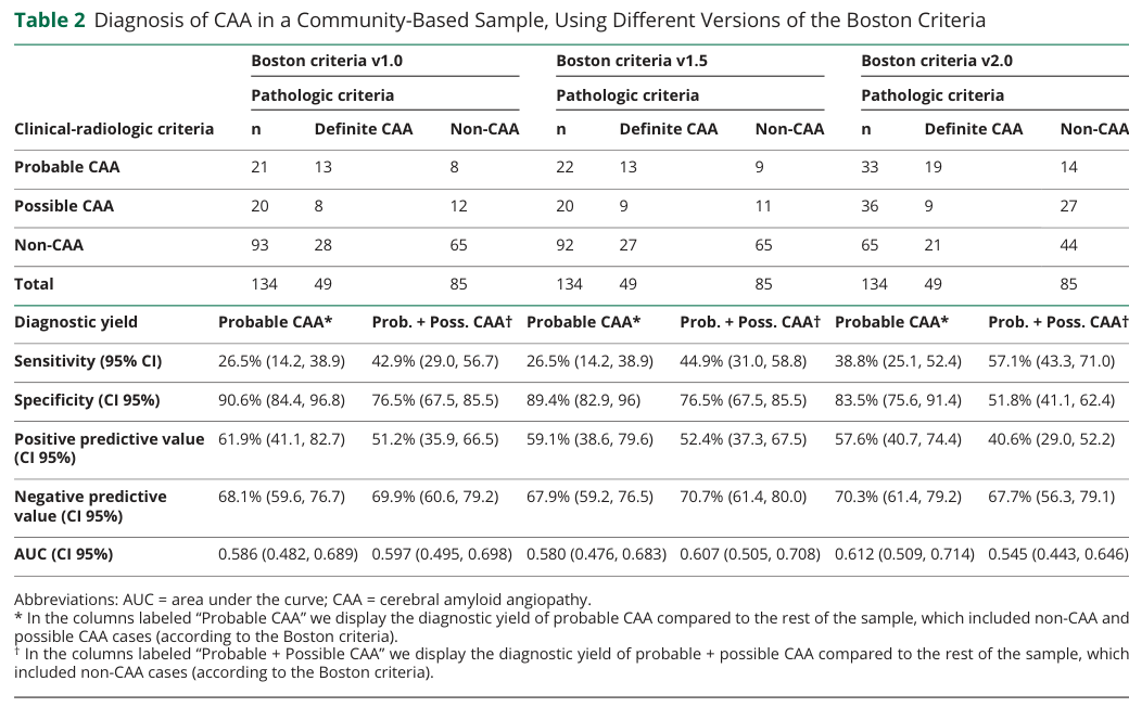
Reference Validation
Checked with linkml-reference-validator 0.3.0rc3.
| Outcome | Count |
|---|---|
| References checked | 13 |
| Resolved | 13 |
| Unresolved (possible confabulation) | 0 |
| Unverifiable | 0 |
| References weighed for topical relevance | 13 |
| On topic | 6 |
| Off topic | 0 |
All extracted references resolved successfully.
Term Validation
Checked with linkml-term-validator 0.4.5, through the ols: adapter.
| Outcome | Count |
|---|---|
| Terms checked | 24 |
| Resolved | 0 |
| Unresolved (possible confabulation) | 0 |
| Obsolete | 0 |
| Unverifiable | 24 |
Prefixes with no resolver
Terms carrying these prefixes were not checked either way, because no configured ontology covers them. An unrecognised prefix may name an ontology this run could not reach as easily as one that does not exist, so nothing here is evidence of fabrication: HP, GO, CL, UBERON, MAXO.
No term could be looked up either way, so nothing here was confirmed or contradicted.