Cataract 33
Non-syndromic early-onset cataract caused by pathogenic variants in BFSP1, which encodes filensin, one of the two lens-specific intermediate filament proteins that co-assemble with BFSP2 (CP49) into the beaded filament of lens fiber cells. Both biallelic (autosomal recessive) and heterozygous (autosomal dominant) families are reported. Recessive families carry frameshifting exon deletions or compound heterozygous alleles and present with juvenile or developmental cataract, in one family with microcornea; dominant families carry missense, splice-site or frameshift alleles and present with congenital nuclear, total, lamellar punctate or posterior sutural pulverulent cataract. Mouse filensin knockouts show that the beaded filament is dispensable for initial fiber cell differentiation but required to maintain fiber cell architecture and optical clarity with age.
Ask OpenScientist
Ask a research question about Cataract 33. OpenScientist will conduct autonomous deep research using the Disorder Mechanisms Knowledge Base and PubMed literature (typically 10-30 minutes).
Do not include personal health information in your question. Questions and results are cached in your browser's local storage.
Inheritance
2Show evidence (2 references)
Show evidence (2 references)
Pathophysiology
4Show evidence (4 references)
Show evidence (2 references)
Show evidence (2 references)
Show evidence (3 references)
Pathograph
Phenotypes
6Show evidence (3 references)
Show evidence (1 reference)
Show evidence (1 reference)
Show evidence (2 references)
Show evidence (1 reference)
Show evidence (1 reference)
Genetic Associations
1Show evidence (7 references)
Medical Actions
1Diagnosis
1Show evidence (2 references)
Animal Models
1Show evidence (1 reference)
Source YAML
click to showname: Cataract 33
creation_date: "2026-10-13T00:00:00Z"
category: Mendelian
synonyms:
- CTRCT33
- BFSP1-related cataract
- filensin-related cataract
description: >-
Non-syndromic early-onset cataract caused by pathogenic variants in BFSP1,
which encodes filensin, one of the two lens-specific intermediate filament
proteins that co-assemble with BFSP2 (CP49) into the beaded filament of lens
fiber cells. Both biallelic (autosomal recessive) and heterozygous (autosomal
dominant) families are reported. Recessive families carry frameshifting exon
deletions or compound heterozygous alleles and present with juvenile or
developmental cataract, in one family with microcornea; dominant families
carry missense, splice-site or frameshift alleles and present with congenital
nuclear, total, lamellar punctate or posterior sutural pulverulent cataract. Mouse
filensin knockouts show that the beaded filament is dispensable for initial
fiber cell differentiation but required to maintain fiber cell architecture
and optical clarity with age.
disease_term:
preferred_term: cataract 33
term:
id: MONDO:0012665
label: cataract 33
parents:
- Early-onset non-syndromic cataract
inheritance:
- name: Autosomal Recessive
description: >-
Biallelic BFSP1 loss-of-function alleles segregate with cataract in
consanguineous Indian and Pakistani families (exon 6 and exons 3-5
deletions) and in a compound heterozygous proband.
inheritance_term:
preferred_term: Autosomal recessive inheritance
term:
id: HP:0000007
label: Autosomal recessive inheritance
evidence:
- reference: PMID:17225135
reference_title: "Autosomal recessive juvenile onset cataract associated with mutation in BFSP1."
supports: SUPPORT
evidence_source: HUMAN_CLINICAL
snippet: "A genome wide scan in a consanguineous family of Indian origin with autosomal recessive developmental cataracts was performed by two-point linkage analysis with 382 microsatellite markers."
explanation: First BFSP1 family, segregating recessive developmental cataract.
- reference: PMID:41553577
reference_title: "Novel mega-deletion in BFSP1 causing autosomal recessive juvenile cataract in a Pakistani consanguineous family."
supports: SUPPORT
evidence_source: HUMAN_CLINICAL
snippet: "Sanger sequencing and PCR were performed for all participants of the family which revealed the absence of the exons (3, 4, 5) in affected individuals and their presence in unaffected parents and siblings confirming the segregation of the mutation as autosomal recessive in the family under study."
explanation: Second consanguineous family confirming recessive segregation.
- name: Autosomal Dominant
description: >-
Heterozygous BFSP1 missense, splice-site and frameshift variants segregate
with congenital cataract in several Chinese and Australian families.
inheritance_term:
preferred_term: Autosomal dominant inheritance
term:
id: HP:0000006
label: Autosomal dominant inheritance
evidence:
- reference: PMID:24379646
reference_title: "A novel beaded filament structural protein 1 (BFSP1) gene mutation associated with autosomal dominant congenital cataract in a Chinese family."
supports: SUPPORT
evidence_source: HUMAN_CLINICAL
snippet: "This is the first report of autosomal dominant congenital cataract being associated with a mutation in BFSP1, highlighting the important role of BFSP1 for physiological lens function and optical properties."
explanation: Five-generation family with heterozygous D348N establishes dominant inheritance.
- reference: PMID:36161833
reference_title: "Pathogenic genetic variants identified in Australian families with paediatric cataract."
supports: SUPPORT
evidence_source: HUMAN_CLINICAL
snippet: "A novel BFSP1 c.1124delA p.(Glu375GlyfsTer2) frameshift variant segregated in an autosomal dominant manner in family CSA182 (figure 1G)."
explanation: Independent dominant family with a heterozygous frameshift allele.
pathophysiology:
- name: BFSP1 Loss of Function
biological_scale: MOLECULAR
description: >-
Recessive alleles are exon deletions predicted to shift the reading frame
(exon 6; exons 3-5) or truncate the tail domain; dominant alleles include
tail-domain missense (D348N, G602R), a splice-site variant and a tail-domain
frameshift. In HEK293T cells the D348N mutant was expressed at lower levels
than wild type. Whether dominant alleles act by haploinsufficiency or by
interfering with filament co-assembly has not been resolved; mouse
heterozygotes for a null allele do develop mild light scattering.
genes:
- preferred_term: BFSP1
term:
id: hgnc:1040
label: BFSP1
molecular_functions:
- preferred_term: structural constituent of eye lens
term:
id: GO:0005212
label: structural constituent of eye lens
modifier: DECREASED
evidence:
- reference: PMID:17225135
reference_title: "Autosomal recessive juvenile onset cataract associated with mutation in BFSP1."
supports: SUPPORT
evidence_source: HUMAN_CLINICAL
snippet: "Direct sequencing revealed a 3343 bp deletion including exon 6 (c.736-1384_c.957-66 del) predicted to result in a shift of the open reading frame."
explanation: Recessive frameshifting deletion predicted to abolish normal filensin.
- reference: PMID:41553577
reference_title: "Novel mega-deletion in BFSP1 causing autosomal recessive juvenile cataract in a Pakistani consanguineous family."
supports: SUPPORT
evidence_source: HUMAN_CLINICAL
snippet: "The large deletion potentially forced the junction of exon 2 with exon 6 at mRNA level and resulting in the shift of open reading frame."
explanation: Second recessive frameshifting deletion of BFSP1.
- reference: PMID:26694549
reference_title: "Sporadic and Familial Congenital Cataracts: Mutational Spectrum and New Diagnoses Using Next-Generation Sequencing."
supports: SUPPORT
evidence_source: HUMAN_CLINICAL
snippet: "In our patient (Family 46), the missense mutation affects the rod domain and the frameshift is predicted to lead to loss of part of the tail domain, highlighting the critical nature of these regions in the functions of BFSP1 required for lens clarity"
explanation: Compound heterozygous rod-domain missense and tail-truncating alleles.
- reference: PMID:37667037
reference_title: "D348N Mutation of BFSP1 Gene in Congenital Cataract: it Does Matter."
supports: SUPPORT
evidence_source: IN_VITRO
snippet: "BFSP1 mutation significantly decreases the expression of BFSP1 protein, weakened the ability of cell proliferation and increased apoptosis."
explanation: >-
Reduced D348N protein expression in transfected HEK293T cells; a non-lens
overexpression system, so the proliferation and apoptosis readouts are not
taken as lens mechanism.
downstream:
- target: Beaded Filament Assembly Failure
causal_link_type: DIRECT
evidence:
- reference: PMID:14638724
reference_title: "Targeted deletion of the lens fiber cell-specific intermediate filament protein filensin."
supports: SUPPORT
evidence_source: MODEL_ORGANISM
snippet: "Disruption of the filensin gene successfully blocked production of filensin mRNA, reduced levels of filensin's assembly partner CP49, and prevented the assembly of beaded filaments."
explanation: Filensin loss in mouse directly prevents beaded filament assembly.
directness: INDIRECT
- name: Beaded Filament Assembly Failure
biological_scale: CELLULAR
description: >-
Filensin and CP49 do not form filaments alone; they co-assemble into the
lens-specific beaded filament. Without filensin, CP49 levels fall and no
beaded filaments form in fiber cells.
cell_types:
- preferred_term: lens fiber cell
term:
id: CL:0011004
label: lens fiber cell
biological_processes:
- preferred_term: intermediate filament organization
term:
id: GO:0045109
label: intermediate filament organization
modifier: DECREASED
evidence:
- reference: PMID:15777782
reference_title: "Seeing is believing! The optical properties of the eye lens are dependent upon a functional intermediate filament cytoskeleton."
supports: SUPPORT
evidence_source: OTHER
snippet: "Whilst CP49 self-assembles into thick bundles of filaments, filensin only forms short fibrils, but when combined together they form filaments."
explanation: Review summarizing that filensin and CP49 must co-assemble to form filaments.
quote_role: REVIEW_SYNTHESIS
- reference: PMID:14638724
reference_title: "Targeted deletion of the lens fiber cell-specific intermediate filament protein filensin."
supports: SUPPORT
evidence_source: MODEL_ORGANISM
snippet: "The lens fiber cell-specific intermediate filament protein filensin is essential for beaded filament assembly."
explanation: Knockout mouse conclusion that filensin is required for beaded filament assembly.
downstream:
- target: Loss of Fiber Cell Architecture and Long-Range Order
causal_link_type: DIRECT
evidence:
- reference: PMID:18326727
reference_title: "Resisting the effects of aging: a function for the fiber cell beaded filament."
supports: SUPPORT
evidence_source: MODEL_ORGANISM
snippet: "The beaded filament is not required for the generation of the differentiated fiber cell phenotype but is required to maintain that differentiated state and the long range order that characterizes the lens at the tissue level."
explanation: Filensin and phakosin knockout lenses lose fiber cell order after differentiation.
- name: Loss of Fiber Cell Architecture and Long-Range Order
biological_scale: TISSUE
description: >-
In filensin-null mouse lenses, fiber cells elongate and lose organelles
normally but later lose their differentiated phenotype and the long-range
stacking needed for clarity. Beaded filament loss also alters lens size,
shape, stiffness and the refractive index gradient.
cell_types:
- preferred_term: lens fiber cell
term:
id: CL:0011004
label: lens fiber cell
evidence:
- reference: PMID:18326727
reference_title: "Resisting the effects of aging: a function for the fiber cell beaded filament."
supports: SUPPORT
evidence_source: MODEL_ORGANISM
snippet: "Subsequent to those stages, however, fiber cells undergo loss of the differentiated fiber cell phenotype and loss of the long-range stacking that characterizes fiber cells and that has been considered essential for clarity."
explanation: Direct morphological observation in filensin and phakosin knockout mice.
- reference: PMID:15777782
reference_title: "Seeing is believing! The optical properties of the eye lens are dependent upon a functional intermediate filament cytoskeleton."
supports: SUPPORT
evidence_source: OTHER
snippet: "Our results suggest that the lens-specific beaded filaments are the key cytoskeletal element in organising and maintaining lens fibre cell architecture and are a key factor in determining the optical properties of the lens."
explanation: Review of Bfsp1 and Bfsp2 knockout work.
quote_role: REVIEW_SYNTHESIS
downstream:
- target: Lens Light Scattering and Loss of Transparency
causal_link_type: DIRECT
evidence:
- reference: PMID:34087204
reference_title: "Deletion of beaded filament proteins or the C-terminal end of Aquaporin 0 causes analogous abnormal distortion aberrations in mouse lens."
supports: SUPPORT
evidence_source: MODEL_ORGANISM
snippet: "Our study reveals that loss of single or both beaded filament proteins significantly affect lens refractive index gradient, transparency and focusing ability in an age-dependent manner"
explanation: Filensin knockout lenses lose transparency and focusing ability with age.
- name: Lens Light Scattering and Loss of Transparency
biological_scale: TISSUE
conforms_to: cataract_lens_opacification#Loss of Lens Refractive Transparency and Light Scattering
description: >-
Light scattering begins after fetal development and worsens with age in
filensin-null mice; heterozygous mice show a milder, later scattering. In
patients this corresponds to congenital, infantile or juvenile lens
opacity.
cell_types:
- preferred_term: lens fiber cell
term:
id: CL:0011004
label: lens fiber cell
biological_processes:
- preferred_term: maintenance of lens transparency
term:
id: GO:0036438
label: maintenance of lens transparency
modifier: DECREASED
evidence:
- reference: PMID:14638724
reference_title: "Targeted deletion of the lens fiber cell-specific intermediate filament protein filensin."
supports: SUPPORT
evidence_source: MODEL_ORGANISM
snippet: "Filensin knockouts began to show evidence of light-scattering by 2 months and worsened with age."
explanation: Progressive light scattering in homozygous filensin knockout mice.
- reference: PMID:14638724
reference_title: "Targeted deletion of the lens fiber cell-specific intermediate filament protein filensin."
supports: SUPPORT
evidence_source: MODEL_ORGANISM
snippet: "Heterozygous animals exhibited an intermediate phenotype, showing a reduction in filensin transcript and moderate light-scattering at 5 months."
explanation: Single-allele loss suffices for milder light scattering, relevant to dominant human alleles.
- reference: PMID:19587458
reference_title: "Functions of the intermediate filament cytoskeleton in the eye lens."
supports: SUPPORT
evidence_source: OTHER
snippet: "Evidence generated in studies in both mice and humans suggests a critical role for these proteins and their filamentous polymers in establishing the optical properties of the eye lens and in maintaining its transparency."
explanation: Review linking beaded filament proteins to lens transparency in mice and humans.
quote_role: REVIEW_SYNTHESIS
downstream:
- target: Developmental cataract
causal_link_type: DIRECT
- target: Juvenile cataract
causal_link_type: DIRECT
- target: Nuclear cataract
causal_link_type: DIRECT
- target: Lamellar cataract
causal_link_type: DIRECT
- target: Sutural pulverulent cataract
causal_link_type: DIRECT
phenotypes:
- category: Ophthalmologic
name: Developmental cataract
description: >-
Cataract present at birth or in early childhood, reported in both recessive
and dominant families; morphology ranges from nuclear to total opacity.
phenotype_term:
preferred_term: Congenital cataract
term:
id: HP:0000519
label: Developmental cataract
evidence:
- reference: PMID:17225135
reference_title: "Autosomal recessive juvenile onset cataract associated with mutation in BFSP1."
supports: SUPPORT
evidence_source: HUMAN_CLINICAL
snippet: "A genome wide scan in a consanguineous family of Indian origin with autosomal recessive developmental cataracts was performed by two-point linkage analysis with 382 microsatellite markers."
explanation: Recessive family with developmental cataract.
- reference: PMID:24379646
reference_title: "A novel beaded filament structural protein 1 (BFSP1) gene mutation associated with autosomal dominant congenital cataract in a Chinese family."
supports: SUPPORT
evidence_source: HUMAN_CLINICAL
snippet: "To identify the disease-causing mutation in a five-generation Chinese family affected with bilateral congenital nuclear cataract."
explanation: Dominant family with bilateral congenital cataract.
- reference: PMID:31842807
reference_title: "Novel mutations identified in Chinese families with autosomal dominant congenital cataracts by targeted next-generation sequencing."
supports: SUPPORT
evidence_source: HUMAN_CLINICAL
snippet: "(c) The patient (III3) in Family 3 showed a total cataract."
explanation: >-
Family 3 carries heterozygous BFSP1 G602R; this affected member had a
total cataract.
- category: Ophthalmologic
name: Juvenile cataract
description: >-
Cataract developing in childhood or adolescence. The recessive Pakistani
family was ascertained for juvenile cataract; the original Indian family's
report is titled juvenile onset, but its abstract describes the cataract
only as developmental.
phenotype_term:
preferred_term: Juvenile cataract
term:
id: HP:0001118
label: Juvenile cataract
evidence:
- reference: PMID:41553577
reference_title: "Novel mega-deletion in BFSP1 causing autosomal recessive juvenile cataract in a Pakistani consanguineous family."
supports: SUPPORT
evidence_source: HUMAN_CLINICAL
snippet: "Objective of the study was to delineate the underlying causative genetic defect for autosomal recessive juvenile cataract (ARJC) in an affected consanguineous multiplex, multigenerational Pakistani consanguineous family."
explanation: Recessive BFSP1 family ascertained for juvenile cataract.
- category: Ophthalmologic
name: Nuclear cataract
description: Bilateral congenital nuclear opacity in the five-generation dominant D348N family.
phenotype_term:
preferred_term: Congenital nuclear cataract
term:
id: HP:0100018
label: Nuclear cataract
evidence:
- reference: PMID:24379646
reference_title: "A novel beaded filament structural protein 1 (BFSP1) gene mutation associated with autosomal dominant congenital cataract in a Chinese family."
supports: SUPPORT
evidence_source: HUMAN_CLINICAL
snippet: "To identify the disease-causing mutation in a five-generation Chinese family affected with bilateral congenital nuclear cataract."
explanation: Nuclear morphology in the dominant family.
- category: Ophthalmologic
name: Lamellar cataract
description: Progressive lamellar punctate cataract was reported in a proband with a heterozygous BFSP1 splice-site variant (c.625+3A>G).
phenotype_term:
preferred_term: Lamellar punctate cataract
term:
id: HP:0007971
label: Lamellar cataract
evidence:
- reference: PMID:28450710
reference_title: "Targeted Exome Sequencing of Congenital Cataracts Related Genes: Broadening the Mutation Spectrum and Genotype-Phenotype Correlations in 27 Chinese Han Families."
supports: SUPPORT
evidence_source: HUMAN_CLINICAL
snippet: "(K) Photograph of proband in family 19 showed a lamellar punctate cataract."
explanation: Family 19 carries the BFSP1 c.625+3A>G variant.
- reference: PMID:36161833
reference_title: "Pathogenic genetic variants identified in Australian families with paediatric cataract."
supports: SUPPORT
evidence_source: HUMAN_CLINICAL
snippet: "Sutural, often pulverulent-like, cataracts are the most frequently reported phenotype with BFSP2 variants while the range of phenotypes observed for BFSP1 variants is wider and includes nuclear, lamellar and cortical cataracts."
explanation: Summarizes the morphological range reported for BFSP1 variants.
quote_role: BACKGROUND
- category: Ophthalmologic
name: Sutural pulverulent cataract
description: Posterior sutural cataract with a pulverulent appearance in all affected members of a dominant frameshift family.
phenotype_term:
preferred_term: Posterior sutural pulverulent cataract
term:
id: HP:0010695
label: Sutural cataract
evidence:
- reference: PMID:36161833
reference_title: "Pathogenic genetic variants identified in Australian families with paediatric cataract."
supports: SUPPORT
evidence_source: HUMAN_CLINICAL
snippet: "A posterior sutural cataract phenotype with a pulverulent appearance (figure 2B) was consistently observed across all affected individuals in the family."
explanation: Family CSA182 carrying BFSP1 c.1124delA.
- category: Ophthalmologic
name: Microcornea
description: >-
Two affected brothers with compound heterozygous BFSP1 variants had
cataract with microcornea. No other BFSP1 family reports it, and no
mechanism links filensin loss to corneal size, so it is not wired to the
lens mechanism.
phenotype_term:
preferred_term: Microcornea
term:
id: HP:0000482
label: Microcornea
evidence:
- reference: PMID:26694549
reference_title: "Sporadic and Familial Congenital Cataracts: Mutational Spectrum and New Diagnoses Using Next-Generation Sequencing."
supports: SUPPORT
evidence_source: HUMAN_CLINICAL
snippet: "This family had two affected sons with cataract and microcornea from a consanguineous family."
explanation: Microcornea observed in the compound heterozygous BFSP1 family.
genetic:
- name: BFSP1
gene_term:
preferred_term: BFSP1
term:
id: hgnc:1040
label: BFSP1
association: Causative
relationship_type: CAUSATIVE
notes: >-
Reported alleles (NM_001195): recessive c.736-1384_c.957-66del (exon 6
deletion), a homozygous 15.4 kb exons 3-5 deletion, and compound
heterozygous c.1492del p.(Ser498Leufs*24) with c.812T>C p.(Ile271Thr);
dominant c.1042G>A p.(Asp348Asn), c.1804G>C p.(Gly602Arg), c.625+3A>G and
c.1124del p.(Glu375Glyfs*2). Two Chinese families linked to 20p12.2-p11.23
were sequenced for BFSP1 without a mutation being found, so linkage to this
region is not by itself evidence of BFSP1 cataract.
evidence:
- reference: PMID:17225135
reference_title: "Autosomal recessive juvenile onset cataract associated with mutation in BFSP1."
supports: SUPPORT
evidence_source: HUMAN_CLINICAL
snippet: "This is the first report of a mutation in the BFSP1 gene associated with human inherited cataracts."
explanation: First BFSP1 cataract family.
- reference: PMID:41553577
reference_title: "Novel mega-deletion in BFSP1 causing autosomal recessive juvenile cataract in a Pakistani consanguineous family."
supports: SUPPORT
evidence_source: HUMAN_CLINICAL
snippet: "Whole-exome sequencing (WES) in an affected family member revealed a homozygous, intragenic 15.40 kb mega-deletion NG_012423.2; g.17485448_17500744del (chr20: 17,485,448-17,500,744-GRCh37/hg19) including three consecutive exons 3, 4, and 5, encompassing two domains of beaded filament structural protein 1 (BFSP1)."
explanation: Homozygous exon 3-5 deletion in a recessive family.
- reference: PMID:26694549
reference_title: "Sporadic and Familial Congenital Cataracts: Mutational Spectrum and New Diagnoses Using Next-Generation Sequencing."
supports: SUPPORT
evidence_source: HUMAN_CLINICAL
snippet: "The proband was found to be compound heterozygous for mutations in BFSP1 (c.1492del p.(Ser498Leufs*24) and c.812T>C p.(Ile271Thr))."
explanation: Compound heterozygous recessive proband.
- reference: PMID:24379646
reference_title: "A novel beaded filament structural protein 1 (BFSP1) gene mutation associated with autosomal dominant congenital cataract in a Chinese family."
supports: SUPPORT
evidence_source: HUMAN_CLINICAL
snippet: "Direct sequencing revealed a heterozygous G>A transversion at c.1042 of the coding sequence in exon 7 of BFSP1 (c.1042G>A) in all affected members, which resulted in the substitution of a wild-type aspartate to an asparagine (D348N)."
explanation: Heterozygous missense segregating in a dominant family.
- reference: PMID:31842807
reference_title: "Novel mutations identified in Chinese families with autosomal dominant congenital cataracts by targeted next-generation sequencing."
supports: SUPPORT
evidence_source: HUMAN_CLINICAL
snippet: "Pedigree of Family 3, all patients carried the heterozygous mutation c.1804G > C in BFSP1."
explanation: Heterozygous G602R segregating in a dominant family.
- reference: PMID:28450710
reference_title: "Targeted Exome Sequencing of Congenital Cataracts Related Genes: Broadening the Mutation Spectrum and Genotype-Phenotype Correlations in 27 Chinese Han Families."
supports: SUPPORT
evidence_source: HUMAN_CLINICAL
snippet: "(G) Reverse sequencing showed c.625 + 3A > G mutation of BFSP1gene in patients from family 19."
explanation: Heterozygous splice-site variant in family 19.
- reference: PMID:36161833
reference_title: "Pathogenic genetic variants identified in Australian families with paediatric cataract."
supports: SUPPORT
evidence_source: HUMAN_CLINICAL
snippet: "A novel BFSP1 c.1124delA p.(Glu375GlyfsTer2) frameshift variant segregated in an autosomal dominant manner in family CSA182 (figure 1G)."
explanation: Heterozygous frameshift in a dominant family.
animal_models:
- name: Bfsp1 (filensin) knockout mouse
species: Mouse
genotype: Bfsp1 exon 1 targeted deletion, homozygous and heterozygous, on wild-type Bfsp2 background
publication: PMID:14638724
modeled_mechanisms:
- target: Lens Light Scattering and Loss of Transparency
relationship: RECAPITULATES
fidelity: MODERATE
description: Null and heterozygous mice develop age-dependent lens light scattering after normal fetal lens development.
limitations: >-
Mice develop scattering postnatally and progressively, whereas some human
families have congenital cataract; human missense and splice alleles are
not modelled.
evidence:
- reference: PMID:14638724
reference_title: "Targeted deletion of the lens fiber cell-specific intermediate filament protein filensin."
supports: SUPPORT
evidence_source: MODEL_ORGANISM
snippet: "Filensin knockouts began to show evidence of light-scattering by 2 months and worsened with age."
explanation: Knockout reproduces lens opacity.
diagnosis:
- name: Targeted cataract gene panel sequencing
description: >-
Clinical appearance does not distinguish BFSP1 cataract from other inherited
cataracts; every reported BFSP1 allele after the first was found by
sequencing panels of known cataract genes with Sanger confirmation and
family segregation.
diagnosis_term:
preferred_term: genetic testing
term:
id: NCIT:C15709
label: Genetic Testing
evidence:
- reference: PMID:28450710
reference_title: "Targeted Exome Sequencing of Congenital Cataracts Related Genes: Broadening the Mutation Spectrum and Genotype-Phenotype Correlations in 27 Chinese Han Families."
supports: SUPPORT
evidence_source: HUMAN_CLINICAL
snippet: "Targeted next-generation sequencing in inherited congenital cataract patients provided significant diagnostic information."
explanation: Panel sequencing study that identified the BFSP1 c.625+3A>G family.
- reference: PMID:31842807
reference_title: "Novel mutations identified in Chinese families with autosomal dominant congenital cataracts by targeted next-generation sequencing."
supports: SUPPORT
evidence_source: HUMAN_CLINICAL
snippet: "Thirty-eight genes associated with cataract were captured and sequenced in 5 typical nonsyndromic congenital cataract probands by targeted next-generation sequencing (NGS), and the results were confirmed by Sanger sequencing."
explanation: Panel sequencing with Sanger confirmation identified the BFSP1 G602R family.
treatments:
- name: Cataract Surgery
description: >-
Removal of the opacified lens with optical correction is the standard
management of visually significant congenital and juvenile cataract; no
BFSP1-specific therapy exists.
therapeutic_modality: SURGERY
treatment_term:
preferred_term: cataract surgery
term:
id: NCIT:C157809
label: Cataract Surgery
target_phenotypes:
- preferred_term: Developmental cataract
term:
id: HP:0000519
label: Developmental cataract
notes: >-
No GeneReviews chapter covers BFSP1-related cataract. Two other Chinese
families linked to chromosome 20p12.2-p11.23 (PMID:17167408, PMID:18958302)
had no BFSP1 mutation and are not part of this entry. A review has raised the
possibility that heterozygous carriers of recessive alleles are predisposed
to early age-related nuclear cataract (PMID:17490642); this has not been
tested.
References & Deep Research
Deep Research
1Deep research results are used as seeds for research; they do not undergo the same validation as the main records and may contain errors. How we use deep research.
Evaluations and curation notes (1)
Create: Cataract_33 · 2026-10-13T04:18:20Z · View source
Created the BFSP1 (filensin) cataract 33 entry from MONDO:0012665 and the stub. Evidence comes from PubMed searches plus a falcon (Edison) deep-research report (research/Cataract_33-deep-research-falcon.md; 7/7 references resolved, its needs_review flag came only from the template text 'if available' attached to the MONDO ID). just preflight-dr could not run because the local MONDO build is absent; the report was checked by reading it and is about BFSP1 throughout. Both recessive families (exon 6 and exons 3-5 deletions, plus one compound heterozygote with microcornea) and dominant families (D348N, G602R, c.625+3A>G, c.1124del) are covered as two inheritance blocks rather than subtypes, since no source shows the modes differ in mechanism. The pathophysiology chain (loss of BFSP1, beaded filament assembly failure, loss of fiber cell order, light scattering) rests on filensin knockout mouse papers. The falcon report says the original recessive family had cortical opacities and normal corneal diameter; the cached PMID:17225135 record is abstract-only, so those points were not curated. Two 20p-linked families without BFSP1 mutations are cited as REFUTE / notes so they are not mistaken for this disease. Validation: just validate, validate-terms, count-verified-snippets (36/36), check-entity-refs, check-causal-targets, check-genereviews (no chapter), validate-disorders.
Question: You are an expert researcher providing comprehensive, well-cited information.
Provide detailed information focusing on: 1. Key concepts and definitions with current understanding 2. Recent developments and latest research (prioritize 2023-2024 sources) 3. Current applications and real-world implementations 4. Expert opinions and analysis from authoritative sources 5. Relevant statistics and data from recent studies
Format as a comprehensive research report with proper citations. Include URLs and publication dates where available. Always prioritize recent, authoritative sources and provide specific citations for all major claims.
Disease Characteristics Research Template
Target Disease
- Disease Name: Cataract 33
- MONDO ID: MONDO:0012665 (if available)
- Category: Mendelian
Research Objectives
Please provide a comprehensive research report on Cataract 33 covering all of the disease characteristics listed below. This report will be used to populate a disease knowledge base entry. Be thorough and cite primary literature (PMID preferred) for all claims.
For each section, suggested databases/resources are listed. These are the first places you should search for information on each topic.
1. Disease Information
Search first: OMIM, Orphanet, ICD-10/ICD-11, MeSH, PubMed
- What is the disease? Provide a concise overview.
- What are the key identifiers? (OMIM, Orphanet, ICD-10/ICD-11, MeSH, Mondo)
- What are the common synonyms and alternative names?
- Is the information derived from individual patients (e.g., EHR) or aggregated disease-level resources?
2. Etiology
- Disease Causal Factors: What are the primary causes? (genetic, environmental, infectious, mechanistic)
- Risk Factors:
Search first: PubMed, Cochrane Library, UpToDate, clinical guidelines, ClinVar, ClinGen, GWAS Catalog, PheGenI, CTD, CDC, WHO, epidemiological databases
- Genetic risk factors (causal variants, susceptibility loci, modifier genes)
- Environmental risk factors (toxins, lifestyle, occupational exposures, age, sex, family history)
- Protective Factors:
Search first: PubMed, Cochrane Library, clinical trial databases, GWAS Catalog, gnomAD, WHO, CDC, nutrition databases
- Genetic protective factors (protective variants, modifier alleles)
- Environmental protective factors (diet, lifestyle, exposures that reduce risk)
- Gene-Environment Interactions: How do genetic and environmental factors interact to influence disease?
Search first: CTD, PubMed, PheGenI, GxE databases
3. Phenotypes
Search first: HPO (Human Phenotype Ontology), OMIM, Orphanet, PubMed, clinicaltrials.gov, MedDRA, SNOMED CT, DECIPHER, LOINC
For each phenotype, provide: - Phenotype type: symptoms, clinical signs, physical manifestations, behavioral changes, or laboratory abnormalities
For symptoms/signs: HPO, OMIM, Orphanet, PubMed For behavioral changes: HPO, DSM, RDoC (Research Domain Criteria), PubMed For laboratory abnormalities: LOINC, SNOMED CT, LabTests Online, PubMed - Phenotype characteristics: Search first: OMIM, Orphanet, HPO, PubMed - Age of symptom onset (neonatal, childhood, adult-onset, late-onset) - Symptom severity (mild, moderate, severe, variable) - Symptom progression (stable, progressive, episodic, fluctuating) - Frequency among affected individuals (percentage or qualitative) - Quality of life impact: Effects on daily functioning and well-being (per-phenotype when possible) Search first: EQ-5D database, SF-36, WHO QOL databases, PubMed - Suggest HPO (Human Phenotype Ontology) terms for each phenotype
4. Genetic/Molecular Information
- Causal Genes: Gene mutations or chromosomal abnormalities responsible for disease (gene symbols, OMIM IDs)
Search first: OMIM, ClinVar, HGMD, Ensembl, NCBI Gene
- Pathogenic Variants:
- Affected genes (gene symbols, HGNC IDs) > Search first: OMIM, NCBI Gene, Ensembl, HGNC, UniProt, GeneCards
- Variant classification (pathogenic, likely pathogenic, VUS per ACMG/AMP guidelines) > Search first: ClinVar, ClinGen, ACMG/AMP guidelines, VarSome
- Variant type/class (missense, frameshift, nonsense, splice-site, structural)
- Allele frequency in population databases > Search first: gnomAD, 1000 Genomes, ExAC, TOPMed, dbSNP
- Somatic vs germline origin > Search first: COSMIC (somatic), ClinVar, ICGC, TCGA
- Functional consequences (loss of function, gain of function, dominant negative)
- Modifier Genes: Genes that modify disease severity or expression
- Epigenetic Information: DNA methylation, histone modifications, chromatin changes affecting disease
Search first: ENCODE, Roadmap Epigenomics, MethBase, DiseaseMeth
- Chromosomal Abnormalities: Large-scale genetic changes (aneuploidy, translocations, inversions)
Search first: DECIPHER, ClinVar, ECARUCA, UCSC Genome Browser
5. Environmental Information
- Environmental Factors: Non-genetic contributing factors (toxins, radiation, pollution, occupational exposure)
Search first: CTD (Comparative Toxicogenomics Database), TOXNET, PubMed, EPA databases
- Lifestyle Factors: Behavioral factors (smoking, diet, exercise, alcohol consumption)
Search first: CDC databases, WHO, PubMed, NHANES
- Infectious Agents: If applicable, pathogens causing or triggering disease (bacteria, viruses, fungi, parasites)
Search first: NCBI Taxonomy, ViPR, BV-BRC, MicrobeDB, GIDEON
6. Mechanism / Pathophysiology
Present this section as an ordered causal chain first, then the detail below. Open with a numbered sequence of mechanistic steps running from the initiating lesion (mutation, exposure, infection) to the clinical manifestation, one step per line, each naming what it causes next. State the causal verb explicitly ("leads to", "results in") and say where a step is inferred rather than demonstrated. Where the mechanism branches, show the branch. The categories below are a checklist of what to cover within those steps, not the organizing structure — a step may draw on several of them, and a category may contribute to several steps.
- Molecular Pathways: Specific signaling cascades or biochemical pathways involved (Wnt, MAPK, mTOR, PI3K-AKT, etc.)
Search first: KEGG, Reactome, WikiPathways, PathBank, BioCyc
- Cellular Processes: Cell-level mechanisms (apoptosis, autophagy, cell cycle dysregulation, inflammation, etc.)
Search first: Gene Ontology (GO), Reactome, KEGG, PubMed
- Protein Dysfunction: How protein structure or function is altered (misfolding, aggregation, loss of function, gain of function)
Search first: UniProt, PDB (Protein Data Bank), InterPro, Pfam, AlphaFold
- Metabolic Changes: Alterations in metabolic processes (energy metabolism, lipid metabolism, amino acid metabolism)
Search first: KEGG, BioCyc, HMDB (Human Metabolome Database), BRENDA
- Immune System Involvement: Role of immune response (autoimmunity, immunodeficiency, chronic inflammation)
Search first: ImmPort, Immunome Database, IEDB, Gene Ontology
- Tissue Damage Mechanisms: How tissues/ are injured (oxidative stress, ischemia, fibrosis, necrosis)
Search first: PubMed, Gene Ontology, Reactome
- Biochemical Abnormalities: Specific molecular defects (enzyme deficiencies, receptor dysfunction, ion channel defects)
Search first: BRENDA, UniProt, KEGG, OMIM, PubMed
- Epigenetic Changes: DNA methylation, histone modifications affecting gene expression in disease
Search first: ENCODE, Roadmap Epigenomics, MethBase, DiseaseMeth
- Molecular Profiling (if available):
- Transcriptomics/gene expression changes > Search first: GEO (Gene Expression Omnibus), ArrayExpress, GTEx, Human Cell Atlas, SRA
- Proteomics findings > Search first: PRIDE, ProteomeXchange, Human Protein Atlas, STRING, BioGRID
- Metabolomics signatures > Search first: MetaboLights, Metabolomics Workbench, HMDB, METLIN
- Lipidomics alterations > Search first: LIPID MAPS, SwissLipids, LipidHome, Metabolomics Workbench
- Genomic structural features > Search first: UCSC Genome Browser, Ensembl, NCBI, dbVar, DGV
- Advanced Technologies (if applicable):
- Single-cell analysis findings (cell-type specific mechanisms, cellular heterogeneity) > Search first: Human Cell Atlas, Single Cell Portal, GEO, CELLxGENE
- Spatial transcriptomics findings > Search first: GEO, Spatial Research, Vizgen, 10x Genomics data
- Multi-omics integration results > Search first: TCGA, ICGC, cBioPortal, LinkedOmics, PubMed
- Functional genomics screens (CRISPR, RNAi) > Search first: DepMap, GenomeRNAi, PubMed, BioGRID ORCS
For each mechanism, describe: - The causal chain from initial trigger to clinical manifestation - Which mechanisms are upstream vs downstream - What cell types and biological processes are involved - Suggest GO terms for biological processes and CL terms for cell types
7. Anatomical Structures Affected
- Organ Level:
- Primary organs directly affected
- Secondary organ involvement (complications, secondary effects)
- Body systems involved (cardiovascular, nervous, digestive, respiratory, endocrine, etc.)
Search first: Uberon, FMA (Foundational Model of Anatomy), OMIM, HPO, ICD-11, MeSH, SNOMED CT
- Tissue and Cell Level:
- Specific tissue types affected (epithelial, connective, muscle, nervous)
- Specific cell populations targeted (with Cell Ontology terms)
Search first: Uberon, Human Protein Atlas, Cell Ontology, Human Cell Atlas, CellMarker, PanglaoDB
- Subcellular Level:
- Cellular compartments involved (mitochondria, nucleus, ER, lysosomes) (with GO Cellular Component terms)
Search first: Gene Ontology (Cellular Component), UniProt, Human Protein Atlas
- Localization:
- Specific anatomical sites (with UBERON terms) > Search first: FMA, Uberon, NeuroNames (for brain), SNOMED CT
- Lateralization (unilateral, bilateral, asymmetric) > Search first: HPO, clinical literature, imaging databases
8. Temporal Development
- Onset:
- Typical age of onset (congenital, pediatric, adult, geriatric)
- Onset pattern (acute, subacute, chronic, insidious)
Search first: OMIM, Orphanet, HPO, PubMed
- Progression:
- Disease stages (early, intermediate, advanced, end-stage) > Search first: Cancer Staging Manual (AJCC), WHO classifications, PubMed
- Progression rate (rapid, slow, variable)
- Disease course pattern (episodic, relapsing-remitting, progressive, stable)
- Disease duration (self-limited, chronic lifelong)
Search first: Disease registries, longitudinal cohort databases, natural history studies, PubMed, Orphanet, OMIM
- Patterns:
- Remission patterns (spontaneous, treatment-induced) > Search first: Clinical trial databases, disease registries, PubMed
- Critical periods (time windows of vulnerability or opportunity for intervention) > Search first: PubMed, developmental biology databases, clinical guidelines
9. Inheritance and Population
- Epidemiology:
- Prevalence (cases per 100,000 at given time)
- Incidence (new cases per 100,000 per year)
Search first: Orphanet, CDC, WHO, GBD (Global Burden of Disease), national registries, SEER, disease registries
- For Genetic Etiology:
- Inheritance pattern (AD, AR, X-linked, mitochondrial, multifactorial, polygenic) > Search first: OMIM, Orphanet, ClinVar, GTR (Genetic Testing Registry)
- Penetrance (complete, incomplete, age-dependent) > Search first: ClinVar, OMIM, PubMed, ClinGen
- Expressivity (variable, consistent) > Search first: OMIM, ClinVar, PubMed
- Genetic anticipation (increasing severity in successive generations) > Search first: OMIM, PubMed (especially for repeat expansion disorders)
- Germline mosaicism > Search first: ClinVar, OMIM, genetic counseling literature, PubMed
- Founder effects (population-specific mutations) > Search first: gnomAD, population genetics databases, PubMed
- Consanguinity role > Search first: OMIM, population studies, genetic counseling resources
- Carrier frequency > Search first: gnomAD, carrier screening databases, GeneReviews, GTR
- Population Demographics:
- Affected populations (ethnic or demographic groups with higher prevalence) > Search first: gnomAD, 1000 Genomes, PAGE Study, PubMed, population registries
- Geographic distribution (endemic areas, regional variation) > Search first: WHO, CDC, GBD, Orphanet, geographic epidemiology databases
- Geographic distribution of specific variants
- Sex ratio (male:female) > Search first: Disease registries, OMIM, PubMed, epidemiological databases
- Age distribution of affected individuals > Search first: CDC, disease registries, SEER, Orphanet
10. Diagnostics
- Clinical Tests:
- Laboratory tests (blood, urine, tissue chemistry, specific enzyme assays) > Search first: LOINC, LabTests Online, PubMed
- Biomarkers (proteins, metabolites, genetic markers, circulating biomarkers) > Search first: FDA Biomarker List, BEST (Biomarkers, EndpointS, and other Tools), PubMed
- Imaging studies (X-ray, CT, MRI, PET, ultrasound) > Search first: RadLex, DICOM, Radiopaedia, imaging databases
- Functional tests (pulmonary function, cardiac stress tests) > Search first: LOINC, clinical guidelines, PubMed
- Electrophysiology (EEG, EMG, ECG, nerve conduction studies) > Search first: LOINC, clinical neurophysiology databases, PubMed
- Biopsy findings (histopathology, immunohistochemistry) > Search first: SNOMED CT, College of American Pathologists resources, PubMed
- Pathology findings (microscopic examination) > Search first: SNOMED CT, Digital Pathology databases, PubMed
- Genetic Testing:
Search first: GTR (Genetic Testing Registry), GeneReviews, ClinGen
- Overview of recommended genetic testing approach
- Whole genome sequencing (WGS) utility > Search first: GTR, ClinVar, GEL (Genomics England), gnomAD
- Whole exome sequencing (WES) utility > Search first: GTR, ClinVar, OMIM, GeneMatcher
- Gene panels (which panels, which genes) > Search first: GTR, ClinVar, laboratory-specific databases
- Single gene testing > Search first: GTR, ClinVar, OMIM, GeneReviews
- Chromosomal microarray (CMA) > Search first: DECIPHER, ClinVar, dbVar, ECARUCA
- Karyotyping > Search first: Chromosome Abnormality Database, ClinVar, cytogenetics resources
- FISH > Search first: ClinVar, cytogenetics databases, PubMed
- Mitochondrial DNA testing > Search first: MITOMAP, MSeqDR, ClinVar, GTR
- Repeat expansion testing > Search first: GTR, ClinVar, repeat expansion databases, PubMed
- Omics-Based Diagnostics (if applicable):
- RNA sequencing / transcriptomics > Search first: GEO, ArrayExpress, GTEx, RNA-seq databases
- Proteomics > Search first: PRIDE, ProteomeXchange, FDA Biomarker database
- Metabolomics > Search first: MetaboLights, Metabolomics Workbench, HMDB
- Epigenomics > Search first: GEO, ENCODE, Roadmap Epigenomics, MethBase
- Liquid biopsy > Search first: COSMIC, ClinVar, liquid biopsy databases, PubMed
- Clinical Criteria:
- Standardized diagnostic criteria (DSM, ICD, society guidelines) > Search first: DSM-5, ICD-11, clinical society guidelines, UpToDate
- Differential diagnosis (other conditions to rule out, with distinguishing features) > Search first: DynaMed, UpToDate, clinical decision support systems
- Screening:
- Screening methods for asymptomatic individuals (newborn screening, carrier screening, cascade screening) > Search first: ACMG recommendations, CDC newborn screening, GTR
11. Outcome/Prognosis
- Survival and Mortality:
- Survival rate (5-year, 10-year, overall) > Search first: SEER, cancer registries, disease-specific registries, PubMed
- Life expectancy (with and without treatment if applicable) > Search first: Orphanet, disease registries, actuarial databases, PubMed
- Mortality rate > Search first: CDC, WHO, GBD, national mortality databases
- Disease-specific mortality (deaths directly attributable to disease) > Search first: Disease registries, CDC Wonder, GBD, PubMed
- Morbidity and Function:
- Morbidity (disease-related disability and health impacts) > Search first: GBD, WHO, disability databases, PubMed
- Disability outcomes (long-term functional impairments) > Search first: ICF (International Classification of Functioning), disability registries
- Quality of life measures (EQ-5D, SF-36, PROMIS, disease-specific tools) > Search first: EQ-5D database, SF-36, PROMIS, PubMed
- Disease Course:
- Complications (secondary problems: infections, organ failure, etc.) > Search first: ICD codes, disease registries, clinical databases, PubMed
- Recovery potential (likelihood and extent of recovery, with vs without treatment) > Search first: Natural history studies, rehabilitation databases, PubMed
- Prediction:
- Prognostic factors (age, disease severity, biomarkers, treatment response) > Search first: Prognostic models databases, clinical calculators, PubMed
- Prognostic biomarkers (molecular markers predicting disease course) > Search first: FDA Biomarker database, PubMed, cancer prognostic databases
12. Treatment
- Pharmacotherapy:
- Pharmacological treatments (drug names, drug classes, mechanisms of action) > Search first: DrugBank, RxNorm, ATC classification, DailyMed, FDA databases
- Pharmacogenomics (how genetic variants affect drug metabolism, efficacy, toxicity) > Search first: PharmGKB, CPIC (Clinical Pharmacogenetics), FDA Table of PGx Biomarkers
- Advanced Therapeutics:
- Gene therapy (viral vectors, CRISPR, gene replacement, gene editing) > Search first: ClinicalTrials.gov, FDA gene therapy database, ASGCT resources
- Cell therapy (stem cell transplant, CAR-T, cellular therapeutics) > Search first: ClinicalTrials.gov, FDA cell therapy database, FACT standards
- RNA-based therapies (ASOs, siRNA, mRNA therapies) > Search first: ClinicalTrials.gov, FDA approvals, PubMed
- Targeted therapies (treatments directed at specific molecular targets) > Search first: My Cancer Genome, OncoKB, ClinicalTrials.gov, FDA approvals
- Immunotherapies (checkpoint inhibitors, monoclonal antibodies) > Search first: Cancer Immunotherapy Database, FDA approvals, ClinicalTrials.gov
- Surgical and Interventional:
- Surgical interventions (types of surgery, timing, outcomes) > Search first: CPT codes, surgical registries, clinical guidelines, PubMed
- Supportive and Rehabilitative:
- Supportive care (symptom management, pain control, nutrition) > Search first: Clinical guidelines, Cochrane Library, PubMed
- Rehabilitation (physical therapy, occupational therapy, speech therapy) > Search first: Rehabilitation medicine databases, clinical guidelines, PubMed
- Experimental:
- Experimental treatments in clinical trials (with NCT identifiers if available) > Search first: ClinicalTrials.gov, EU Clinical Trials Register, WHO ICTRP
- Treatment Outcomes:
- Treatment response rates > Search first: Clinical trial databases, FDA reviews, systematic reviews, PubMed
- Side effects and adverse events > Search first: FDA Adverse Event Reporting System (FAERS), MedWatch, PubMed
- Treatment Strategy:
- Treatment algorithms (clinical pathways, decision trees) > Search first: Clinical practice guidelines, NCCN Guidelines, UpToDate
- Combination therapies > Search first: ClinicalTrials.gov, treatment guidelines, PubMed
- Personalized medicine approaches (genotype-guided treatment) > Search first: My Cancer Genome, CIViC, PharmGKB, precision medicine databases
For each treatment, suggest NCIT (NCI Thesaurus) clinical-intervention terms where applicable.
13. Prevention
- Prevention Levels:
- Primary prevention (preventing disease occurrence: vaccination, risk factor modification) > Search first: CDC, WHO, USPSTF recommendations, Cochrane Library
- Secondary prevention (early detection and treatment: screening programs, early intervention) > Search first: USPSTF, CDC screening guidelines, WHO
- Tertiary prevention (preventing complications in those with disease) > Search first: Clinical guidelines, disease management protocols, PubMed
- Immunization: Vaccine strategies (if applicable)
Search first: CDC vaccine schedules, WHO immunization, FDA vaccine database
- Screening and Early Detection:
- Screening programs (population-based: newborn screening, cancer screening) > Search first: CDC screening programs, USPSTF, cancer screening databases
- Genetic screening (carrier screening, preimplantation genetic diagnosis, prenatal testing) > Search first: ACMG recommendations, ACOG guidelines, GTR
- Risk stratification (identifying high-risk individuals for targeted prevention) > Search first: Risk prediction models, clinical calculators, PubMed
- Behavioral Interventions: Lifestyle modifications to reduce risk
Search first: CDC, WHO, behavioral intervention databases, Cochrane Library
- Counseling: Genetic counseling (risk assessment, family planning guidance)
Search first: NSGC resources, ACMG guidelines, GeneReviews
- Public Health:
- Public health interventions (sanitation, vector control, health education) > Search first: CDC, WHO, public health databases, PubMed
- Environmental interventions (reducing environmental risk factors) > Search first: EPA databases, WHO environmental health, PubMed
- Prophylaxis: Preventive medications or procedures
Search first: Clinical guidelines, FDA approvals, PubMed
14. Other Species / Natural Disease
- Taxonomy: Species affected (with NCBI Taxon identifiers)
Search first: NCBI Taxonomy
- Breed: Specific breeds affected (with VBO identifiers if applicable)
Search first: VBO (Vertebrate Breed Ontology)
- Gene: Orthologous genes in other species (with NCBI Gene IDs)
Search first: NCBI Gene
- Natural Disease:
- Naturally occurring disease in other species (companion animals, wildlife) > Search first: OMIA (Online Mendelian Inheritance in Animals), VetCompass, PubMed
- Veterinary relevance and importance in animal health > Search first: OMIA, veterinary databases, PubMed
- Comparative Biology:
- Comparative pathology (similarities and differences across species) > Search first: OMIA, comparative pathology databases, PubMed
- Evolutionary conservation of disease mechanisms > Search first: HomoloGene, OrthoMCL, Alliance of Genome Resources
- Transmission (if applicable):
- Zoonotic potential > Search first: CDC zoonotic diseases, WHO zoonoses, GIDEON
- Cross-species susceptibility > Search first: NCBI Taxonomy, veterinary databases, PubMed
15. Model Organisms
- Model Types:
- Model organism type (mammalian, invertebrate, cellular, in vitro) > Search first: Alliance of Genome Resources, model organism databases
- Specific model systems (mouse, rat, zebrafish, Drosophila, C. elegans, yeast, cell lines, organoids, iPSCs) > Search first: MGI, RGD, ZFIN, FlyBase, WormBase, SGD, ATCC, Cellosaurus
- Induced models (drug treatment, surgical intervention, environmental manipulation) > Search first: MGI, model organism databases, PubMed
- Genetic Models:
- Types available (knockout, knock-in, transgenic, conditional, humanized) > Search first: MGI, IMPC, KOMP, EuMMCR, IMSR
- Model Characteristics:
- Phenotype recapitulation (how well model reproduces human disease features) > Search first: Model organism databases, comparative studies, PubMed
- Model limitations (aspects of human disease not captured) > Search first: Model organism databases, PubMed, review articles
- Applications:
- Research applications (what aspects of disease can be studied) > Search first: Model organism databases, PubMed
- Resources:
- Model databases > Search first: MGI, RGD, ZFIN, FlyBase, WormBase, IMSR, EMMA, MMRRC
Citation Requirements
- Cite primary literature (PMID preferred) for all mechanistic and clinical claims
- Prioritize recent reviews and landmark papers
- Include direct quotes from abstracts where possible to support key statements
- Distinguish evidence source types: human clinical, model organism, in vitro, computational
Output Format
Structure your response as a comprehensive narrative organized by the sections above. For each section, provide: - Factual content with specific details (numbers, percentages, gene names, variant nomenclature) - Ontology term suggestions (HPO, GO, CL, UBERON, CHEBI, NCIT, MONDO) where applicable - Evidence citations with PMIDs - Direct quotes from abstracts to support key claims - Clear indication when information is not available or not applicable for this disease
This report will be used to populate a disease knowledge base entry with: - Pathophysiology descriptions with causal chains - Gene/protein annotations (HGNC, GO terms) - Phenotype associations (HP terms) with frequencies - Cell type involvement (CL terms) - Anatomical locations (UBERON terms) - Chemical entities (CHEBI terms) - Treatment annotations (NCIT terms) - Evidence items with PMIDs and exact abstract quotes - Epidemiology, prognosis, diagnostic, and prevention information - Animal model descriptions with phenotype recapitulation details
Cataract 33: disease-characteristics research report
Scope and evidence. Cataract 33 (MONDO:0012665) is a rare, BFSP1-associated inherited lens opacity. The strongest subtype-defining evidence is a 2007 study of autosomal-recessive, juvenile-onset cataract in an Indian family. Later reports associate other BFSP1 variants with autosomal-dominant pediatric cataracts of different morphologies. Findings from congenital cataract generally should not be interpreted as measured frequencies, risks, or treatment effects for Cataract 33 specifically. The evidence summarized here comes from published families, experimental systems, and aggregated disease resources—not individual electronic health records. (OpenTargets Search: Cataract 33, ramachandran2007autosomalrecessivejuvenile pages 1-2, li2019novelmutationsidentified pages 1-2, jones2022pathogenicgeneticvariants pages 4-4)
1. Disease information and identifiers
Cataract 33 denotes hereditary opacification of the crystalline lens associated with BFSP1, which encodes beaded filament structural protein 1, also called filensin or CP115. Its defining recessive presentation was developmental/juvenile cataract with predominantly cortical opacity; the broader gene-associated spectrum includes congenital total and posterior sutural/pulverulent cataracts. Thus, “congenital cataract” is not an accurate onset description for every affected person. BFSP2 encodes the distinct partner protein phakinin/CP49 and should not be substituted for BFSP1. (shiels2024throughthecatmap pages 11-12, ramachandran2007autosomalrecessivejuvenile pages 1-2, ramachandran2007autosomalrecessivejuvenile pages 2-4, li2019novelmutationsidentified pages 1-2, jones2022pathogenicgeneticvariants pages 4-4)
| Identifier or term | Evidence-calibrated entry |
|---|---|
| MONDO | MONDO:0012665, cataract 33. |
| Causal gene | BFSP1; the 2019 primary report labels its gene-level OMIM entry 611391. This is a gene identifier, not a verified disease-entry identifier. |
| Useful names | Cataract 33; BFSP1-associated cataract; filensin-associated hereditary cataract; autosomal-recessive juvenile-onset cataract for the original family. |
| Orphanet, subtype-specific OMIM, ICD-11, MeSH | A verified Cataract 33-specific identifier was not established from the retrieved evidence. Do not invent one. Broad congenital/developmental cataract coding may be used clinically, but does not encode the molecular subtype. |
| ICD-10 | Q12.0, congenital cataract, is documented in a pediatric cataract clinical cohort; its applicability depends on actual onset and should not automatically be assigned to juvenile-onset cases. |
The disease–gene and MONDO association comes from an aggregated Open Targets record; the individual-family phenotype and segregation evidence come from primary studies. (OpenTargets Search: Cataract 33, ramachandran2007autosomalrecessivejuvenile pages 1-2, li2019novelmutationsidentified pages 1-2)
2. Etiology, risk, protection, and environment
Established causal factor: a familial homozygous 3,343-bp deletion encompassing BFSP1 exon 6 segregated with recessive juvenile cataract. The investigators examined 19 family members—11 affected and eight unaffected—and identified the deletion in affected relatives; it was absent in 50 ethnically matched controls, or 100 control chromosomes. The maximum two-point linkage LOD was 5.40 at D20S860 in a chromosome-20q interval containing BFSP1. Consanguinity was important to this family’s recessive segregation, but is not itself a molecular lesion. (ramachandran2007autosomalrecessivejuvenile pages 1-2, ramachandran2007autosomalrecessivejuvenile pages 4-7)
Other published BFSP1 candidates segregate with dominant cataract; their interpretation must be variant-specific. A 2024 expert genetics review also discusses common variation near BFSP1 in age-related nuclear cataract, but that association concerns a different, complex trait and does not establish an exposure or susceptibility factor for Mendelian Cataract 33. No protective BFSP1 allele, validated modifier gene in human patients, genotype-specific dietary or environmental protection, or measured BFSP1–environment interaction was established. Smoking, ultraviolet exposure, diabetes, steroids, radiation, and maternal infection may matter in the differential diagnosis of cataract, but none was shown to cause or prevent this specific inherited condition in the cited pedigrees. (shiels2024throughthecatmap pages 11-12, ramachandran2007autosomalrecessivejuvenile pages 1-2, li2019novelmutationsidentified pages 8-10)
3. Phenotypes and their consequences
| Phenotype; suggested HPO search term | Type, timing, and evidence | Frequency and functional effect |
|---|---|---|
| Cataract; Cataract / Juvenile cataract | Clinical sign. In the original recessive family, opacities were generally documented by age five, with one documented at age two. Early-childhood onset is supported; onset at birth in this family is not established. | Cataract was the ascertainment phenotype in the 11 affected members; 11/19 is a pedigree proportion, not population prevalence. Opacity can obstruct sight and require surgery. |
| Cortical cataract; Cortical cataract | Clinical sign. The three affected relatives still available for native-lens examination had fluffy, cotton-like cortical opacities, sometimes with anterior cortical grape-like cysts. | Observed in three examined phakic affected relatives; the others had already undergone surgery, so 3/11 is not a defensible phenotype frequency. |
| Reduced visual acuity; Decreased visual acuity / Visual impairment | Clinical functional manifestation. Family records include preoperative vision as poor as hand motion or light perception and variable acuities after intervention. | No rigorously estimated BFSP1-specific percentage. Impairs learning, mobility, and everyday visual tasks according to severity; subtype-specific quality-of-life scales were not reported. |
| Total cataract; Total cataract | Congenital clinical sign in the separate 2019 Chinese family carrying heterozygous p.Gly602Arg. | Five affected relatives were recorded as having total cataract from birth; this is a separate genotype and should not be pooled with the 2007 family. |
| Posterior sutural/pulverulent cataract; Sutural cataract / Punctate cataract | Pediatric clinical sign reported in a 2022 Australian family with a heterozygous BFSP1 frameshift. | Consistent among the reported affected relatives, but a reliable numeric penetrance or frequency is unavailable. |
The first family’s clinical examination reported normal corneal diameter and axial length and no other detected ocular abnormality; that observation does not prove that every BFSP1 case lacks extracocular disease. Older relatives with nuclear sclerosis beginning around age 50 were treated as unaffected for the recessive linkage analysis, not as additional proven Cataract 33 cases. The original paper’s Figure 2 directly illustrates three cortical cases and one older nuclear case. Suggested HPO labels require verification against the current ontology release before assigning exact HP identifiers. (ramachandran2007autosomalrecessivejuvenile pages 1-2, ramachandran2007autosomalrecessivejuvenile pages 2-4, ramachandran2007autosomalrecessivejuvenile media 82382e04, ramachandran2007autosomalrecessivejuvenile media 07d9e375, li2019novelmutationsidentified pages 4-6, jones2022pathogenicgeneticvariants pages 4-4)
4. Genetics and molecular findings
| BFSP1 finding | Human evidence and interpretation |
|---|---|
| c.736-1384_c.957-66del, 3,343 bp encompassing exon 6; reported predicted p.Thr246fs | Homozygous in affected members of the consanguineous Indian family and heterozygous in relatives predicted to be carriers. Removes exon 6, shifts reading frame, and predicts the first 245 normal amino acids, six new amino acids, premature termination, and loss of the normal protein’s final 414 amino acids. Strong recessive segregation evidence; RNA decay and protein behavior were predicted, not measured. The notation is the original authors’ and should be re-normalized against a named reference transcript before clinical reporting. |
| c.625+3A>G | Reported as a heterozygous splice-region candidate in family 19 of a 2017 Chinese cataract sequencing study. An experimental demonstration of aberrant BFSP1 splicing was not established from the available evidence. |
| NM_001195.3:c.1804G>C, p.Gly602Arg | Heterozygous in a 2019 Chinese dominant-congenital-cataract family: five affected and four unaffected relatives supported segregation. Reported ExAC frequency 0.000066. Its residue was relatively poorly conserved and computational predictions conflicted; the investigators described suspected pathogenicity. Do not elevate this result to a verified pathogenic ClinVar/ACMG classification without current review. |
| NM_001195.5:c.1124del, p.(Glu375GlyfsTer2) | A heterozygous frameshift segregating with dominantly inherited posterior sutural/pulverulent pediatric cataract in Australian family CSA182. The 2022 investigators included this family among those with a disease-causing likely pathogenic variant. The study screened 37 families and confirmed likely pathogenic variants across several genes in eight; those are cohort-wide figures, not a BFSP1 diagnostic yield. |
These are constitutional/germline family findings, not evidence for a somatic cancer variant. A reliable subtype-wide carrier frequency, current gnomAD allele count for each variant, verified ClinVar assertion, HGNC numeric ID, founder effect, repeat expansion, aneuploidy, or separate disease-causing chromosome rearrangement was not established. The exon-6 intragenic deletion is the relevant structural variant; do not reinterpret its chromosome-20 linkage interval as a chromosomal deletion. Dominant frameshift inheritance also means one cannot assume that haploinsufficiency explains every BFSP1 allele. (ramachandran2007autosomalrecessivejuvenile pages 4-7, ramachandran2007autosomalrecessivejuvenile pages 7-8, li2019novelmutationsidentified pages 1-2, li2019novelmutationsidentified pages 8-10, jones2022pathogenicgeneticvariants pages 3-3, jones2022pathogenicgeneticvariants pages 4-4, zhai2017targetedexomesequencing pages 1-2)
5. Environmental, lifestyle, and infectious information
No toxin, diet, occupational exposure, infectious agent, vaccine-preventable pathogen, or behavioral trigger has been demonstrated as necessary or sufficient for BFSP1-associated disease. Exposures relevant to other pediatric or age-related cataracts should be assessed when clinically indicated, rather than entered as established Cataract 33 causes. No disease-specific metabolite, circulating biomarker, or chemopreventive ChEBI entity was established. (ramachandran2007autosomalrecessivejuvenile pages 1-2, li2019novelmutationsidentified pages 8-10)
6. Mechanism and pathophysiology
Ordered causal chain—supported steps and inferences:
- A biallelic BFSP1 exon-6 deletion leads to an out-of-frame predicted transcript and premature stop; the alternate branch, RNA degradation, is inferred rather than demonstrated in patients. A surviving truncated-protein branch is likewise hypothetical. (ramachandran2007autosomalrecessivejuvenile pages 4-7, ramachandran2007autosomalrecessivejuvenile pages 7-8)
- Reduced functional filensin leads to deficient assembly or maintenance of filensin–phakinin/BFSP2 beaded filaments in lens fiber cells; this direction is supported by Bfsp1-deletion mouse observations, whereas direct measurement of filaments in the affected human family was unavailable. (ramachandran2007autosomalrecessivejuvenile pages 1-2, ramachandran2007autosomalrecessivejuvenile pages 7-8)
- Impaired beaded-filament cytoskeleton leads to altered fiber-cell support, packing, biomechanics, and transparency. Mouse perturbation of the partner filament protein CP49/Bfsp2, especially together with Tmod1, demonstrates fiber-cell disorder, altered compressive properties, and light scattering; the precise contribution of each intermediate to a given human BFSP1 allele remains inferred. (gokhin2012tmod1andcp49 pages 1-2, gokhin2012tmod1andcp49 pages 13-14, gokhin2012tmod1andcp49 pages 12-13)
- Increased lens light scattering results in childhood cortical opacity and impaired visual acuity; sustained obstruction during visual development can lead to secondary deprivation amblyopia. The original family establishes the opacity and visual impairment, but not a quantified amblyopia rate or direct molecular-to-optical measurements. (ramachandran2007autosomalrecessivejuvenile pages 1-2, ramachandran2007autosomalrecessivejuvenile pages 2-4, ramachandran2007autosomalrecessivejuvenile pages 4-7)
- Alternative allele branch: heterozygous missense, splice-region, and truncating alleles are associated with congenital total, lamellar, or sutural cataract in separate families; a shared dominant-negative, gain-of-function, or haploinsufficiency mechanism has not been demonstrated across them. (li2019novelmutationsidentified pages 8-10, jones2022pathogenicgeneticvariants pages 4-4, zhai2017targetedexomesequencing pages 1-2)
Molecular context. Filensin is a lens-enriched intermediate-filament-like cytoskeletal protein that associates with BFSP2/phakinin and α-crystallin-containing complexes; the principal supported process is beaded-filament organization, not a demonstrated disease-specific Wnt, MAPK, PI3K–AKT, or mTOR cascade. In experimental mouse lenses, Tmod1-linked spectrin–actin organization interacts functionally with the beaded-filament network, but TMOD1 is a mechanistic partner, not a validated human Cataract 33 modifier. No disease-specific enzyme deficiency, inflammatory/immune pathway, mitochondrial lesion, oxidative-stress measurement, or metabolomic/lipidomic signature was established. Suggested annotation concepts are GO:0005882, intermediate filament and GO:0005856, cytoskeleton, with a lens fiber-cell differentiation/organization biological-process term selected after ontology lookup; assign specific GO process and CL identifiers only after database validation. (shiels2024throughthecatmap pages 11-12, ramachandran2007autosomalrecessivejuvenile pages 1-2, ramachandran2007autosomalrecessivejuvenile pages 7-8, gokhin2012tmod1andcp49 pages 1-2, gokhin2012tmod1andcp49 pages 12-13)
Profiling and advanced technologies—context, not patient data. A 2022 embryonic-chicken lens epithelial-versus-fiber multi-omics study included BFSP1 among genes associated with differentiation-related methylation, accessibility, and expression changes. Across the experiment, investigators found 7,621 differentially methylated regions; methylation versus expression had r = −0.37, and methylation versus chromatin accessibility r = −0.86. These are genome-wide correlations in chick development, not human Cataract 33 epigenetic lesions or BFSP1-specific effect sizes. No disease-specific single-cell, spatial-transcriptomic, proteomic, CRISPR-screen, or patient multi-omics diagnostic signature was established. (disatham2022changesindna pages 2-3, disatham2022changesindna pages 1-2)
7. Anatomy and localization
The primary organ is the eye’s crystalline lens; in the original family the visible lesion was chiefly the lens cortex, with some anterior cortical cyst-like opacities. Differentiating and mature lens fiber cells are the mechanistically implicated cells; the anterior lens epithelium supplies differentiating fiber cells but was not demonstrated to be the primary damaged cell population. The affected subcellular structures are cytoplasmic and membrane-associated beaded filaments. No primary brain, kidney, skin, or systemic organ lesion was established. Corneal dimensions and axial lengths were normal in the examined family. Suggested ontology concepts: UBERON:0000963, lens of camera-type eye, pending release verification; lens cortex and lens fiber cell should be mapped to appropriate current UBERON/CL entries rather than assigned unverified numbers. Laterality should be recorded patient by patient; avoid assigning a numerical bilateral frequency from incomplete clinical records. (ramachandran2007autosomalrecessivejuvenile pages 1-2, ramachandran2007autosomalrecessivejuvenile pages 2-4, ramachandran2007autosomalrecessivejuvenile pages 7-8, ramachandran2007autosomalrecessivejuvenile media 82382e04)
8. Temporal development
For the original recessive phenotype, this is a persistent, early-childhood/developmental cataract, often first documented at five years and in one child at two years; the paper describes maturation of the opacities in early childhood. The separate dominant p.Gly602Arg family instead reported opacities from birth. There is no validated Cataract 33 staging system, subtype-wide annual progression rate, spontaneous-remission frequency, or lifetime natural-history curve. Removal of an opaque lens corrects the optical obstruction but does not reverse the genotype; timely visual rehabilitation matters most when opacity threatens developing vision. Older heterozygous carriers’ nuclear sclerosis does not establish age-dependent penetrance for the Mendelian subtype. (ramachandran2007autosomalrecessivejuvenile pages 2-4, ramachandran2007autosomalrecessivejuvenile pages 7-8, li2019novelmutationsidentified pages 4-6)
9. Inheritance and population
Autosomal recessive inheritance is firmly documented for the original homozygous exon-6 deletion. Autosomal dominant segregation is reported for certain other BFSP1 variants; the evidential strength differs among alleles. Although the 2007 linkage model assumed complete penetrance, that is an analysis assumption, not a measured population penetrance estimate. Cataract morphology and onset differ among the described families, supporting gene-level phenotypic variability. No subtype-specific incidence per 100,000/year, prevalence per 100,000, population carrier frequency, male:female ratio, anticipation, germline mosaicism, or founder mutation has been established. Reported Indian, Chinese, and Australian families demonstrate geographical occurrence, not differential population risk. Broad congenital-cataract prevalence estimates are not a valid substitute for Cataract 33 epidemiology. (ramachandran2007autosomalrecessivejuvenile pages 1-2, ramachandran2007autosomalrecessivejuvenile pages 2-4, li2019novelmutationsidentified pages 1-2, li2019novelmutationsidentified pages 8-10, jones2022pathogenicgeneticvariants pages 1-2, jones2022pathogenicgeneticvariants pages 4-4)
10. Diagnosis, differential diagnosis, and screening
Clinical evaluation: a pediatric ophthalmologist assesses the red reflex, dilated lens and anterior segment by slit lamp, visual acuity or age-appropriate visual behavior, refraction, intraocular pressure, and fundus where visible. The original investigators used slit-lamp examination, Snellen acuity, tonometry, fundus examination, and pediatric retinoscopy. Ultrasound/A-scan can support biometry or evaluation when opacity prevents a satisfactory posterior view; in the original family A-scan determined intraocular-lens power, not BFSP1 genotype. Clinical appearance alone does not distinguish reliably among the many hereditary cataract genes. (ramachandran2007autosomalrecessivejuvenile pages 1-2, ramachandran2007autosomalrecessivejuvenile pages 2-4, li2019novelmutationsidentified pages 1-2)
Molecular workflow: document phenotype and pedigree; offer a comprehensive pediatric-cataract NGS panel containing BFSP1 and BFSP2 and other established crystallin, gap-junction, membrane, and developmental genes, or use WES/WGS when the panel is unrevealing or the phenotype suggests a broader disorder. Assess exon-level copy-number changes and structural breakpoints: sequencing that detects only single-nucleotide variants could miss the defining 3,343-bp deletion. Confirm candidate findings, test familial segregation, and interpret each allele under current ACMG/AMP and laboratory standards; use RNA studies where a splice-region candidate needs functional clarification. WGS can help characterize structural/noncoding variants; neither WES nor WGS has a demonstrated Cataract 33-specific diagnostic-yield estimate. Targeted NGS was used in the Chinese families, and genome sequencing in the Australian families. (ramachandran2007autosomalrecessivejuvenile pages 1-2, ramachandran2007autosomalrecessivejuvenile pages 4-7, li2019novelmutationsidentified pages 1-2, jones2022pathogenicgeneticvariants pages 1-2, jones2022pathogenicgeneticvariants pages 4-4, zhai2017targetedexomesequencing pages 1-2)
Differential diagnosis: BFSP2, crystallin, GJA3/GJA8, MIP, and other hereditary cataracts; metabolic, infectious, traumatic, developmental, and syndromic cataracts according to history and examination. Routine blood enzymes, electrophysiology, biopsy, karyotype, FISH, mitochondrial sequencing, and repeat-expansion testing are not established as subtype-specific tests; deploy alternatives when a different suspected cause warrants them. There is no validated liquid-biopsy or epigenomic assay for Cataract 33. Targeted testing and cascade examination/genotyping may identify relatives at risk; a normal neonatal red reflex cannot definitively exclude the original juvenile-onset phenotype. (shiels2024throughthecatmap pages 11-12, ramachandran2007autosomalrecessivejuvenile pages 1-2, li2019novelmutationsidentified pages 1-2, jones2022pathogenicgeneticvariants pages 1-2)
11. Outcomes and prognosis
The principal morbidity is potentially substantial visual impairment, with risk of lasting visual-development consequences when a dense childhood opacity is untreated. In the original family, many affected relatives had already undergone surgery; individual outcomes varied, so neither a gene-specific surgical-response percentage nor a quality-of-life score can be calculated. No Cataract 33-specific mortality, reduced life expectancy, systemic failure, or disease-specific survival statistic was identified. Prognosis is plausibly affected by opacity density and timing of detection, surgery, optical correction, and amblyopia care, but genotype-specific prognostic biomarkers and validated risk models are unavailable. Cataract recurrence as a genetically affected native lens is not relevant once that lens is removed; postoperative visual-axis opacification or glaucoma are distinct treatment complications requiring follow-up, not established primary BFSP1 phenotypes. (ramachandran2007autosomalrecessivejuvenile pages 1-2, ramachandran2007autosomalrecessivejuvenile pages 2-4, ramachandran2007autosomalrecessivejuvenile media 07d9e375)
12. Treatment and current applications
Established management treats the opacity and visual consequences, not the causal allele. Observe an opacity only if its visual significance allows; otherwise perform pediatric cataract removal/lensectomy with age- and anatomy-appropriate intraocular lens or aphakic optical correction, followed by refraction, amblyopia management, and long-term surveillance. In the original family, two examined relatives underwent small-incision cataract surgery with intraocular-lens implantation. Suggested NCIT intervention labels for verification are Cataract Extraction, Intraocular Lens Implantation, Visual Rehabilitation, and Genetic Counseling; do not attach unverified NCIT codes. (ramachandran2007autosomalrecessivejuvenile pages 1-2, ramachandran2007autosomalrecessivejuvenile pages 2-4)
Contemporary, non-genotype-specific evidence: a 2024 prospective Kinshasa cohort of 91 children with bilateral congenital or juvenile cataracts reported improvement in WHO visual-acuity category in 93/182 eyes (51.1%), unchanged category in 63/182 (34.6%), and secondary cataract in 26.37% of operated eyes. These are real-world pediatric-care figures, not outcomes measured in Cataract 33. Registered pediatric cataract studies include NCT00212134 (Infant Aphakia Treatment Study), NCT01297153 (bilateral surgery: aphakia versus pseudophakia), and NCT03908359 (minimally invasive versus traditional surgery); no BFSP1-specific intervention trial was found in the trial search. No proven BFSP1-directed pharmacotherapy, pharmacogenomic protocol, gene editing, cell therapy, RNA therapy, or approved molecularly targeted treatment was established. (shiels2024throughthecatmap pages 22-23)
13. Prevention and counseling
Primary: there is no vaccine, medication, dietary measure, or exposure avoidance proven to prevent a pathogenic BFSP1 genotype from causing cataract. Genetic counseling, family-variant testing, and informed reproductive planning can clarify familial risk; in a recessive family where both parents are confirmed carriers of the same pathogenic allele, the standard Mendelian probability for each pregnancy is 25% biallelic affected, 50% carrier, and 25% neither inherited familial allele. These probabilities do not apply unchanged to a confirmed dominant family. (ramachandran2007autosomalrecessivejuvenile pages 1-2, ramachandran2007autosomalrecessivejuvenile pages 4-7, jones2022pathogenicgeneticvariants pages 4-4)
Secondary: newborn eye examination and age-appropriate follow-up of at-risk children can detect early lens opacity; a family with juvenile onset requires observation beyond birth. Tertiary: prompt assessment of visually significant opacity, optical correction, amblyopia care, and postsurgical monitoring seek to limit avoidable visual disability. Prenatal or preimplantation testing is conceptually possible once the familial variant is rigorously established and appropriate counseling is provided; no subtype-specific screening program was identified. (ramachandran2007autosomalrecessivejuvenile pages 1-2, ramachandran2007autosomalrecessivejuvenile pages 2-4, li2019novelmutationsidentified pages 1-2)
14. Other species and natural disease
The cited comparative evidence concerns experimental mouse lens phenotypes, not a confirmed naturally occurring veterinary condition caused by the orthologous BFSP1 allele. No defensible breed-specific VBO term, OMIA condition, animal carrier frequency, cross-species transmission, or zoonotic risk was established. A Mendelian lens-cytoskeleton defect is not infectious or zoonotic. Suggested taxonomic identifiers: human NCBI Taxon 9606, laboratory mouse 10090; ortholog-specific NCBI Gene identifiers require database verification before knowledge-base ingestion. (ramachandran2007autosomalrecessivejuvenile pages 1-2, ramachandran2007autosomalrecessivejuvenile pages 7-8, gokhin2012tmod1andcp49 pages 1-2)
15. Model organisms and experimental systems
Mouse: targeted deletion of Bfsp1/filensin was reported to prevent lens-fiber beaded-filament formation; lens light scattering increased with age, supporting a role in maintaining transparency rather than initiating lens formation. Bfsp2/CP49 knockout is a related-pathway model, not an exact BFSP1 allele model. In a separate mouse experiment, removing Tmod1, CP49, or both tested cytoskeletal cooperation; combined loss produced greater fiber-cell disorganization and optical effects than could be inferred from either network alone. These models illuminate filament organization and biomechanics but do not reproduce the specific human 3,343-bp deletion, all human inheritance modes, or the complete clinical course. (ramachandran2007autosomalrecessivejuvenile pages 7-8, gokhin2012tmod1andcp49 pages 1-2, gokhin2012tmod1andcp49 pages 13-14, gokhin2012tmod1andcp49 pages 12-13)
Other systems: embryonic-chicken lens-cell multi-omics profiles normal differentiation rather than a disease model. Human stem-cell-derived lentoid bodies are an emerging general cataract-model platform, but a validated patient-derived BFSP1 Cataract 33 organoid or allele-specific rescue experiment was not established in the retrieved evidence. Relevant model-resource suggestions are MGI/IMPC for mouse alleles and GEO for lens-expression datasets; accession numbers need independent verification. (disatham2022changesindna pages 2-3, disatham2022changesindna pages 1-2)
Selected primary and recent sources; abstract excerpts
- Ramachandran RD, Perumalsamy V, Hejtmancik JF. Human Genetics 121:475–482, published online 16 January 2007. DOI: https://doi.org/10.1007/s00439-006-0319-6. Original abstract: “Direct sequencing revealed a 3343 bp deletion including exon 6 ... predicted to result in a shift of the open reading frame.” This is the key human clinical/genetic source for the recessive phenotype. The corresponding PubMed record should be checked for its PMID before database import rather than assigning an unverified number. (ramachandran2007autosomalrecessivejuvenile pages 1-2)
- Zhai Y et al. Scientific Reports 7, April 2017. DOI: https://doi.org/10.1038/s41598-017-01182-9. Human family sequencing, including a heterozygous BFSP1 splice-region candidate; experimental splice validation was not established here. (zhai2017targetedexomesequencing pages 1-2, zhai2017targetedexomesequencing pages 2-4)
- Li S et al. BMC Medical Genetics 20:196, December 2019. DOI: https://doi.org/10.1186/s12881-019-0933-5. Original abstract calls the reported alleles “five potential causative mutations,” appropriately qualifying the human clinical/computational p.Gly602Arg finding. (li2019novelmutationsidentified pages 1-2, li2019novelmutationsidentified pages 8-10)
- Jones JL et al. BMJ Open Ophthalmology 7:e001064, accepted 5 August 2022. DOI: https://doi.org/10.1136/bmjophth-2022-001064. Its abstract states that likely pathogenic findings included “BFSP1 p.(Glu375GlyfsTer2).” This is human genome-sequencing evidence for a dominant, different-morphology family. (jones2022pathogenicgeneticvariants pages 1-2, jones2022pathogenicgeneticvariants pages 4-4)
- Gokhin DS et al. PLoS ONE 7:e48734, November 2012. DOI: https://doi.org/10.1371/journal.pone.0048734. The abstract concludes that spectrin–actin and beaded filaments show “a novel functional synergy critical for regulating lens fiber cell geometry, transparency, and mechanical stiffness.” This is experimental mouse, not patient evidence. (gokhin2012tmod1andcp49 pages 1-2)
- Disatham J et al. Epigenetics & Chromatin 15:8, March 2022. DOI: https://doi.org/10.1186/s13072-022-00440-z. Original abstract: “Whole genome bisulfite sequencing identified 7621 genomic loci” differing between lens-cell states. This is embryonic-chick developmental multi-omics, not disease-specific human epigenetics. (disatham2022changesindna pages 2-3, disatham2022changesindna pages 1-2)
- Shiels A. Genes 15:785, June 2024. DOI: https://doi.org/10.3390/genes15060785. This expert review, rather than a new Cataract 33 case series, places BFSP1 within the now broad inherited-cataract gene landscape and distinguishes recessive BFSP1 discovery from dominant BFSP2 discovery. (shiels2024throughthecatmap pages 11-12, shiels2024throughthecatmap pages 23-25)
Knowledge-base ingestion caution: retain the exact evidence distinctions—human recessive deletion versus separately reported dominant alleles; directly observed versus predicted molecular effects; patient data versus mouse/chick models; gene-level versus disease-level identifiers. Exact PMIDs, HGNC/HP/GO/CL/NCIT codes, present-day ClinVar classifications, and currently indexed population allele frequencies were not independently verified in the retrieved records and should remain unset rather than guessed. (ramachandran2007autosomalrecessivejuvenile pages 1-2, ramachandran2007autosomalrecessivejuvenile pages 4-7, li2019novelmutationsidentified pages 8-10, jones2022pathogenicgeneticvariants pages 4-4, disatham2022changesindna pages 2-3)
References
-
(OpenTargets Search: Cataract 33): Open Targets Query (Cataract 33, 41 results). Buniello, A. et al. (2025). Open Targets Platform: facilitating therapeutic hypotheses building in drug discovery. Nucleic Acids Research.
-
(ramachandran2007autosomalrecessivejuvenile pages 1-2): Ramya Devi Ramachandran, Ramya Devi Ramachandran, Vijayalakshmi Perumalsamy, and J. Hejtmancik. Autosomal recessive juvenile onset cataract associated with mutation in bfsp1. Human Genetics, 121:475-482, Jan 2007. URL: https://doi.org/10.1007/s00439-006-0319-6, doi:10.1007/s00439-006-0319-6. This article has 127 citations and is from a peer-reviewed journal.
-
(li2019novelmutationsidentified pages 1-2): Shan Li, Jianfei Zhang, Yixuan Cao, Yi You, and Xiuli Zhao. Novel mutations identified in chinese families with autosomal dominant congenital cataracts by targeted next-generation sequencing. BMC Medical Genetics, Dec 2019. URL: https://doi.org/10.1186/s12881-019-0933-5, doi:10.1186/s12881-019-0933-5. This article has 18 citations and is from a peer-reviewed journal.
-
(jones2022pathogenicgeneticvariants pages 4-4): Johanna L Jones, Bennet J McComish, Sandra E Staffieri, Emmanuelle Souzeau, Lisa S Kearns, James E Elder, Jac C Charlesworth, David A Mackey, Jonathan B Ruddle, Deepa Taranath, John Pater, Theresa Casey, Jamie E Craig, and Kathryn P Burdon. Pathogenic genetic variants identified in australian families with paediatric cataract. BMJ Open Ophthalmology, 7:e001064, Aug 2022. URL: https://doi.org/10.1136/bmjophth-2022-001064, doi:10.1136/bmjophth-2022-001064. This article has 18 citations and is from a peer-reviewed journal.
-
(shiels2024throughthecatmap pages 11-12): Alan Shiels. Through the cat-map gateway: a brief history of cataract genetics. Genes, 15:785, Jun 2024. URL: https://doi.org/10.3390/genes15060785, doi:10.3390/genes15060785. This article has 25 citations.
-
(ramachandran2007autosomalrecessivejuvenile pages 2-4): Ramya Devi Ramachandran, Ramya Devi Ramachandran, Vijayalakshmi Perumalsamy, and J. Hejtmancik. Autosomal recessive juvenile onset cataract associated with mutation in bfsp1. Human Genetics, 121:475-482, Jan 2007. URL: https://doi.org/10.1007/s00439-006-0319-6, doi:10.1007/s00439-006-0319-6. This article has 127 citations and is from a peer-reviewed journal.
-
(ramachandran2007autosomalrecessivejuvenile pages 4-7): Ramya Devi Ramachandran, Ramya Devi Ramachandran, Vijayalakshmi Perumalsamy, and J. Hejtmancik. Autosomal recessive juvenile onset cataract associated with mutation in bfsp1. Human Genetics, 121:475-482, Jan 2007. URL: https://doi.org/10.1007/s00439-006-0319-6, doi:10.1007/s00439-006-0319-6. This article has 127 citations and is from a peer-reviewed journal.
-
(li2019novelmutationsidentified pages 8-10): Shan Li, Jianfei Zhang, Yixuan Cao, Yi You, and Xiuli Zhao. Novel mutations identified in chinese families with autosomal dominant congenital cataracts by targeted next-generation sequencing. BMC Medical Genetics, Dec 2019. URL: https://doi.org/10.1186/s12881-019-0933-5, doi:10.1186/s12881-019-0933-5. This article has 18 citations and is from a peer-reviewed journal.
-
(ramachandran2007autosomalrecessivejuvenile media 82382e04): Ramya Devi Ramachandran, Ramya Devi Ramachandran, Vijayalakshmi Perumalsamy, and J. Hejtmancik. Autosomal recessive juvenile onset cataract associated with mutation in bfsp1. Human Genetics, 121:475-482, Jan 2007. URL: https://doi.org/10.1007/s00439-006-0319-6, doi:10.1007/s00439-006-0319-6. This article has 127 citations and is from a peer-reviewed journal.
-
(ramachandran2007autosomalrecessivejuvenile media 07d9e375): Ramya Devi Ramachandran, Ramya Devi Ramachandran, Vijayalakshmi Perumalsamy, and J. Hejtmancik. Autosomal recessive juvenile onset cataract associated with mutation in bfsp1. Human Genetics, 121:475-482, Jan 2007. URL: https://doi.org/10.1007/s00439-006-0319-6, doi:10.1007/s00439-006-0319-6. This article has 127 citations and is from a peer-reviewed journal.
-
(li2019novelmutationsidentified pages 4-6): Shan Li, Jianfei Zhang, Yixuan Cao, Yi You, and Xiuli Zhao. Novel mutations identified in chinese families with autosomal dominant congenital cataracts by targeted next-generation sequencing. BMC Medical Genetics, Dec 2019. URL: https://doi.org/10.1186/s12881-019-0933-5, doi:10.1186/s12881-019-0933-5. This article has 18 citations and is from a peer-reviewed journal.
-
(ramachandran2007autosomalrecessivejuvenile pages 7-8): Ramya Devi Ramachandran, Ramya Devi Ramachandran, Vijayalakshmi Perumalsamy, and J. Hejtmancik. Autosomal recessive juvenile onset cataract associated with mutation in bfsp1. Human Genetics, 121:475-482, Jan 2007. URL: https://doi.org/10.1007/s00439-006-0319-6, doi:10.1007/s00439-006-0319-6. This article has 127 citations and is from a peer-reviewed journal.
-
(jones2022pathogenicgeneticvariants pages 3-3): Johanna L Jones, Bennet J McComish, Sandra E Staffieri, Emmanuelle Souzeau, Lisa S Kearns, James E Elder, Jac C Charlesworth, David A Mackey, Jonathan B Ruddle, Deepa Taranath, John Pater, Theresa Casey, Jamie E Craig, and Kathryn P Burdon. Pathogenic genetic variants identified in australian families with paediatric cataract. BMJ Open Ophthalmology, 7:e001064, Aug 2022. URL: https://doi.org/10.1136/bmjophth-2022-001064, doi:10.1136/bmjophth-2022-001064. This article has 18 citations and is from a peer-reviewed journal.
-
(zhai2017targetedexomesequencing pages 1-2): Yi Zhai, Jinyu Li, Wangshu Yu, Sha Zhu, Yinhui Yu, Menghan Wu, Guizhen Sun, Xiaohua Gong, and Ke Yao. Targeted exome sequencing of congenital cataracts related genes: broadening the mutation spectrum and genotype–phenotype correlations in 27 chinese han families. Scientific Reports, Apr 2017. URL: https://doi.org/10.1038/s41598-017-01182-9, doi:10.1038/s41598-017-01182-9. This article has 60 citations and is from a peer-reviewed journal.
-
(gokhin2012tmod1andcp49 pages 1-2): David S. Gokhin, Roberta B. Nowak, Nancy E. Kim, Ernest E. Arnett, Albert C. Chen, Robert L. Sah, John I. Clark, and Velia M. Fowler. Tmod1 and cp49 synergize to control the fiber cell geometry, transparency, and mechanical stiffness of the mouse lens. PLoS ONE, 7:e48734, Nov 2012. URL: https://doi.org/10.1371/journal.pone.0048734, doi:10.1371/journal.pone.0048734. This article has 82 citations and is from a peer-reviewed journal.
-
(gokhin2012tmod1andcp49 pages 13-14): David S. Gokhin, Roberta B. Nowak, Nancy E. Kim, Ernest E. Arnett, Albert C. Chen, Robert L. Sah, John I. Clark, and Velia M. Fowler. Tmod1 and cp49 synergize to control the fiber cell geometry, transparency, and mechanical stiffness of the mouse lens. PLoS ONE, 7:e48734, Nov 2012. URL: https://doi.org/10.1371/journal.pone.0048734, doi:10.1371/journal.pone.0048734. This article has 82 citations and is from a peer-reviewed journal.
-
(gokhin2012tmod1andcp49 pages 12-13): David S. Gokhin, Roberta B. Nowak, Nancy E. Kim, Ernest E. Arnett, Albert C. Chen, Robert L. Sah, John I. Clark, and Velia M. Fowler. Tmod1 and cp49 synergize to control the fiber cell geometry, transparency, and mechanical stiffness of the mouse lens. PLoS ONE, 7:e48734, Nov 2012. URL: https://doi.org/10.1371/journal.pone.0048734, doi:10.1371/journal.pone.0048734. This article has 82 citations and is from a peer-reviewed journal.
-
(disatham2022changesindna pages 2-3): Joshua Disatham, Lisa Brennan, Xiaodong Jiao, Zhiwei Ma, J. Fielding Hejtmancik, and Marc Kantorow. Changes in dna methylation hallmark alterations in chromatin accessibility and gene expression for eye lens differentiation. Epigenetics & Chromatin, Mar 2022. URL: https://doi.org/10.1186/s13072-022-00440-z, doi:10.1186/s13072-022-00440-z. This article has 22 citations and is from a peer-reviewed journal.
-
(disatham2022changesindna pages 1-2): Joshua Disatham, Lisa Brennan, Xiaodong Jiao, Zhiwei Ma, J. Fielding Hejtmancik, and Marc Kantorow. Changes in dna methylation hallmark alterations in chromatin accessibility and gene expression for eye lens differentiation. Epigenetics & Chromatin, Mar 2022. URL: https://doi.org/10.1186/s13072-022-00440-z, doi:10.1186/s13072-022-00440-z. This article has 22 citations and is from a peer-reviewed journal.
-
(jones2022pathogenicgeneticvariants pages 1-2): Johanna L Jones, Bennet J McComish, Sandra E Staffieri, Emmanuelle Souzeau, Lisa S Kearns, James E Elder, Jac C Charlesworth, David A Mackey, Jonathan B Ruddle, Deepa Taranath, John Pater, Theresa Casey, Jamie E Craig, and Kathryn P Burdon. Pathogenic genetic variants identified in australian families with paediatric cataract. BMJ Open Ophthalmology, 7:e001064, Aug 2022. URL: https://doi.org/10.1136/bmjophth-2022-001064, doi:10.1136/bmjophth-2022-001064. This article has 18 citations and is from a peer-reviewed journal.
-
(shiels2024throughthecatmap pages 22-23): Alan Shiels. Through the cat-map gateway: a brief history of cataract genetics. Genes, 15:785, Jun 2024. URL: https://doi.org/10.3390/genes15060785, doi:10.3390/genes15060785. This article has 25 citations.
-
(zhai2017targetedexomesequencing pages 2-4): Yi Zhai, Jinyu Li, Wangshu Yu, Sha Zhu, Yinhui Yu, Menghan Wu, Guizhen Sun, Xiaohua Gong, and Ke Yao. Targeted exome sequencing of congenital cataracts related genes: broadening the mutation spectrum and genotype–phenotype correlations in 27 chinese han families. Scientific Reports, Apr 2017. URL: https://doi.org/10.1038/s41598-017-01182-9, doi:10.1038/s41598-017-01182-9. This article has 60 citations and is from a peer-reviewed journal.
-
(shiels2024throughthecatmap pages 23-25): Alan Shiels. Through the cat-map gateway: a brief history of cataract genetics. Genes, 15:785, Jun 2024. URL: https://doi.org/10.3390/genes15060785, doi:10.3390/genes15060785. This article has 25 citations.
Artifacts
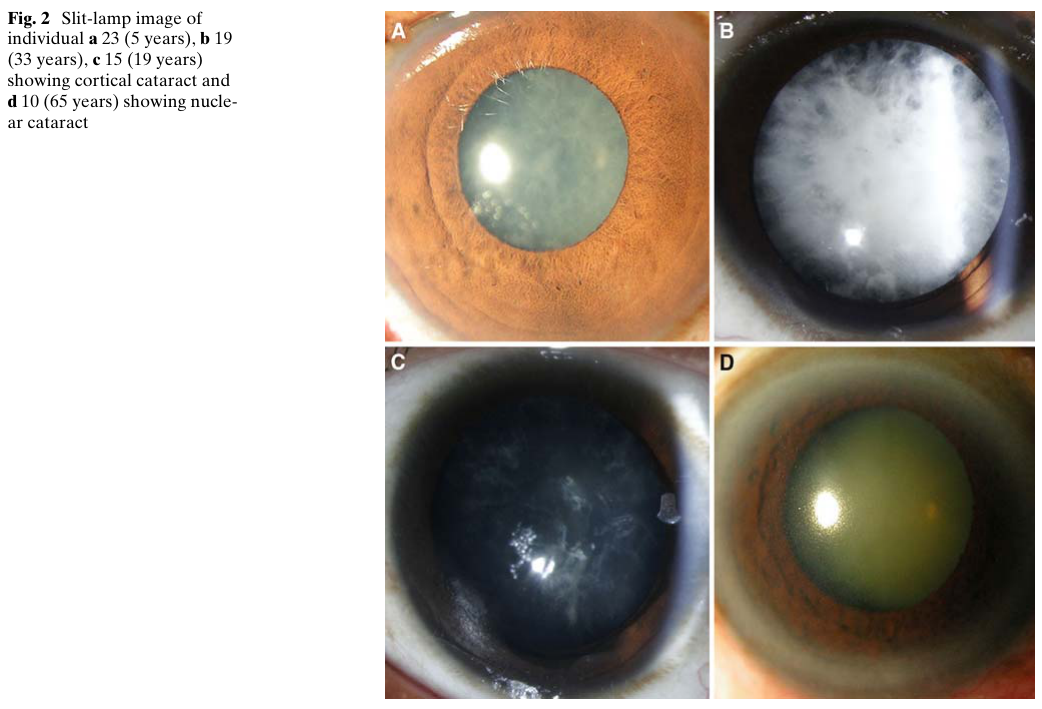
Reference Validation
Checked with linkml-reference-validator 0.3.0.
| Outcome | Count |
|---|---|
| References checked | 7 |
| Resolved | 7 |
| Unresolved (possible confabulation) | 0 |
| Unverifiable | 0 |
| References weighed for topical relevance | 7 |
| On topic | 3 |
| Off topic | 0 |
All extracted references resolved successfully.
Term Validation
Checked with linkml-term-validator 0.4.5, through the ols: adapter.
| Outcome | Count |
|---|---|
| Terms checked | 4 |
| Resolved | 4 |
| Unresolved (possible confabulation) | 0 |
| Obsolete | 0 |
| Unverifiable | 0 |
| Terms whose name was checked | 1 |
| Terms named correctly | 0 |
| Terms named as a different term | 1 |
Terms the report names something else
These identifiers resolve, so nothing about them looks wrong, and the ontology calls them something unrelated to what the report calls them. That usually means the identifier is not the one the sentence needs:
MONDO:0012665(3 mentions) - the report calls it "if available"; MONDO calls it cataract 33